. 2012 Jun 29;36(6):986-1002.
doi: 10.1016/j.immuni.2012.05.016. Epub 2012 Jun 7.

The receptor Ly108 functions as a SAP adaptor-dependent on-off switch for T cell help to B cells and NKT cell development

Affiliations

The receptor Ly108 functions as a SAP adaptor-dependent on-off switch for T cell help to B cells and NKT cell development

Robin Kageyama et al. Immunity. .

Abstract

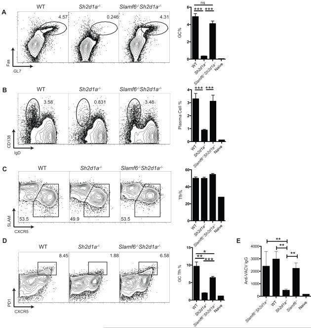
Humans and mice deficient in the adaptor protein SAP (Sh2d1a) have a major defect in humoral immunity, resulting from a lack of T cell help for B cells. The role of SAP in this process is incompletely understood. We found that deletion of receptor Ly108 (Slamf6) in CD4(+) T cells reversed the Sh2d1a(-/-) phenotype, eliminating the SAP requirement for germinal centers. This potent negative signaling by Ly108 required immunotyrosine switch motifs (ITSMs) and SHP-1 recruitment, resulting in high amounts of SHP-1 at the T cell:B cell synapse, limiting T cell:B cell adhesion. Ly108-negative signaling was important not only in CD4(+) T cells; we found that NKT cell differentiation was substantially restored in Slamf6(-/-)Sh2d1a(-/-) mice. The ability of SAP to regulate both positive and negative signals in T cells can explain the severity of SAP deficiency and highlights the importance of SAP and SHP-1 competition for Ly108 ITSM binding as a rheostat for the magnitude of T cell help to B cells.


Figures

Figure 1
Figure 1. Ly108 expression and function in an acute viral infection
Ly108 expression on splenic (a) effector (Th1) (CXCR5lo), Tfh (CXCR5hi), and GC Tfh (CXCR5hiPD1hi) CD44hi CD4+ T cells eight days post LCMV infection, and (b) Naïve B cells, plasma cells (CD19+CD138hiIgDlo), and germinal center B cells (CD19+GL7hiFashi) 8 days after LCMV infection. (c-d) CD4+ T cells (c) and B cells (d) from uninfected wild type (WT) and Slamf6−/− mice. (e) Slamf6+/+ and Slamf6−/− mice or (f) Slamf6−/− and SAP−/− mice were infected with LCMV and splenocytes were analyzed for GC B cells 8 days later. (g) WT, Slamf6+/-, and Slamf6−/− mice were infected with LCMV and virus specific serum IgG was measured day 30 post infection. (h) Slamf6−/− and SAP−/− mice were infected with LCMV and virus specific serum IgG was measured day 8 post infection. (a-h) Data are representative of 2 or more independent experiments. N = 4 or more per group. *** P < 0001. Error bars are SEM.
Figure 2
Figure 2. Loss of Ly108 eliminates the SAP expression requirement for germinal center development
(a-d) Wild type, Sh2d1a−/− and Slamf6−/−Sh2d1a−/− mice were infected with LCMV. Splenocytes were analyzed day 8 after infection. (a) Germinal center B cells (FashiGL7hi) and (b) plasma cells (IgDloCD138hi) are shown as a % of total B cells (CD19+CD4). (c) Tfh cells (CXCR5+SLAMlo) and (d) GC Tfh cells (CXCR5+PD1hi) are shown as a % of activated CD4+ T cells (CD44hiCD4+). Data are representative of 3 (a-c) or 2 (d) independent experiments. N = 5 per group. (e) Wild type, Sh2d1a−/−, Slamf6−/−, and Slamf6−/−Sh2d1a−/− mice were infected with vaccinia virus. Serum antibody titers were analyzed day 8 after infection. * P < 0.05, ** P < 0.005, *** P < 0001. n.s., no statistically significant difference (P > 0.05). Error bars are SEM.
Figure 3
Figure 3. Loss of Ly108 reverses the SAP requirement in CD4+ T cells for T cell help to B cells
(a-d) WT, Sh2d1a−/−, or Slamf6−/−Sh2d1a−/− CD45.1+ SM TCR transgenic CD4+ T cells were transferred into Sh2d1a−/− recipient mice subsequently infected with LCMV. Splenocytes were analyzed day 8 after infection. (a) Germinal center B cells (FashiGL7hi) and (b) plasma cells (CD138+IgDlo) shown as % of total B cells (CD19+CD4). (c) Tfh cells (CXCR5+) and (d) GC Tfh cells (CXCR5+PD1hi) shown as a % of SM (CD45.1+CD4+B220). (e-g) Slamf6−/−Sh2d1a−/− SM CD4+ T cells (CD45.1+) were transduced with Ly108-1, Ly108-2, SAP or GFP vector in vitro, transferred into Sh2d1a−/− recipient mice, and mice were infected with LCMV. (e) Germinal center B cells (FashiGL7hi) are shown 8 days after infection, as a % of total B cells. (f) Plasma cell (CD19+ IgDloCD138+) frequencies, as a % of total B cells. (g) MFI of Ly108 expression on Slamf6−/−Sh2d1a−/− SM CD4+ T cells transduced with empty vector (GFP) or Ly108 vector (Ly108-2), in comparison to endogenous Ly108 expression on WT SM cells. Data are representative of 4 (a-c,e) or 2 (d,f,g) independent experiments. N = 4 or more mice per group. * P < 0.05, ** P < 0.005, *** P < 0001. Error bars are SEM.
Figure 4
Figure 4. Slamf6−/−Sh2d1a−/− CD4+ T cells form stable conjugates with B cells, and ITSM phosphotyrosine motifs are required for inhibitory signals transmitted by Ly108 in vivo
(a-b) Conjugation efficiency of WT, Sh2d1a−/−, and Slamf6−/− Sh2d1a−/− SM CD4+ T cells with B cells pulsed with cognate peptide (LCMV gp66-77). (a) Representative flow cytometry plots, gated on CD4+ T cells. (b) Mean frequency of CD4+CD19+ conjugates in total CD4+ events. N = 2. (c-g) Roles of Ly108 tyrosines in vivo. (c-e) Slamf6−/−Sh2d1a−/− SM CD4+ T cells were transduced with GFP, Ly108-2 (“Ly108”), Ly108-Y3 mutant, or Ly108-AllF mutant RV and transferred into Sh2d1a−/− recipient mice. An additional group received Sh2d1a−/− SM cells transduced with RV-GFP. Mice were infected with LCMV and B cell responses in spleen were analyzed 8 days following infection. (c) Representative germinal center B cell FACS plots are shown, gated on total B cells (CD19+CD4). (d) Quantitation of GC B cells as gated in (c). (e) Quantitation of the plasma cell response. (f-g) Slamf6−/−Sh2d1a−/− SM -Ly108-Y1 mutant or - Ly108-Y2 mutant CD4+ T cells were transduced with RV-GFP, -Ly108-2 (“Ly108”), and transferred into Sh2d1a−/− recipient mice subsequently infected with LCMV. (f) Representative germinal center B cell FACS plots are shown, gated on total B cells (CD19+CD4), analyzed at day 8 after infection. (g) Quantitation of GC B cells as gated in (f). Data are representative of 2 independent experiments. N = 4 or more per group. * P < 0.05, ** P < 0.005 Error bars are SEM.
Figure 5
Figure 5. Ly108 recruits SHP-1 to the T:B immunological synapse
(a) Activated WT and Sh2d1a−/− AND TCR transgenic (PCC-specific) CD4+ T cells were lysed without stimulation or post-incubation with LPS-activated B cells that were untreated or pulsed with PCC peptide. Lysates were immunoprecipated for Ly108 and blotted for SHP-1 and Ly108. Total cell lysates (TCL) were examined for pERK activation and total ERK protein levels. NP = no peptide. (b-d) WT, Sh2d1a−/−, and Slamf6−/− Sh2d1a−/− SM CD4+ T cell conjugates with LPS-activated B cells pulsed with cognate peptide (LCMV gp66-77) were stained with Hoechst (blue), and antibodies to CD4 (white) and SHP-1 (green). (b) Cells were examined from the side as a confocal projection in the x-y plane (top row), at 45° (middle row), and 90° (en face, bottom row) rotations in the y-z plane. (c) Representative immunofluorescence images of SHP-1 localization at the immune synapse. (d) Quantification of SHP-1 localization at the immune synapse. Data represent two independent experiments with over 40 conjugates scored / genotype for each experiment. (e) Activated Slamf6−/− and Slamf6−/−Sh2d1a−/− SM CD4+ T cells expressing Ly108-GFP or Ly108-AllF-GFP (‘Ly108-AllF’) constructs were incubated with activated B cells pulsed with cognate peptide (LCMV gp66-77). Lysates were immunoprecipated for Ly108 and blotted for SHP-1 and Ly108. (f-h) Slamf6−/− and Slamf6−/−Sh2d1a−/− SM CD4+ T cells were transfected with either Ly108-GFP (‘WTLy108’) or Ly108-AllF-GFP, and conjugated to WT B cell targets pulsed with cognate peptide (LCMV gp66-77). Cells were stained with Hoechst (blue) and antibodies against SHP-1 (red). Green is Ly108-GFP fluorescence. (f) Quantitation of SHP-1 localization at the synapse. (g) Representative immunofluorescence images of cells expressing Ly108-GFP. (h) Representative immunofluorescence images of cells expressing Ly108-AllF-GFP. Further examples are shown in Figure S4. Data for each experiment depicted is representative of two or more experiments.
Figure 6
Figure 6. Inhibition of SHP-1 reverses the adhesion defect of SAP-deficient CD4+ T cells
(a-c) Activated WT, Sh2d1a−/−, Slamf6−/−, and Slamf6−/−Sh2d1a−/− SM CD4+ T cells were incubated with activated WT B cells pulsed with cognate peptide (LCMV gp66-77. 0, 0.01, or 1.0 μg/ml), in the presence or absence of SSG, followed by flow cytometry. (a) Representative flow cytometry plots, gated on CD4+ T cells. (b-c) Mean frequency of T:B conjugates in total CD4+ events, in the (b) absence or (c) presence of SSG. (d) CD4+ T cells incubated with activated Slamf6−/− B cells pulsed with cognate peptide (LCMV gp66-77. 0, 0.01, or 1.0 μg/ml). Data are shown from one of two experiments with equivalent results.
Figure 7
Figure 7. Ly108 provides both positive and negative signals for NKT cell development
WT, Slamf6−/−, Sh2d1a−/− and Slamf6−/−Sh2d1a−/− (‘DKO’) mice were analyzed for NKT cells. (a) Frequencies of splenic NKT cells, gated as shown in (c). (b) Absolute numbers of splenic NKT cells. (c) Representative flow cytometry plots of splenic NKT cells, gated on total CD4+ cells. Data are representative of 2 independent experiments. (d) Representative flow cytometry plots of thymic NKT cells, gated on total live CD8 cells. (e) Quantitation of (d), combining two independent experiments. (f) Developmental profiles of thymic NKT cells, gated as described in (d), from 1 of 2 representative experiments. (g) Representative flow cytometry plots of liver NKT cells, gated on total live CD19 mononuclear cells. (h) Quantitation of (g), combining two independent experiments. (i) Developmental profiles of liver NKT cells, gated as described in (g), from 1 of 2 representative experiments. ** P < 0.005, *** P < 0.0005. Error bars are SEM.

Comment in

References

    1. Allen CDC, Okada T, Cyster JG. Germinal-center organization and cellular dynamics. Immunity. 2007;27:190–202. - PMC - PubMed
    1. Barber DL, Wherry EJ, Masopust D, Zhu B, Allison JP, Sharpe AH, Freeman GJ, Ahmed R. Restoring function in exhausted CD8 T cells during chronic viral infection. Nature. 2006;439:682–687. - PubMed
    1. Bendelac A, Savage PB, Teyton L. The biology of NKT cells. Annual review of immunology. 2007;25:297–336. - PubMed
    1. Benhnia MR-E-I, McCausland MM, Laudenslager J, Granger SW, Rickert S, Koriazova L, Tahara T, Kubo RT, Kato S, Crotty S. Heavily isotype-dependent protective activities of human antibodies against vaccinia virus extracellular virion antigen B5. J Virol. 2009;83:12355–12367. - PMC - PubMed
    1. Berger SB, Romero X, Ma C, Wang G, Faubion WA, Liao G, Compeer E, Keszei M, Rameh L, Wang N, et al. SLAM is a microbial sensor that regulates bacterial phagosome functions in macrophages. Nat Immunol. 2010;11:920–927. - PMC - PubMed

Publication types

MeSH terms

Substances