

Review
doi: 10.1016/j.ceb.2012.05.005.
Epub 2012 Jun 8.
Centrosome asymmetry and inheritance during animal development
Affiliations
- PMID: 22683192
- PMCID: PMC3425708
- DOI: 10.1016/j.ceb.2012.05.005
Item in Clipboard
Review
Centrosome asymmetry and inheritance during animal development
Curr Opin Cell Biol.
2012 Aug.
Abstract
The centrosome is a subcellular organelle that is responsible for the majority of microtubule organization. Through this ability, the centrosome is involved in cell division, migration, and polarization. Recent studies have revealed intriguing asymmetries between mother and daughter centrioles as well as between mother and daughter centrosomes, and the involvement of such asymmetries in multiple cellular and developmental processes. This review aims to summarize recent discoveries on such asymmetries in centrioles/centrosomes and the potential implication of their inheritance patterns during cell division and development.
Copyright © 2012 Elsevier Ltd. All rights reserved.
Figures
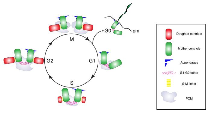
Schematic representation of the centrosome/centriole duplication cycle in animal cells. The centrosome consists of mother (green) and daughter centrioles (red), that are interconnected by S-M tethers and embedded in pericentriolar material (PCM), which anchors microtubules. The cycle can be separated in four slightly overlapping phases. i) Centriole disengagement, ii) centriole duplication, iii) centrosome maturation and iv) centrosome separation followed by spindle assembly. The mother centriole can be distinguished by the presence of appendages. During disengagement centrioles lose their orthogonal arrangement through the removal of the S-M linker. During S phase, procentrioles forms perpendicular to each mother centriole. During S-G2 phases, the daughter centrioles continue to elongate. In late S-phase G2, the centrosome increases in size and the newly formed centriole pairs disconnect through the disassembly of the G1-G2 tether, so that at the onset of G2/M the two centrosomes move to opposite sides of the cell and establish the two mitotic spindle poles. At the end of the cycle, the daughter centrioles acquire appendages and behave as a mother centriole during the subsequent cycle. When cell exit the cell cycle and enter G0, centrioles move to the plasma membrane (pm) and assemble cilia.
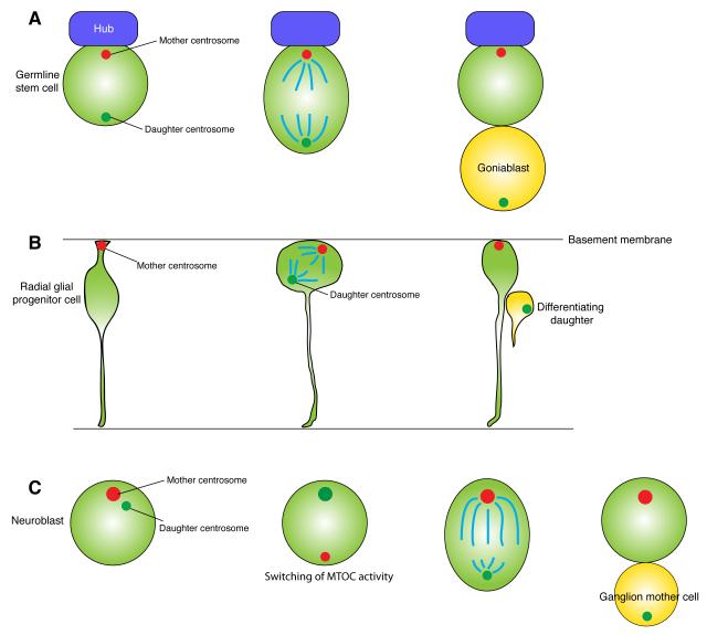
A. Drosophila male germline stem cells (GSCs) inherit the mother centrosome upon division. The mother centrosome is anchored to the interface between the GSC and the hub cells. The hub secretes the signaling ligand (Unpaired, Upd) to specify GSC identity. B. Mouse radial glial progenitor cells inherit the mother centrosome upon division. The mother centrosome is located near the basal membrane throughout the cell cycle, even during interkinetic nuclear migration. This mother centrosome is also involved in primary cilia formation of radial glial progenitor cells. C. Drosophila neuroblasts inherit the daughter centrosome upon division. During each cell cycle, the mother centrosome is inactivated, while the daughter acquires robust microtubule organizing activity.
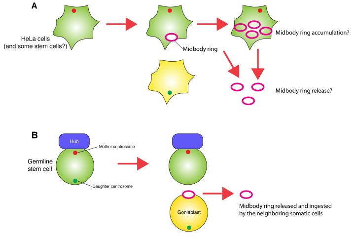
A. The cell with the mother centrosome inherits the mother centrosome and midbody ring. Stem cells and cancer cells were reported to accumulate and/or release the midbody ring (see the text for detail). B. Drosophila male GSCs, which inherit the mother centrosome, exclude the midbody ring upon division. The midbody ring that is inherited by the differentiating gonialblast is eventually released and ingested by neighboring somatic cells.
References
-
- Andersen JS, Wilkinson CJ, Mayor T, Mortensen P, Nigg EA, Mann M. Proteomic characterization of the human centrosome by protein correlation profiling. Nature. 2003;426:570–574. - PubMed
-
- Palazzo RE, Vogel JM, Schnackenberg BJ, Hull DR, Wu X. Centrosome maturation. Curr Top Dev Biol. 2000;49:449–470. - PubMed
-
- Tsou MF, Stearns T. Mechanism limiting centrosome duplication to once per cell cycle. Nature. 2006;442:947–951. - PubMed
-
- Tsou MF, Stearns T. Controlling centrosome number: licenses and blocks. Curr Opin Cell Biol. 2006;18:74–78. - PubMed
Publication types
MeSH terms
Grants and funding
LinkOut - more resources
Full Text Sources
