. 2012 Jul 6;11(1):100-9.
doi: 10.1016/j.stem.2012.05.018. Epub 2012 Jun 7.

Direct reprogramming of mouse and human fibroblasts into multipotent neural stem cells with a single factor

Affiliations

Direct reprogramming of mouse and human fibroblasts into multipotent neural stem cells with a single factor

Karen L Ring et al. Cell Stem Cell. .

Abstract

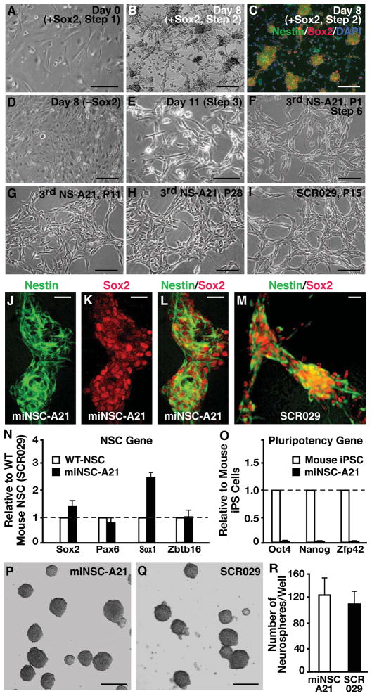
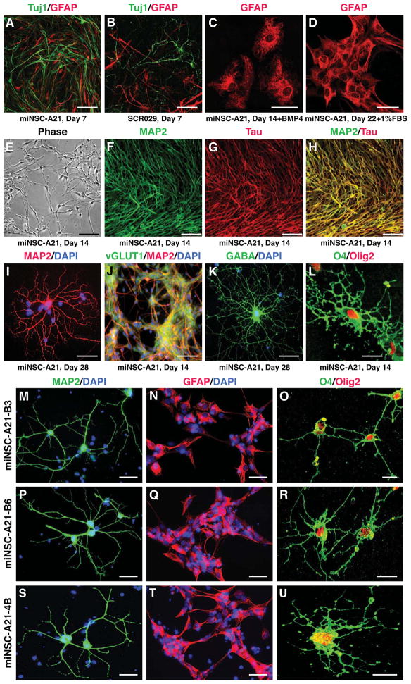
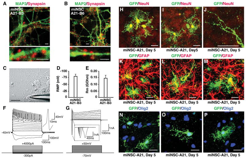
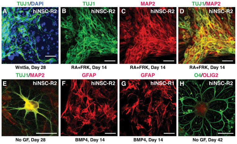
The generation of induced pluripotent stem cells (iPSCs) and induced neuronal cells (iNCs) from somatic cells provides new avenues for basic research and potential transplantation therapies for neurological diseases. However, clinical applications must consider the risk of tumor formation by iPSCs and the inability of iNCs to self-renew in culture. Here we report the generation of induced neural stem cells (iNSCs) from mouse and human fibroblasts by direct reprogramming with a single factor, Sox2. iNSCs express NSC markers and resemble wild-type NSCs in their morphology, self-renewal, ability to form neurospheres, and gene expression profiles. Cloned iNSCs differentiate into several types of mature neurons, as well as astrocytes and oligodendrocytes, indicating multipotency. Implanted iNSCs can survive and integrate in mouse brains and, unlike iPSC-derived NSCs, do not generate tumors. Thus, self-renewable and multipotent iNSCs without tumorigenic potential can be generated directly from fibroblasts by reprogramming.


Figures

Figure 1
Figure 1. Generation and characterization of iNSCs from mouse fibroblasts
(A) Phase-contrast image of MEFs after overnight treatment with Sox2 retrovirus in fibroblast medium. (B) Sox2-infected cells in NSC medium with growth factors generate networks and colonies on gelatin-coated glass coverslips by 8 days after infection. (C) Sox2-transformed colonies are positive for the NSC markers Nestin and Sox2. (D) Fibroblasts cultured in NSC medium with growth factors but without Sox2 retroviral transduction do not generate colonies or networks. (E) Sox2-transduced cells after 11 days have drastically different morphology from their fibroblast counterparts. (F) After three rounds of neurosphere generation, reprogrammed cells take on the characteristic bipolar NSC morphology. (G) After multiple passages as a monolayer, NSC-like cells are a morphologically homogenous population. (H) Morphology of NSC-like cells stays the same over prolonged passaging, and reprogrammed cells can proliferate over 28 passages. (I) The morphology of NSC-like cells is similar to that of wild-type cortical-derived NSCs such as the commercial cell line SCR029 (Millipore). (J–M) For the miNSC-A21 cell line, expression of Nestin and Sox2 is similar to that of brain-derived wild-type NSCs as revealed by immunostaining. (N and O) qRT-PCR reveals that miNSC-A21 express typical NSC markers (N), but do not express pluripotency related genes (O). Error bars denote standard deviation of triplicate reactions. (P–R) In suspension culture, miNSC-A21 generates neurospheres similar to wild-type NSCs and with similar efficiency (n=3). Scale bars = 50 μm in A and D–I; scale bars = 100 μm in B and C; scale bars = 50 μm in J–M; scale bars = 100 μm in P and Q. See also Figures S1 and S2 and Tables S1, S2, and S4.
Figure 2
Figure 2. Multipotency of miNSCs in vitro
(A and B) Like wild-type NSCs, miNSC-A21 can differentiate into Tuj1+ neurons and GFAP+ astrocytes by 7 days in culture after growth factor withdrawal. (C and D) miNSC-A21 can robustly generate GFAP+ astrocytes by 14 days in vitro in the presence of BMP4 or FBS. (E) miNSC-A21 can generate mature looking neurons and neuronal networks by 14 days in culture without growth factors. (F–H) Neurons derived from miNSC-A21 stain positive for the mature neuronal markers MAP2 and Tau. (I) miNSC-A21 can differentiate into mature arborized neurons by 28 days in vitro. (J and K) miNSC-A21 can differentiate into subtypes of neurons, including excitatory vGluT1+ neurons (J) and inhibitory GABA+ neurons (K). (L) miNSC-A21 can generate O4+ and Olig2+ oligodendrocytes by 14 days in culture. (M–U) After 14 days in culture, subcloned lines B3 (M–O), B6 (P–R), and 4B (S–U) of miNSC-A21 can differentiate into MAP2+ mature neurons (M, P, S), GFAP+ astrocytes (N, Q, T), and O4+/Olig2+ oligodendrocytes (O, R, U). Scale bars = 50 μm in A, B, E, and J; scale bars = 25 μm in C and D; scale bars = 75 μm in F–H; scale bars = 10 μm in I, K, L, and M–U. See also Figure S3.
Figure 3
Figure 3. miNSC-derived functional neurons in vitro and multipotency of miNSCs in vivo
(A and B) Neurons derived from subclones miNSC-A21-B3 or miNSC-A21-B6 at 14 days in culture express MAP2 (green) and Synapsin (red), a presynaptic marker of mature neurons. (C) A patched neuron derived from miNSC-A21-B3 at 17 days in culture. (D and E) Whole-cell capacitance and membrane resistance of neurons derived from miNSC-A21 were determined from a transient 5-mV hyperpolarizing step from a holding potential of −70 mV. (F) Current-clamp recordings of neurons derived from miNSC-A21 at −40mV reveal action potentials with stepwise current injection. (G) Voltage-clamp recordings of neurons derived from miNSC-A21 reveal both fast inactivating inward and outward currents indicating functional voltage-dependent Na+ and K+ channels. (H–P) GFP-labeled miNSC-A21 were grown in suspension cultures for one day to generate small neurospheres and then microinjected into the cortex of P2–3 wild-type pups. Five days after transplantation, mouse brains were collected, fixed, sectioned, and immunostained. (H–J) Immunostainings reveal that miNSC-A21 can differentiate into NeuN+ neurons (H and I) with mature looking dendritic spines (J) in vivo. (K–M) miNSC-A21 can also differentiate into GFAP+ astrocytes in vivo. (N–P) miNSC-A21 can also differentiate into Olig2+ oligodendrocytes in vivo. Scale bars = 2 μm in A and B; scale bar = 10 μm in C; scale bars = 10 μm in H–P. See also Table S3.
Figure 4
Figure 4. Multipotency of hiNSCs in vitro
(A) hiNSCs can differentiate into TUJ1+ immature neurons in the presence of the signaling protein WNT5A by 28 days in culture. (B) The addition of retinoic acid (RA) and forskolin (FRK) to neuronal differentiation conditions pushed hiNSCs to differentiate into TUJ1+ neurons by 14 days in vitro. (C and D) hiNSCs can generate TUJ1+/MAP2+ neurons by 14 days in the presence of RA and FRK. (E) hiNSC can generate mature looking neurons that are MAP2+ by 28 days in vitro in hNSC medium without growth factors. (F) hiNSCs can also generate GFAP+ astrocytes in the presence of BMP4 by 14 days. (G) A separate hiNSC line can also robustly generate GFAP+ astrocytes at 14 days in vitro. (H) hiNSCs can generate O4+/OLIG2+ oligodendrocytes by 40 days in culture in hNSC medium lacking growth factors. Scale bars = 20 μm in A–D; scale bars = 10 μm in E–H. See also Figure S4.

Similar articles

Cited by

References

    1. Ambasudhan R, Talantova M, Coleman R, Yuan X, Zhu S, Lipton SA, Ding S. Direct reprogramming of adult human fibroblasts to functional neurons under defined conditions. Cell Stem Cell. 2011;9:113–118. - PMC - PubMed
    1. Brennand KJ, Simone A, Jou J, Gelboin-Burkhart C, Tran N, Sangar S, Li Y, Mu Y, Chen G, Yu D, et al. Modelling schizophrenia using human induced pluripotent stem cells. Nature. 2011;473:221–225. - PMC - PubMed
    1. Bylund M, Andersson E, Novitch BG, Muhr J. Vertebrate neurogenesis is counteracted by Sox1–3 activity. Nat Neurosci. 2003;6:1162–1168. - PubMed
    1. Caiazzo M, Dell’Anno MT, Dvoretskova E, Lazarevic D, Taverna S, Leo D, Sotnikova TD, Menegon A, Roncaglia P, Colciago G, et al. Direct generation of functional dopaminergic neurons from mouse and human fibroblasts. Nature. 2011;476:224–227. - PubMed
    1. Dimos JT, Rodolfa KT, Niakan KK, Weisenthal LM, Mitsumoto H, Chung W, Croft GF, Saphier G, Leibel R, Goland R, et al. Induced pluripotent stem cells generated from patients with ALS can be differentiated into motor neurons. Science. 2008;321:1218–1221. - PubMed

Publication types

Substances