. 2012 Jul 27;47(2):291-305.
doi: 10.1016/j.molcel.2012.05.004. Epub 2012 Jun 7.

A death effector domain chain DISC model reveals a crucial role for caspase-8 chain assembly in mediating apoptotic cell death

Affiliations

A death effector domain chain DISC model reveals a crucial role for caspase-8 chain assembly in mediating apoptotic cell death

Laura S Dickens et al. Mol Cell. .

Abstract

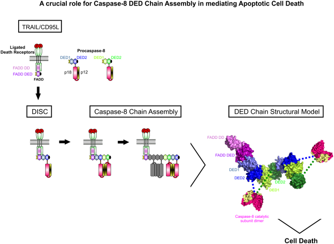
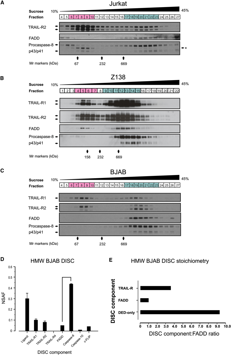
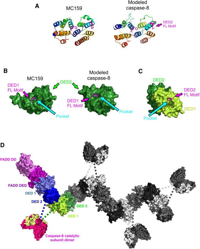
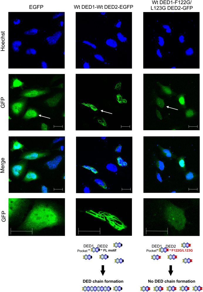
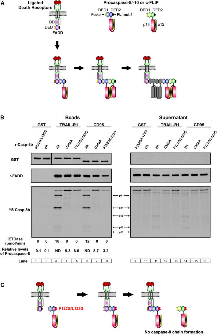
Formation of the death-inducing signaling complex (DISC) is a critical step in death receptor-mediated apoptosis, yet the mechanisms underlying assembly of this key multiprotein complex remain unclear. Using quantitative mass spectrometry, we have delineated the stoichiometry of the native TRAIL DISC. While current models suggest that core DISC components are present at a ratio of 1:1, our data indicate that FADD is substoichiometric relative to TRAIL-Rs or DED-only proteins; strikingly, there is up to 9-fold more caspase-8 than FADD in the DISC. Using structural modeling, we propose an alternative DISC model in which procaspase-8 molecules interact sequentially, via their DED domains, to form a caspase-activating chain. Mutating key interacting residues in procaspase-8 DED2 abrogates DED chain formation in cells and disrupts TRAIL/CD95 DISC-mediated procaspase-8 activation in a functional DISC reconstitution model. This provides direct experimental evidence for a DISC model in which DED chain assembly drives caspase-8 dimerization/activation, thereby triggering cell death.


Figures

None
Graphical abstract
Figure 1
Figure 1
Western Blot and LC-MS/MS Analysis of TRAIL DISC Composition in BJAB, Jurkat, or Z138 Cells (A) Schematic for activation and affinity purification of total, HMW, or soluble TRAIL DISC: BJAB, Jurkat, or Z138 cells were stimulated with either biotin-labeled (bTRAIL) or Strep-II-tagged (stTRAIL) TRAIL under conditions that induced maximal DISC formation. Cleared cell lysates (Figure S1) were subjected to one of three protocols (Expt A, Expt B, or Expt C). Expt A allowed the purification of all ligand-bound complexes, and Expt B was used to separate low molecular weight ligand-receptor complexes from high molecular weight (HMW) DISC, while Expt C permitted independent isolation and analysis of lipid raft-associated (LR) fractions and soluble TRAIL DISC (S-DISC). Bead-captured complexes and LR fractions were separated by SDS-PAGE and analyzed by mass spectrometry or western blotting. (B) Cells were stimulated with bTRAIL for 1 hr at 4°C and further incubated at 37°C for 10 (BJAB) or 25 (Jurkat and Z138) min, lysed, and the TRAIL DISC isolated. DISC-containing eluates and cleared lysate supernatants (1% of total input) were separated by SDS-PAGE for western blot of the known components. Film exposures for each protein (DISC and supernatants) are the same across the three cell lines except in the case of FADD, where 10-fold longer film exposures were required for detection of FADD in the DISC compared to supernatants. Asterisk denotes residual signal from caspase-8 antibody. TRAIL stim., TRAIL treated; TRAIL p.lysis, TRAIL added to cleared lysates to capture TRAIL-Rs. (C–E) Graphical analysis of TRAIL DISC mass spectrometry data. Proteins identified by shotgun proteomics of TRAIL DISC purified from BJAB (C), Jurkat (D), or Z138 (E) cells that fulfilled the necessary criteria were plotted according to median protein probability (PP) and median spectral abundance factor (SAF) relative to TRAIL-R2. n = 3 for BJAB and Jurkat, n = 6 for Z138.
Figure 2
Figure 2
TRAIL DISC Is Predominantly Formed in the Soluble Cellular Fraction Rather Than in Lipid Rafts (A) Cullin-3 is associated with TRAIL DISC formed in the epithelial HeLa cell line. HeLa, BJAB, and Z138 cells were stimulated with bTRAIL for 1 hr at 4°C and further incubated at 37°C for 10 (BJAB) or 25 (Z138 and HeLa) min and the TRAIL DISC isolated. DISC-containing eluates were separated by SDS-PAGE for western blot. Blots for the known components of the BJAB and Z138 DISC are shown in Figure 1B. Exposure times for cullin-3 are the same across the three cell lines. (B) Cullin-3 is predominantly found in the LR fraction (green) of hematopoietic cell lines and is not associated with S-DISC (blue). Z138 or BJAB cells were stimulated with bTRAIL (1 hr at 4°C and 25 min at 37°C) and the lysate separated into soluble and lipid raft fractions. Equivalent quantities of the LR fraction or TRAIL DISC isolated from the soluble fraction (S-DISC) were subjected to SDS-PAGE and western blotting for cullin-3. (C) TRAIL DISC is predominantly found in the soluble fraction of hematopoietic cell lines. Samples prepared as in (B) were analyzed for core DISC components by western blotting. Blots shown are representative of n = 3.
Figure 3
Figure 3
Mass Spectrometry Reveals an Alternative Stoichiometry for the TRAIL DISC (A–C) Normalized spectral abundance factor (NSAF) analysis of mass spectrometry data obtained for the TRAIL DISC isolated from total BJAB (A), Jurkat (B), or Z138 (C) lysate (n = 3 for BJAB and Jurkat, n = 6 for Z138; error bars are SEM). (D) Each DISC component was assigned to a category (receptors, FADD or DED-only) and the NSAF values combined. Total NSAF values were corrected to FADD to produce the stoichiometry of the TRAIL DISC from total cellular lysates. (E) Relative total levels of core DISC components in BJAB, Jurkat, and Z138 cells. Equivalent quantities of total cellular protein were subjected to SDS-PAGE and analyzed for core DISC components by western blotting. GAPDH served as loading control.
Figure 4
Figure 4
A High Molecular Weight TRAIL DISC Is Formed in BJAB, Jurkat, and Z138 Cells (A–C) Jurkat (A), Z138 (B), or BJAB (C) cells were treated with bTRAIL for 1 hr at 4°C and further incubated for 10 (BJAB) or 25 (Jurkat and Z138) min at 37°C. DISC was isolated from each fraction of a continuous 10%–45% sucrose density gradient. Fractions highlighted in pink contain low molecular weight ligand-receptor complexes, while blue fractions contain high molecular weight (HMW) DISC. Asterisk denotes residual signal from TRAIL-R2 antibody. Blots shown are representative of n = 3 (BJAB and Z138) or n = 2 (Jurkat). Molecular weight (Mr) markers (kDa) are indicated (n = 3). (D) Normalized spectral abundance factor (NSAF) analysis of LC-MS/MS data obtained for the HMW BJAB TRAIL DISC (Figure 4C, fractions 14–27) (n = 2; error bars, range). (E) Combined NSAF values for HMW DISC components corrected to FADD as in Figure 3D.
Figure 5
Figure 5
Structural Modeling of Procaspase-8 DED Chain Formation within the DISC (A) Published structure for MC159 used by Phyre to structurally model the DEDs of procaspase-8. (B) Surface structure of MC159 DED2 and modeled procaspase-8 DED2 showing the FL motif from DED1 interacting with a pocket on the surface of DED2. (C) Modeled surface structure of caspase-8 DEDs showing the pocket in DED1 that could interact with the FL motif from either FADD or DED2 of another molecule. The interaction interface between adjacent tandem DEDs was modeled using the intramolecular interface between DED1 and DED2. (D) Structural modeling of the interactions that may occur between the DEDs of FADD and caspase-8. Interactions between FADD (structure 2GF5) and procaspase-8 (or between adjacent sets of procaspase-8 DEDs) were modeled using the intramolecular interface between DED1 and DED2, resulting in a DED chain. The colored subunits of the model correspond to the model of the interaction between FADD DED and one dimer of procaspase-8. The extended chain is shown in gray. Catalytic subunit dimers (p182/p102, modeled from structure 3KJQ) are shown to indicate how we would expect antiparallel dimers to form along the length of the DED chain (dotted lines represent the linker between DED2 and p18 subunit of procaspase-8). Note that the helical character of the DED chain is uncertain, since small changes in the interface would significantly change the long-range topology.
Figure 6
Figure 6
Mutation of the FL Motif in Caspase-8 DED2 Prevents Death Effector Filament Formation HeLa cells were transfected with either empty vector (EGFP) or the caspase-8 DED variants DED1-DED2-EGFP or DED1-DED2 F122G/L123G-EGFP for 20 hr before fixing and staining with Hoechst. Cells were imaged using a Zeiss LSM510 confocal microscope, and a representative field for each transfection is shown. Lower panels show enlargement of those areas arrowed in the GFP panels. Scale bar, 20 μm.
Figure 7
Figure 7
A Paradigm-Changing Model for Recruitment of Procaspase-8 and Other DED-Only Proteins to the DISC (A) Upon stimulation by the appropriate death ligand, death receptors recruit FADD via their death domains (DDs). FADD in turn recruits procaspase-8 (or procaspase-10 or c-FLIP) through a DED-mediated interaction. FADD may initially recruit one molecule of procaspase-8 via a single interaction with DED1 of procaspase-8 (for other permutations of this model, see Figure S4). The exposed DED2 FL motif then recruits additional molecules to produce a chain, facilitating dimerization and full activation of caspase-8/10. (B) Mutation of the DED2 FL motif reduces procaspase-8 recruitment and prevents processing in the in vitro DISC reconstitution model. Reconstituted DISC was formed with GST-CD95-IcD or GST-TRAIL-R1-ICD; recombinant FADD; and 35S-labeled IVT wild-type (WT), active site mutant (C360A), or F122G/L123G mutant caspase-8. Reconstituted DISC and supernatants were assessed for GST (loading control), 35S-labeled caspase-8, FADD, and IETDase activity. The relative levels of procaspase-8 recruited to the complex were determined by densitometry (corrected relative to GST-receptor-IcD). (C) Model depicting the potential impact of mutating the FL motif in procaspase-8 DED2. F122G/L123G does not prevent binding of caspase-8 to FADD but limits recruitment of additional procaspase-8 molecules, thus preventing DED chain formation and dimerization/activation of procaspase-8 catalytic subunits within the DISC.

References

    1. Barbero S., Barila D., Mielgo A., Stagni V., Clair K., Stupack D. Identification of a critical tyrosine residue in caspase 8 that promotes cell migration. J. Biol. Chem. 2008;283:13031–13034. - PMC - PubMed
    1. Berglund H., Olerenshaw D., Sankar A., Federwisch M., McDonald N.Q., Driscoll P.C. The three-dimensional solution structure and dynamic properties of the human FADD death domain. J. Mol. Biol. 2000;302:171–188. - PubMed
    1. Bertrand M.J., Vandenabeele P. The Ripoptosome: death decision in the cytosol. Mol. Cell. 2011;43:323–325. - PubMed
    1. Blondeau F., Ritter B., Allaire P.D., Wasiak S., Girard M., Hussain N.K., Angers A., Legendre-Guillemin V., Roy L., Boismenu D. Tandem MS analysis of brain clathrin-coated vesicles reveals their critical involvement in synaptic vesicle recycling. Proc. Natl. Acad. Sci. USA. 2004;101:3833–3838. - PMC - PubMed
    1. Boatright K.M., Renatus M., Scott F.L., Sperandio S., Shin H., Pedersen I.M., Ricci J.E., Edris W.A., Sutherlin D.P., Green D.R., Salvesen G.S. A unified model for apical caspase activation. Mol. Cell. 2003;11:529–541. - PubMed

Publication types

Substances