Review
. 2012 Oct;52(3-4):155-64.
doi: 10.1016/j.molimm.2012.05.004. Epub 2012 Jun 7.

T cell antigen recognition at the cell membrane

Affiliations
Review

T cell antigen recognition at the cell membrane

Jun Huang et al. Mol Immunol. 2012 Oct.

Abstract

T cell antigen receptors (TCRs) on the surface of T cells bind specifically to particular peptide bound major histocompatibility complexes (pMHCs) presented on the surface of antigen presenting cells (APCs). This interaction is a key event in T cell antigen recognition and activation. Most studies have used surface plasmon resonance (SPR) to measure the in vitro binding kinetics of TCR-pMHC interactions in solution using purified proteins. However, these measurements are not physiologically precise, as both TCRs and pMHCs are membrane-associated molecules which are regulated by their cellular environments. Recently, single-molecule förster resonance energy transfer (FRET) and single-molecule mechanical assays were used to measure the in situ binding kinetics of TCR-pMHC interactions on the surface of live T cells. These studies have provided exciting insights into the biochemical basis of T cell antigen recognition and suggest that TCRs serially engage with a small number of antigens with very fast kinetics in order to maximize TCR signaling and sensitivity.


Figures

Figure 1
Figure 1. Important molecules on and underneath either the T cell or APC surface
TCRs recognize antigen pMHCs on the cell membrane with the help of co-receptors and accessory molecules. These molecules play critical roles in the T cell recognition and the function of each of which is discussed in the text. Also depicted are possible regulatory mechanisms of TCR-pMHC interaction by the T cell membrane and intracellular structures.
Figure 2
Figure 2. Schematic of SPR for measuring the in vitro 3D kinetics of TCR-pMHC interactions
Interaction between purified soluble TCRs in solution and immobilized pMHCs on a sensor chip is measured by SPR angle shifts when the mass of the surface layer changes.
Figure 3
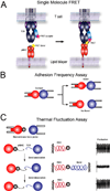
Figure 3. 2D methods for measuring in situ TCR-pMHC interactions
(A) Single-molecule FRET. A TCR on the T cell membrane is labeled with a single chain antibody fragment conjugated with a FRET acceptor, and the peptide on a pMHC anchored to the lipid bilayer is labeled with a FRET donor. The TCR-pMHC interaction brings the donor and acceptor into close distance to trigger FRET. (B) Adhesion frequency assay. A T cell (right) is brought in and out of contact with a pMHC coated RBC (left) to estimate an adhesion frequency using micropipette manipulation. The presence or absence of RBC deformation signifies whether a TCR-pMHC bond is present on T cell retraction. (C) Thermal fluctuation assay. A T cell (right) is brought into close proximity to a pMHC coated bead attached to a RBC (left). Single bond association and dissociation events are detected by monitoring the thermal fluctuation amplitude of the bead that is either restrained by only the RBC or the combination of the RBC and the single TCR-pMHC bond.
Figure 4
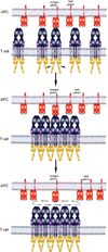
Figure 4. A fast kinetics based serial triggering model
TCR recognition of an antigenic pMHC leads to CD3 ITAM phosphorylation and signaling. The TCR ligation signal promotes the formation of TCR protein islands/clusters on the T cell membrane. The fast kinetics of TCR-pMHC interaction allows the TCRs in the cluster to serially engage with a small number of antigens on the APC surface and maximize T cell signaling and activation.

References

    1. Alam SM, Travers PJ, Wung JL, Nasholds W, Redpath S, Jameson SC, Gascoigne NR. T-cell-receptor affinity and thymocyte positive selection. Nature. 1996;381:616–620. - PubMed
    1. Alegre ML, Frauwirth KA, Thompson CB. T-cell regulation by CD28 and CTLA-4. Nat Rev Immunol. 2001;1:220–228. - PubMed
    1. Aleksic M, Dushek O, Zhang H, Shenderov E, Chen JL, Cerundolo V, Coombs D, van der Merwe PA. Dependence of T cell antigen recognition on T cell receptor-peptide MHC confinement time. Immunity. 2011;32:163–174. - PMC - PubMed
    1. Altschuh D, Dubs MC, Weiss E, Zeder-Lutz G, Van Regenmortel MH. Determination of kinetic constants for the interaction between a monoclonal antibody and peptides using surface plasmon resonance. Biochemistry. 1992;31:6298–6304. - PubMed
    1. Artyomov MN, Lis M, Devadas S, Davis MM, Chakraborty AK. CD4 and CD8 binding to MHC molecules primarily acts to enhance Lck delivery. Proc Natl Acad Sci U S A. 2010;107:16916–16921. - PMC - PubMed

Publication types

MeSH terms