. 2012 Aug;38(3):294-300.
doi: 10.1097/SHK.0b013e31825f5fb2.

Vagal nerve stimulation blocks peritoneal macrophage inflammatory responsiveness after severe burn injury

Affiliations

Vagal nerve stimulation blocks peritoneal macrophage inflammatory responsiveness after severe burn injury

Nicole E Lopez et al. Shock. 2012 Aug.

Abstract

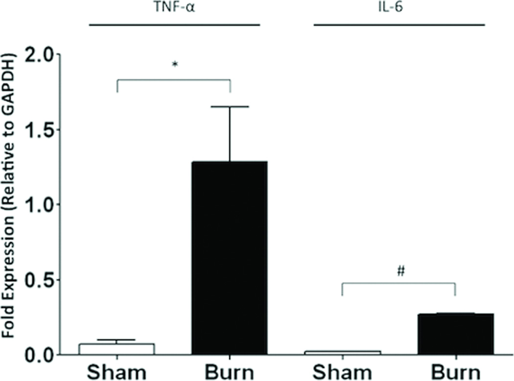
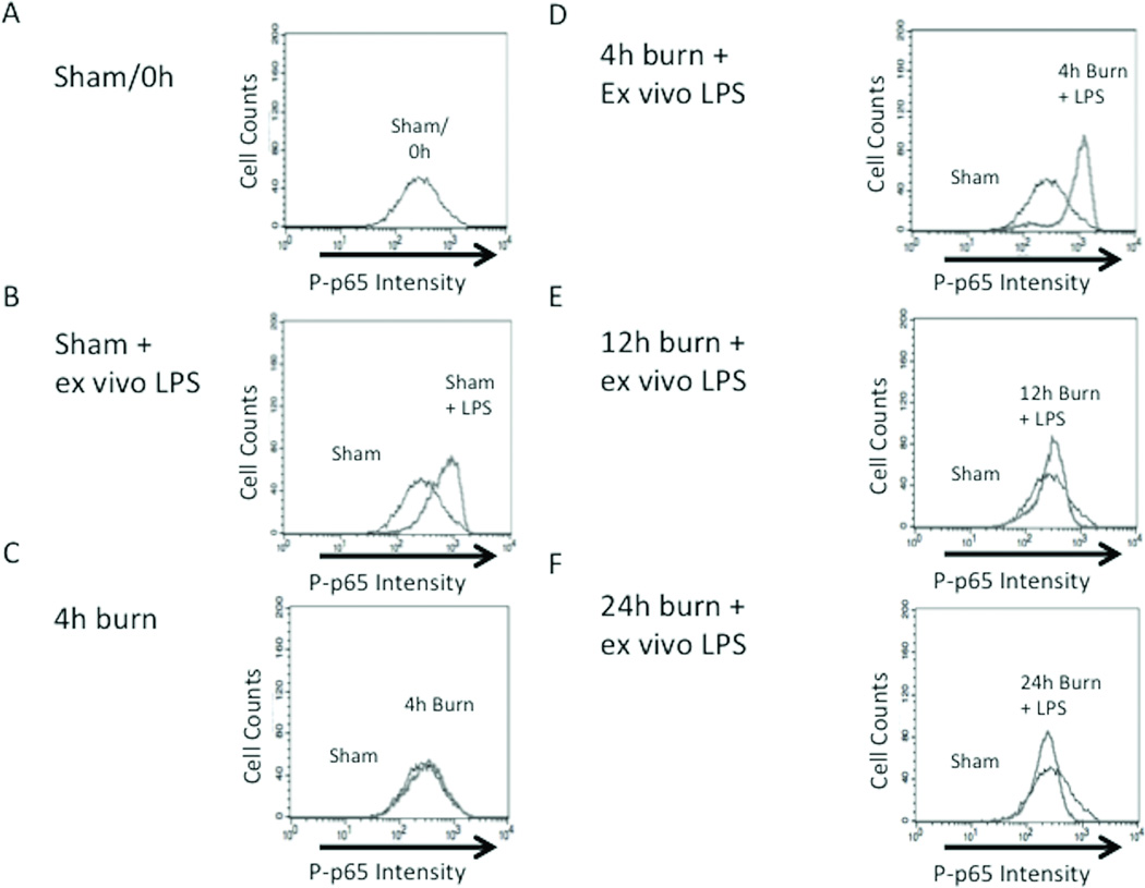
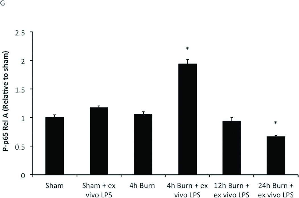
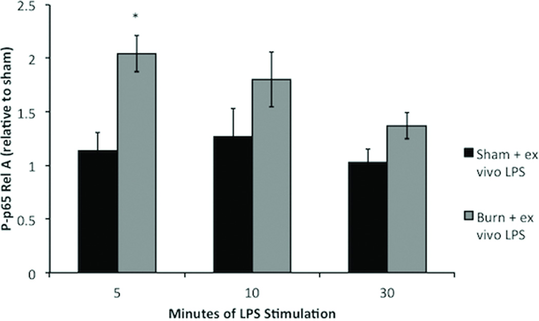
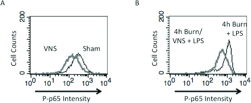
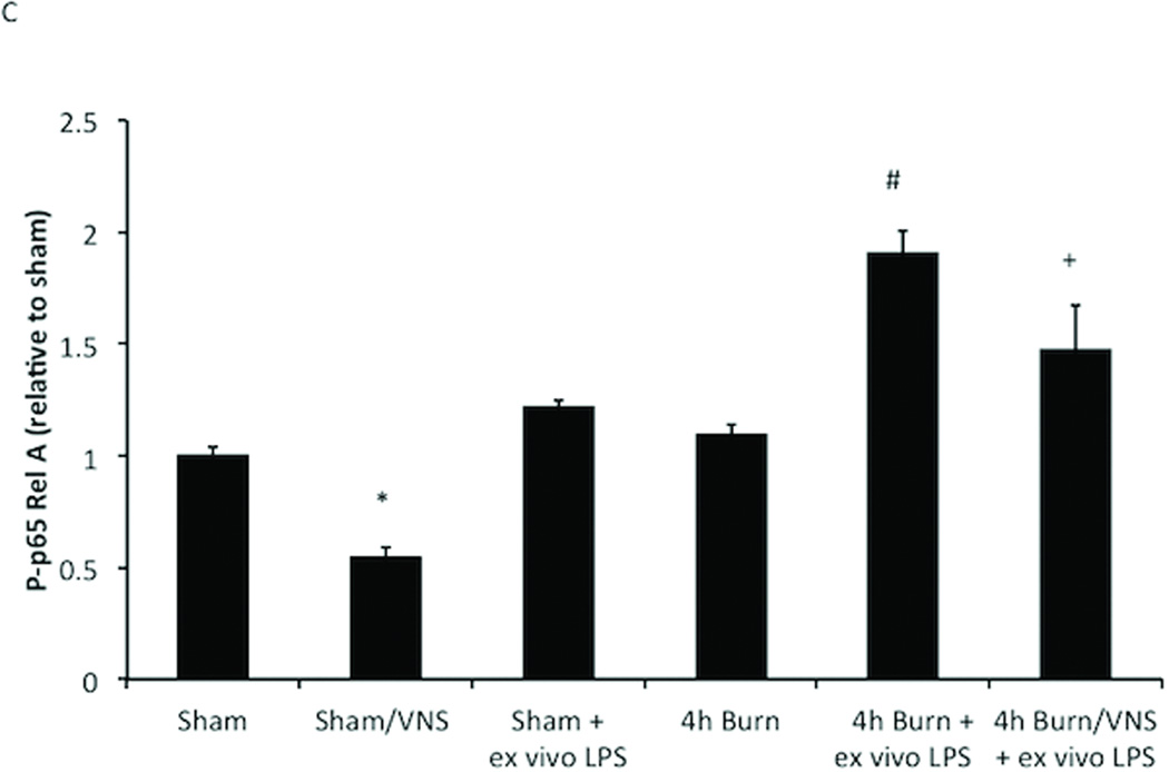
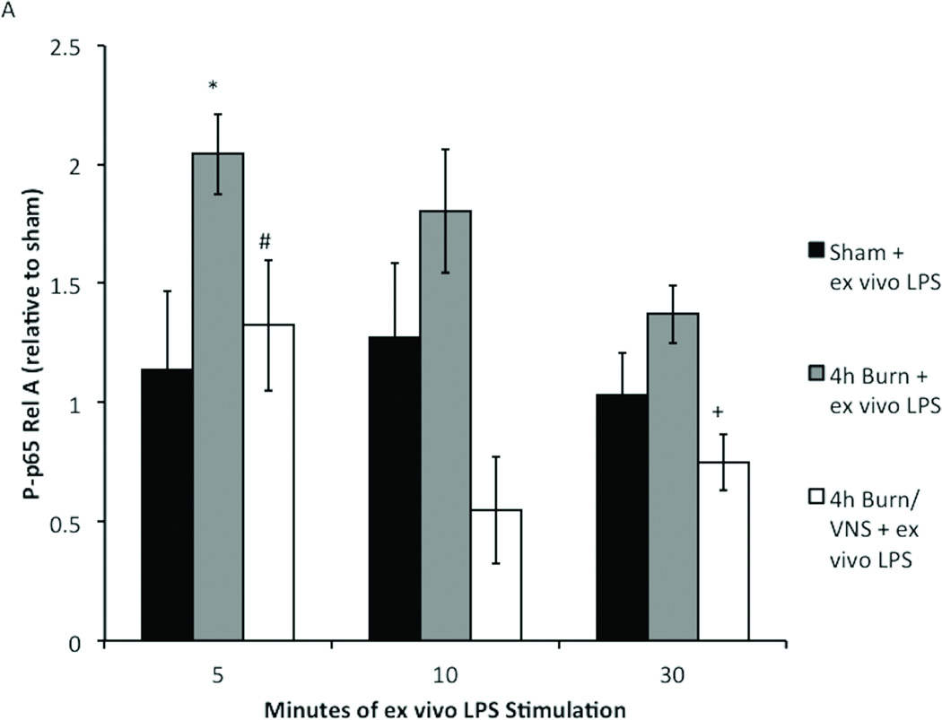
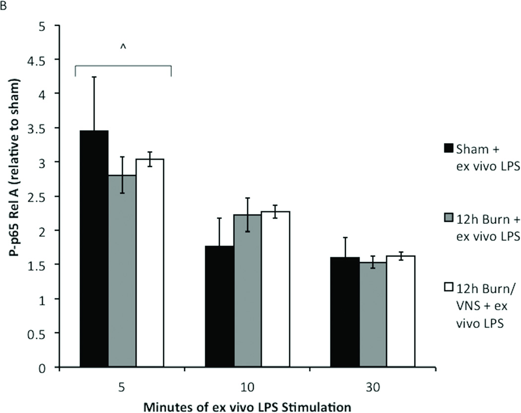
Large surface area burn injuries lead to activation of the innate immune system, which can be blocked by parasympathetic inputs mediated by the vagus nerve. We hypothesized that vagal nerve stimulation (VNS) would alter the inflammatory response of peritoneal macrophages after severe burn injury. Male BALB/c mice underwent right cervical VNS before 30% total body surface area steam burn and were compared with animals subjected to burn alone. Peritoneal macrophages were harvested at several time points following injury and exposed to lipopolysaccharide (LPS) in culture conditions. The inflammatory response of peritoneal macrophages was measured by analyzing changes in nuclear factor κB p65 phosphorylation using flow cytometry. We found that peritoneal macrophages isolated from mice subjected to burn injury were hyperresponsive to LPS challenge, suggesting burn-induced macrophage activation. We identified a protective role for VNS in blocking peritoneal macrophage activation. Analysis of the phosphorylation state of nuclear factor κB pathway mediator, p65 Rel A, revealed a VNS-mediated reduction in p65 phosphorylation levels after exposure to LPS compared with burn alone. In combination, these studies suggest VNS mediates the inflammatory response in peritoneal macrophages by affecting the set point of LPS responsiveness.


Figures

Figure 1
Figure 1. Burn injury increases peritoneal macrophage cytokine expression
Quantitative PRC was performed on peritoneal macrophages isolated by plating 4 hours following burn. Burn injury results in increased TNF-α and IL-6 mRNA expression compared to sham treatment. Results are expressed as cytokine mRNA expression relative to GAPDH± SEM. *p=0.03; #p<0.001.
Figure 2
Figure 2. Determination of the kinetics of macrophage priming after burn injury
Peritoneal macrophages harvested at multiple time-points following sham or burn injury to assess changes in NF-kB Phospho-p65 Rel A expression after in vitro exposure to LPS. Cells stained for NF-kB Phosph-p65 Rel A. Black line represents cells obtained from sham animals with no exposure to LPS, gray line represents comparison groups (A) P-p65 in macrophages from sham animals; no LPS stimulation. (B) Rightward shift of gray peak demonstrating increased NF-KB signaling in sham macrophages after LPS stimulation (C) Burn injury alone does not increase NFKB signaling. (D) Ex vivo LPS stimulation after 4h burn injury significantly increases P-p65. Peritoneal macrophages are no longer hyper-responsive to LPS stimulation (E) 12h and (F) 24h following burn injury. (G) Graph representing data obtained from flow cytometry experiments. The inflammatory responsiveness of peritoneal macrophages peaks at 4 hours after burn injury and decreases to sub-baseline levels by 24 hours post-injury. Error bars represent SEM. *P<0.001 vs. sham, sham+ ex vivo LPS, burn, 12h burn + ex-vivo LPS.
Figure 3
Figure 3. Determination of the kinetics of LPS stimulation in primed macrophages
Macrophages obtained from sham animals show no significant increases in NFKB signaling after 5, 10, or 30 minutes of LPS stimulation. Ex vivo LPS stimulation for 5 minute significantly increases P-p65 Rel A in macrophages harvested 4 hours following burn injury. After 10 or 30 minutes of LPS stimulation phosphorylation returns to baseline Error bars represent SEM. *p<0.001 vs sham + ex vivo LPS (5 minutes).
Figure 4
Figure 4. VNS decreases NF-KB activation in sham animals and negates macrophage hyper-responsiveness following burn injury
(A) VNS treatment of sham animals reduced Phospho-p65 Rel A below baseline of unstimulated sham animals, demonstrating that VNS alters the inflammatory set-point in peritoneal macrophages. (B) Performing VNS prior to severe burn injury reduced the inflammatory response of peritoneal macrophages as measured by LPS induced Phospho-p65 Rel A levels using flow cytometry. (C) Graph representing flow based findings. Error bars represent SEM. * p < 0.005 vs. sham or 4 hour burn mice without VNS; # p < 0.001 vs. sham, VNS only or 4h burn; +p<0.005 vs. 4h burn + ex vivo LPS.
Figure 5
Figure 5. Kinetics of p-65 Rel A phosphorylation are unchanged with VNS and increased time after burn injury
To confirm that our findings were not due to altered kinetics of phosphorylation, macrophages from sham, burn and burn/VNS animals were stimulated with LPS for 5, 10 and 30 minutes. (A) At the 4 hour time point only 5 minute LPS stimulation caused a significant increase in P-p65 in 4h burn macrophages. P-p65 in the 4h burn/VNS group remained lower than both sham and 4h burn after LPS stimulation for 5, 10 and 30 minutes, and was significantly lower than burn at stimulation times of 5 and 30 minutes. (B) Additionally, kinetics of phosphorylation were similar at the 12 hour time point; maximal increase in P-p65 was at 5 minutes. There was no difference in P-p65 Rel A between the groups regardless of LPS exposure time. This suggests that neither macrophage activation following burn injury, nor its inhibition by VNS results from differential kinetics of phosphorylation of p-65 Rel A. Error bars represent SEM. *p<0.05 vs. sham + ex vivo LPS (5 minute); #p<0.05 vs. 4h burn + ex vivo LPS (5 minute); + p=0.03 vs. 4h burn + ex vivo LPS (30 minute); ^p<0.001 vs. 10 and 30 minutes.

Similar articles

Cited by

References

    1. Xiao W, Mindrinos MN, Seok J, Cuschieri J, Cuenca AG, Gao H, Hayden DL, Hennessy L, Moore EE, Minei JP, Bankey PE, Johnson JL, Sperry J, Nathens AB, Billiar TR, West MA, Brownstein BH, Mason PH, Baker HV, Finnerty CC, Jeschke MG, Lopez MC, Klein MB, Gamelli RL, Gibran NS, Arnoldo B, Xu W, Zhang Y, Calvano SE, McDonald-Smith GP, Schoenfeld DA, Storey JD, Cobb JP, Warren HS, Moldawer LL, Herndon DN, Lowry SF, Maier RV, Davis RW, Tompkins RG. A genomic storm in critically injured humans. J Exp Med. 2011;208(13):2581–2590. - PMC - PubMed
    1. Schwacha MG. Macrophages and post-burn immune dysfunction. Burns. 2003;29(1):1–14. - PubMed
    1. Bellingan GJ, Caldwell H, Howie SE, Dransfield I, Haslett C. In vivo fate of the inflammatory macrophage during the resolution of inflammation: inflammatory macrophages do not die locally, but emigrate to the draining lymph nodes. J Immunol. 1996;157(6):2577–2585. - PubMed
    1. Ogle CK, Mao JX, Wu JZ, Ogle JD, Alexander JW. The 1994 Lindberg Award. The production of tumor necrosis factor, interleuki1-1, interleukin-6, and prostaglandin E2 by isolated enterocytes and gut macrophages: effect of lipopolysaccharide and thermal injury. J Burn Care Rehabil. 1994;15(6):470–477. - PubMed
    1. Toth B, Alexander M, Daniel T, Chaudry IH, Hubbard WJ, Schwacha MG. The role of gammadelta T cells in the regulation of neutrophil-mediated tissue damage after thermal injury. J Leukoc Biol. 2004;76(3):545–552. - PubMed

Publication types

MeSH terms