. 2012 Jul;18(7):1118-22.
doi: 10.1038/nm.2819.

Autocrine activation of the MET receptor tyrosine kinase in acute myeloid leukemia

Affiliations

Autocrine activation of the MET receptor tyrosine kinase in acute myeloid leukemia

Alex Kentsis et al. Nat Med. 2012 Jul.

Abstract

Although the treatment of acute myeloid leukemia (AML) has improved substantially in the past three decades, more than half of all patients develop disease that is refractory to intensive chemotherapy. Functional genomics approaches offer a means to discover specific molecules mediating the aberrant growth and survival of cancer cells. Thus, using a loss-of-function RNA interference genomic screen, we identified the aberrant expression of hepatocyte growth factor (HGF) as a crucial element in AML pathogenesis. We found HGF expression leading to autocrine activation of its receptor tyrosine kinase, MET, in nearly half of the AML cell lines and clinical samples we studied. Genetic depletion of HGF or MET potently inhibited the growth and survival of HGF-expressing AML cells. However, leukemic cells treated with the specific MET kinase inhibitor crizotinib developed resistance resulting from compensatory upregulation of HGF expression, leading to the restoration of MET signaling. In cases of AML where MET is coactivated with other tyrosine kinases, such as fibroblast growth factor receptor 1 (FGFR1), concomitant inhibition of FGFR1 and MET blocked this compensatory HGF upregulation, resulting in sustained logarithmic cell killing both in vitro and in xenograft models in vivo. Our results show a widespread dependence of AML cells on autocrine activation of MET, as well as the key role of compensatory upregulation of HGF expression in maintaining leukemogenic signaling by this receptor. We anticipate that these findings will lead to the design of additional strategies to block adaptive cellular responses that drive compensatory ligand expression as an essential component of the targeted inhibition of oncogenic receptors in human cancers.


Conflict of interest statement

Competing Financial Interests

The authors declare competing financial interests: details accompany the full-text HTML version of the paper at http://www.nature.com/naturemedicine/.

Figures

Fig 1
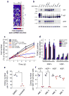
Fig 1. Aberrant HGF expression by AML cells is associated with MET activation and is necessary for cell growth and survival
(a) Heat map of the 30 top-ranking genes in the RNAi screen, whose depletion reduced the growth of OCI/AML-2 cells but not diffuse large B-cell lymphoma (Ly3, Ly10, Ly7, Ly19, K1106), myeloma (KMS12, H929, SKMM1), or T-cell acute lymphoblastic leukemia (Jurkat, CEM) cell lines. Relative cell depletion is represented by a bluered color gradient. The master myeloid transcription factor SPI1 served as the internal positive control; HGF is denoted with an arrowhead. (b) Western blot analysis of lysates of colon carcinoma DLD-1 cells with MET amplification, WI-38 fibroblasts expressing HGF, normal human CD34+ cells, and seven AML cell lines; OCI/AML-2 is duplicated. HGF is detected with an apparent mobility of 90 kDa, corresponding to its intracellular pro-form, while MET is detected as both pro- and mature forms in DLD-1 cells (180 and 140 kDa, respectively), and predominantly as the mature form (140 kDa) in AML cells (arrowhead). (c) Growth of OCI/AML-2 cells is inhibited by transduction of specific shRNAs targeting HGF (h9 and h10) or by treatment with a neutralizing anti-HGF antibody (100 nM), but not by transduction of control shRNA (GFP) or by concomitant rescue with HGF cDNA or recombinant human HGF (0.1 nM). Measurements are normalized to the value for untreated cells at day 7, and shown as means and standard deviations of three biologic replicates. * p < 0.05 versus untreated control. (d) TUNEL analysis of AML cells that express HGF and activate MET (OCI/AML-2, HEL, KG-1) versus those that lack HGF expression (F36P, MOLM-13, K562) as a function of depletion of HGF or MET using RNAi, treatment with the MET kinase inhibitor SU11274 (1 μM) or crizotinib (0.1μM) for 48 hours. Transduction with GFP shRNA and treatment with DMSO served as controls. Values are means and standard deviations of three biologic replicates. * p < 0.05 versus DMSO or GFP shRNA control. (e,f) Methylcellulose colony-forming assays of KG-1 cells (e) in the presence of DMSO control (black box) or crizotinib (0.1μM, red circle), and primary AML specimens (f) with aberrant HGF expression (AML 1, AML 2) versus those lacking HGF (AML 3, AML 4). Individual data points and means (bars) of three biologic replicates are shown. * p < 0.05 versus DMSO control.
Fig 2
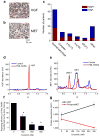
Fig 2. HGF and MET are co-expressed in the leukemic blasts of patients with AML and are induced by leukemogenic transcription factors in primary mouse hematopoietic progenitor cells, conferring susceptibility to MET kinase inhibition
(a,b) Immunohistochemical analysis of diagnostic bone marrow AML biopsy, demonstrating intracellular staining of HGF and pericytoplasmic membrane staining of MET in leukemic blasts, consistent with autocrine activation of MET. Scale bar = 25 μm. (c) Distribution of primary AML specimens that co-express HGF and MET (HGF+) versus those that lack HGF expression (HGF) by immunohistochemistry among patients with a normal karyotype, complex karyotype (complex), and other cytogenetic abnormalities, demonstrating aberrant HGF expression in 58 (42%) of the cases. (d) Abundance of mouse HGF at 7 days after retroviral transduction of mouse hematopoietic progenitors with PML-RARA (red) or vector control (blue), showing induction of HGF expression as measured with nanoimmunoassays. (e) Abundance of MET and phospho-MET (pMET) 7 days after retroviral transduction of mouse hematopoietic progenitors with PML-RARA (red) or vector control (blue), showing activation of MET upon induction of HGF expression. Equal protein loading was confirmed by the use of β2-microglubilin as the loading control. (f) Colony replating efficiency of PML-RARA transformed mouse hematopoietic progenitor cells, as a function of increasing concentration of crizotinib. Values are normalized to the number of colonies in mock-treated cells and plotted as means and standard deviations of three biologic replicates. * p < 0.05 for comparison to DMSO treated cells. (g) Abundance of HGF (black) and phospho-MET (red) in PML-RARA-transformed mouse hematopoietic progenitor cells treated with 0 and 300 nM crizotinib, demonstrating inhibition of MET phosphorylation and upregulation of HGF.
Fig 3
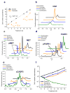
Fig 3. Restoration of leukemic cell growth upon chronic MET kinase inhibitor treatment is due to compensatory upregulation of HGF and MET re-activation, which can be overcome by inhibiting compensatory upregulation of HGF
(a) Kinetics of growth of OCI/AML-2 cells treated with crizotinib (0.1 μM in DMSO) or vehicle (DMSO), demonstrating that acute crizotinib treatment leads to significant reduction in AML cell growth (doubling time of 2.1 versus 12 days, p < 0.05), while with chronic treatment (10 days), the doubling time is 2.0 days. Cells were split and culture medium changed to maintain a constant cell density of 1 million cells ml−1. (b) Abundance of HGF in KG-1 cells treated for 10 days with DMSO (black), 100 nM crizotinib (orange), 20 nM PD173074 (blue), or a combination of crizotinib and PD173074 (green), as measured by quantitative nanoimmunoassay, with β2-microglobulin as the loading control, also see (Supplementary Fig. 16e). (c) MET activation as assessed by the abundance of phospho-MET (pMET) in KG-1 cells treated for 10 days with the indicated drugs, demonstrating maintenance of MET signaling in cells treated with 100 nM crizotinib or 20 nM PD1730734, but not in cells exposed to combination treatment. (d) FGFR1 activation as assessed by abundance of phospho-FGFR1 (pFGFR1) in KG-1 cells treated for 10 days with the indicated drugs, demonstrating lack of effect of crizotinib on FGFR1 activity. (e) Induction of apoptosis as assessed by the abundance of cleaved caspase 3 (cCASP3) in KG-1 cells treated for 10 days demonstrating substantially greater induction of apoptosis in cells treated with combination of 100 nM crizotinib and 20 nM PD173074 as compared to either drug alone. (f) Combined treatment of KG-1 cells with crizotinib and PD173074 leads to sustained logarithmic cell kill as compared to either drug alone. Values are means and standard deviations of three biologic replicates. * p < 0.05 versus either drug alone.
Fig 4
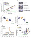
Fig 4. Combined inhibition of MET and FGFR1 blocks compensatory upregulation of HGF, leading to sustained inhibition of MET in KG-1 cells and to near-complete regression of AML in vivo
(a) Bioluminescence measurements of leukemic mice engrafted with luciferase-modified KG-1 cells and treated with vehicle control (black), 50 mg−1 kg−1 crizotinib alone (orange), 25 mg−1 kg−1 PD173074 (blue), or a combination of crizotinib and PD173074 by daily oral gavage (green). Values are means and standard deviations of each treatment group (n = 9). * p < 0.05 versus each remaining group. (b) Bioluminescent photographs of representative mice from each treatment group (blue-to-red color gradient represents increasing bioluminescence intensities). (c,d) Scatter plots of the fraction of human CD45+ KG-1 cells in the peripheral blood (c) and bone marrow (d) of mice after 10 days of treatment, demonstrating near-complete elimination of human AML cells in mice treated with the combination of crizotinib and PD173074. Boxes denote means and standard deviation for each group (n = 9). * p < 0.05 versus vehicle control group. (e,f) Abundance of HGF (e) and phospho-MET (f) in human CD45-selected KG-1 cells isolated from the bone marrow of mice after 10 days of treatment as indicated, demonstrating blockade of compensatory HGF upregulation in response to crizotinib treatment by the combined inhibition of MET and FGFR1, and sustained inhibition of MET activation.

Comment in

References

    1. Grimwade D, et al. The predictive value of hierarchical cytogenetic classification in older adults with acute myeloid leukemia (AML): analysis of 1065 patients entered into the United Kingdom Medical Research Council AML11 trial. Blood. 2001;98:1312–1320. - PubMed
    1. Burnett A, Wetzler M, Lowenberg B. Therapeutic advances in acute myeloid leukemia. J Clin Oncol. 2011;29:487–494. - PubMed
    1. Westbrook TF, Stegmeier F, Elledge SJ. Dissecting cancer pathways and vulnerabilities with RNAi. Cold Spring Harb Symp Quant Biol. 2005;70:435–444. - PubMed
    1. Bernards R, Brummelkamp TR, Beijersbergen RL. shRNA libraries and their use in cancer genetics. Nat Methods. 2006;3:701–706. - PubMed
    1. Ngo VN, et al. A loss-of-function RNA interference screen for molecular targets in cancer. Nature. 2006;441:106–110. - PubMed

Publication types

MeSH terms