. 2012 Sep 1;53(5):1123-38.
doi: 10.1016/j.freeradbiomed.2012.05.036. Epub 2012 Jun 6.

Mitochondrial reactive oxygen species generation triggers inflammatory response and tissue injury associated with hepatic ischemia-reperfusion: therapeutic potential of mitochondrially targeted antioxidants

Affiliations

Mitochondrial reactive oxygen species generation triggers inflammatory response and tissue injury associated with hepatic ischemia-reperfusion: therapeutic potential of mitochondrially targeted antioxidants

Partha Mukhopadhyay et al. Free Radic Biol Med. .

Abstract

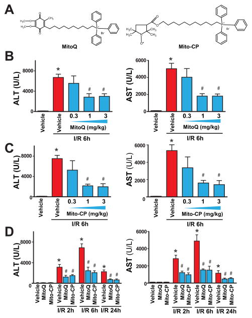
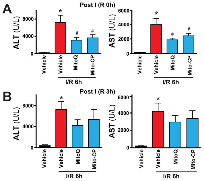
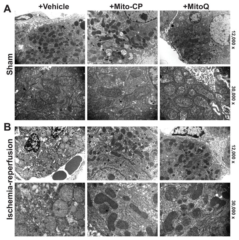
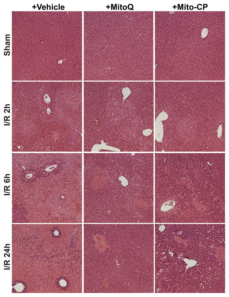
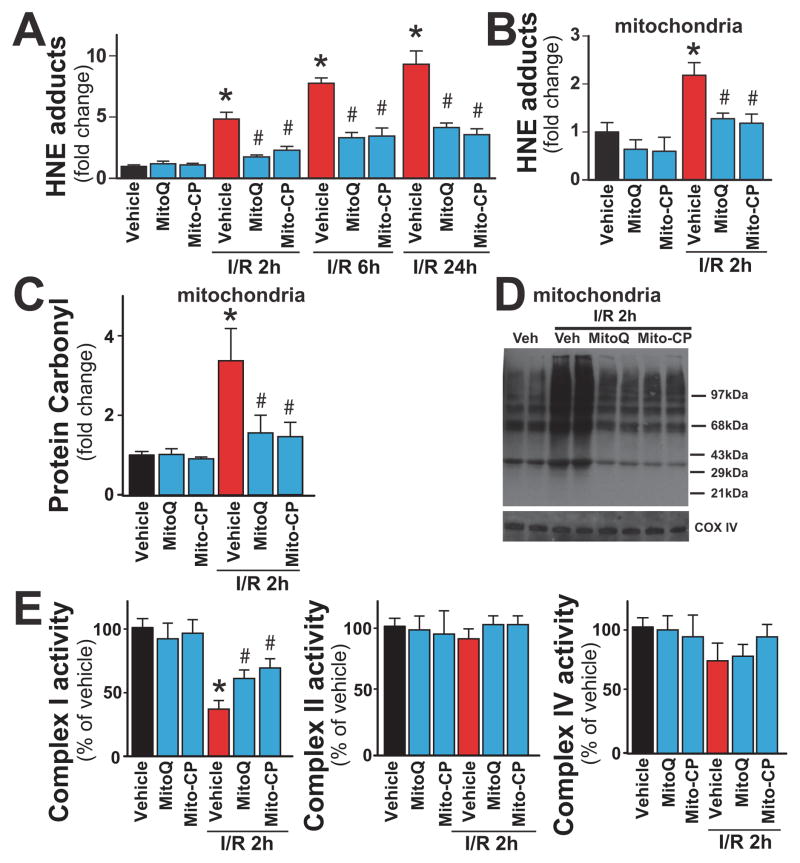
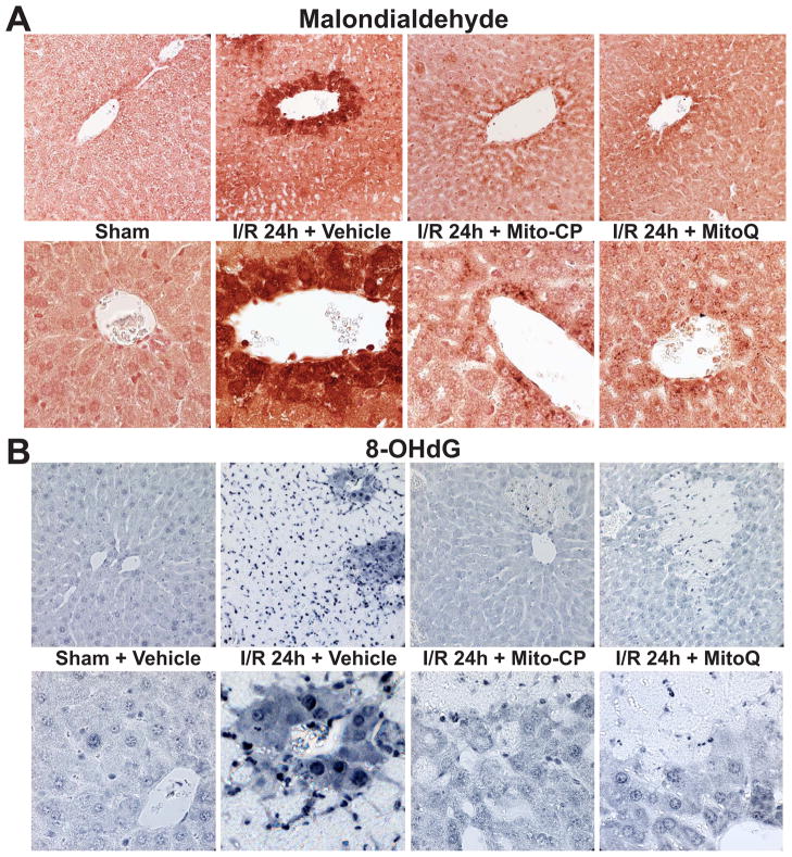
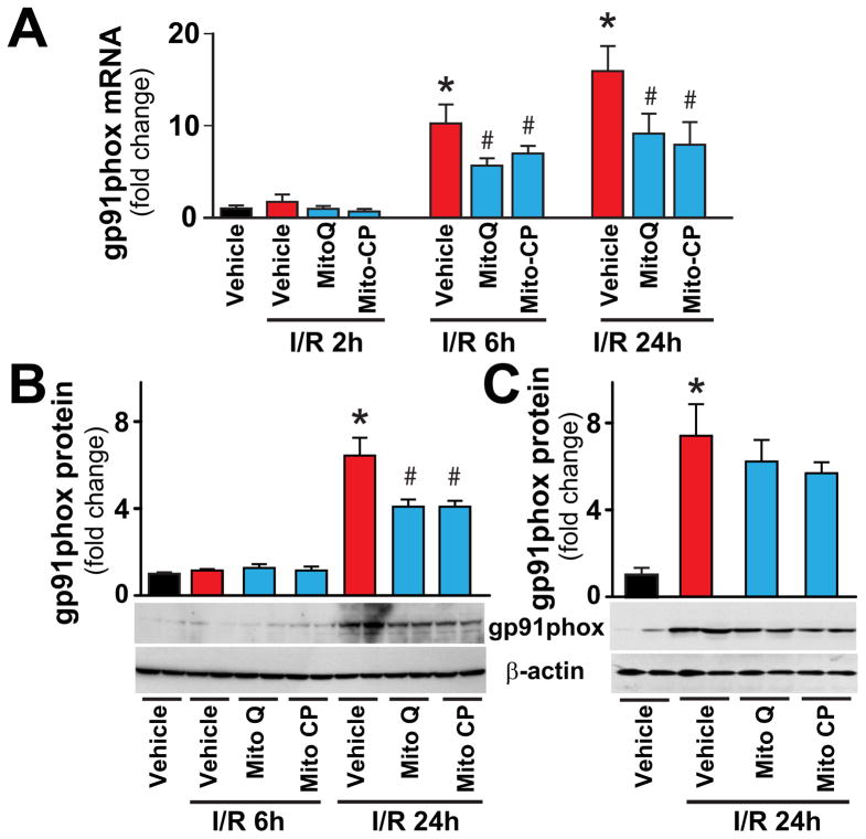
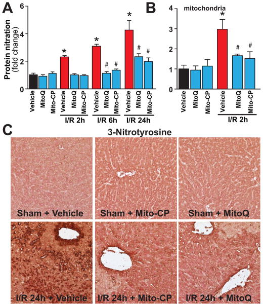
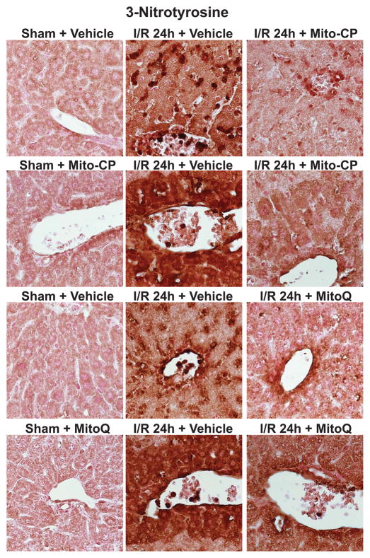
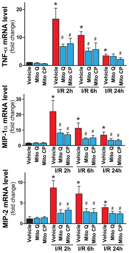
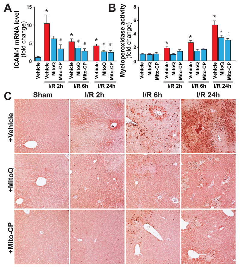
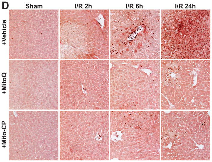
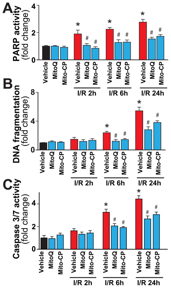
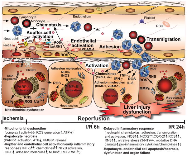
Mitochondrial reactive oxygen species generation has been implicated in the pathophysiology of ischemia-reperfusion (I/R) injury; however, its exact role and its spatial-temporal relationship with inflammation are elusive. Herein we explore the spatial-temporal relationship of oxidative/nitrative stress and inflammatory response during the course of hepatic I/R and the possible therapeutic potential of mitochondrial-targeted antioxidants, using a mouse model of segmental hepatic ischemia-reperfusion injury. Hepatic I/R was characterized by early (at 2 h of reperfusion) mitochondrial injury, decreased complex I activity, increased oxidant generation in the liver or liver mitochondria, and profound hepatocellular injury/dysfunction with acute proinflammatory response (TNF-α, MIP-1α/CCL3, MIP-2/CXCL2) without inflammatory cell infiltration, followed by marked neutrophil infiltration and a more pronounced secondary wave of oxidative/nitrative stress in the liver (starting from 6 h of reperfusion and peaking at 24 h). Mitochondrially targeted antioxidants, MitoQ or Mito-CP, dose-dependently attenuated I/R-induced liver dysfunction, the early and delayed oxidative and nitrative stress response (HNE/carbonyl adducts, malondialdehyde, 8-OHdG, and 3-nitrotyrosine formation), and mitochondrial and histopathological injury/dysfunction, as well as delayed inflammatory cell infiltration and cell death. Mitochondrially generated oxidants play a central role in triggering the deleterious cascade of events associated with hepatic I/R, which may be targeted by novel antioxidants for therapeutic advantage.


Figures

Figure 1
Figure 1. MitoQ and Mito-CP pretreatment dose-dependently attenuates hepatic I/R injury
Panel A: Chemical structures of MitoQ and Mito-CP. Panels B and C: Serum transaminases ALT and AST levels in sham-operated mice treated with vehicle or in mice exposed to 1 h of hepatic ischemia followed by 6 h of reperfusion (I/R 6h) pretreated with vehicle, MitoQ or Mito-CP (0.3, 1 and 3 mg/kg i.p., n=6–21/group). Panel D: Serum ALT and AST levels in sham operated mice treated with vehicle, MitoQ or Mito-CP (n=6–21/group) or in mice exposed to 1 h of hepatic ischemia followed by 2, 6 and 24 hours of reperfusion (I/R 2h, 6 h and 24h) pretreated with vehicle or MitoQ/Mito-CP (3 mg/kg i.p.). The peak damaged occurs at I/R 6h, and MitoQ/Mito-CP is able to attenuate the inflicted injury at any point investigated. *P<0.05 sham control vs. I/R; #P<0.05 I/R vs. corresponding I/R+ MitoQ/Mito-CP.
Figure 2
Figure 2. MitoQ and Mito-CP treatment after ischemia attenuates hepatic I/R injury
Panels A and B: Serum transaminases ALT and AST levels in sham-operated mice or in mice exposed to 1 h of hepatic ischemia followed by 6 h of reperfusion (I/R 6h) treated with vehicle or MitoQ/Mito-CP (3 mg/kg i.p., n=6–10/group) right after the ischemia before the reperfusion (post I (R 0h)) or 3 hours following the ischemia (post I (R 3h)), respectively. *P<0.05 sham control vs. I/R; #P<0.05 I/R vs. corresponding I/R+ MitoQ/Mito-CP. Notably, MitoQ/Mito-CP are able to attenuate the inflicted injury only when administered right after the ischemia, but not 3 hours after.
Figure 3
Figure 3. MitoQ and Mito-CP treatment attenuates I/R injury-induced early mitochondrial injury
Panel A: Representative transmission electron micrographs of the non-ischemic control livers of mice undergoing sham surgery (sham) treated with vehicle reveal normal hepatic tissue with good preservation of hepatocytes and lining cells of sinusoidal venules, as well as normal mitochondrial structure, which are not affected by treatments with MitoQ/Mito-CP (3 mg/kg i.p.). Upper images depict 12,000× and lower 30,000× magnification. Panel B: Post-ischemic liver tissue at 2 hours of reperfusion (I/R 2h) exhibit marked disintegration of ultrastructure (e.g., swelling of mitochondria, vacuolization, nuclear and cytoplasmic degeneration) in most hepatocytes. In some mitochondria the matrix totally disappears and only the outer membrane remains, while in others, the cristae are disorganized because of edema in the matrix (left images). Mito-CP/MitoQ pretreatment (3 mg/kg i.p.) markedly attenuates the morphological and mitochondrial injury induced by 1 h of ischemia followed by 2 hours of reperfusion (I/R 2h) to a similar extent (middle and right panels). Upper images depict 12,000× and lower 30,000× magnification. A similar histological profile was seen in three to five livers/group.
Figure 4
Figure 4. MitoQ and Mito-CP treatment attenuates histological damage at 2, 6 and 24h of reperfusion following 1 hour of ischemia
Hematoxylin and eosin staining of representative liver sections of sham mice treated with vehicle (sham), and mice exposed to 1 hour of ischemia followed by 2, 6 or 24h of reperfusion treated with vehicle or Mito-CP/MitoQ (3 mg/kg i.p.). I/R inflicts marked coagulation necrosis in the liver (lighter staining at 6 and 24 h of reperfusion), which is markedly attenuated by MitoQ/Mito-CP pretreatment. MitoQ/Mito-CP has no effect in control mice exposed to sham surgery. Figure 4 depicts 200x magnification. A similar histological profile was seen in three to five livers/group.
Figure 5
Figure 5. MitoQ and Mito-CP treatment attenuates the I/R-induced increased hepatic and/or mitochondrial oxidative stress and dysfunction
Panels A: HNE adducts (a marker for lipid peroxidation/oxidative stress) are time-dependently increased following I/R injury peaking at 24 hours. MitoQ/Mito-CP pretreatment (3 mg/kg i.p.) attenuates these increases. Panel B: HNE adducts in mitochondrial fraction are increased following I/R 2h and are attenuated with MitoQ/Mito-CP pretreatment (3 mg/kg i.p.). Panels C, D: Carbonyl adducts in mitochondrial fraction measured by ELISA or Oxyblot are increased following I/R 2h and are attenuated with MitoQ/Mito-CP pretreatment (3 mg/kg i.p.), respectively. Panel E: The complex I activity in isolated liver mitochondria is markedly decreased following I/R 2h, which is attenuated with MitoQ/Mito-CP pretreatment (3 mg/kg i.p.). *P<0.05 sham control vs. I/R; #P<0.05 I/R vs. corresponding I/R+MitoQ/Mito-CP. For panels A–C and E n=6–12/group.
Figure 6
Figure 6. MitoQ and Mito-CP treatment attenuates the I/R-induced increased hepatic malondialdehyde formation and oxidative DNA damage
Panel A: Malondialdehyde staining (brown; a marker of lipid peroxidation/oxidative stress) of representative liver sections of sham mice treated with vehicle (sham) or MitoQ/Mito-CP (antioxidants in sham mice had no effects, not shown), and mice exposed to 1 hour of ischemia followed by 24 hours of reperfusion (I/R 24h) treated with vehicle or MitoQ/Mito-CP (3 mg/kg i.p.). 24 hours of I/R triggers marked increase in liver malondialdehyde formation, which is predominantly localized to endothelial cells, perivascular hepatocytes, and infiltrating (or attached to the endothelium) inflammatory cells, and these increases are markedly attenuated by pretreatment with MitoQ/Mito-CP. Minimal staining is seen in the livers of control mice exposed to sham surgery. Slides are counterstained by nuclear fast red. Upper row of images depicts 400× magnification, while the lower one 1000× magnification. A similar histological profile was seen in three to five livers/group. Panel B: 8-OHdG staining (blue; marker of oxidative DNA damage) of representative liver sections of sham mice treated with vehicle (sham) or MitoQ/Mito-CP (antioxidants in sham mice had no effects, not shown) and mice exposed to 1 hour of ischemia followed by 24 hours of reperfusion (I/R 24h) treated with vehicle or MitoQ/Mito-CP. 24 hours of I/R triggers markedly increased 8-OHdG formation in endothelial cells, perivascular hepatocytes and infiltrating inflammatory cells, and these changes are attenuated by pretreatment with MitoQ/Mito-CP. Please note that the necrotic areas are lighter and infiltrated by neutrophils showing intense nuclear staining. Minimal nuclear 8-OHdG staining is seen in the livers of control mice exposed to sham surgery. Upper row of images depicts 400× magnification, while the lower one 1000× magnification. A similar histological profile was seen in three to five livers/group.
Figure 7
Figure 7. MitoQ and Mito-CP treatment attenuates the I/R-induced gp91phox expression
Panels A–C: Real-time PCR and Western blot show significant increase in hepatic NAD(P)H oxidase isoform NOX2/gp91phox mRNA or protein expression level at 24h of reperfusion (I/R 24h). Pretreatment with MitoQ/Mito-CP (Panels A and B), but not post-treatment following 3 hours of reperfusion (Panel C), attenuates the I/R-induced increases. n=6–14/group. *P<0.05 sham control vs. I/R; #P<0.05 I/R vs. corresponding I/R+MitoQ/Mito-CP.
Figure 8
Figure 8. MitoQ and Mito-CP treatment attenuates the I/R-induced increased nitrative stress
Panel A: Protein nitration (3-NT), a marker for nitrative stress, time-dependently increases following I/R injury peaking at 24 hours. MitoQ/Mito-CP pretreatment (3 mg/kg i.p.) attenuates this increase. n=8–12/group. *P<0.05 sham control vs. I/R; #P<0.05 I/R vs. corresponding I/R+MitoQ/Mito-CP. Panel B: Protein nitration in mitochondria is increased at I/R 2h. MitoQ/Mito-CP pretreatment (3 mg/kg i.p.) attenuates this increase. n=4–8/group. *P<0.05 sham control vs. I/R; #P<0.05 I/R vs. corresponding I/R+MitoQ/Mito-CP. Panel C: 3-Nitrotyrosine staining (brown) of representative liver sections of sham mice treated with vehicle (sham), MitoQ/Mito-CP and mice exposed to 1 hour of ischemia followed by 24 hours of reperfusion (I/R 24h) treated with vehicle or MitoQ/Mito-CP. Images depicts 400× magnification. A similar histological profile was seen in three to five livers/group.
Figure 9
Figure 9. MitoQ and Mito-CP treatment attenuates the I/R-induced increased nitrative stress in hepatocytes, endothelial and inflammatory cells
3-Nitrotyrosine staining of representative liver sections of sham mice treated with vehicle (sham), MitoQ/Mito-CP and mice exposed to 1 hour of ischemia followed by 24 hours of reperfusion treated with vehicle (I/R 24h) or MitoQ/Mito-CP. 24 hours of I/R (middle images) triggers markedly increased 3-NT formation in endothelial cells, perivascular hepatocytes and infiltrating inflammatory cells, and these changes are attenuated by pretreatment with MitoQ/Mito-CP. Images depict 1000× magnification. A similar histological profile was seen in three to five livers/group.
Figure 10
Figure 10. MitoQ and Mito-CP treatment attenuate the I/R-induced increased acute pro-inflammatory response
Real-time PCR shows significant increase in hepatic pro-inflammatory cytokine TNF-α, chemokines MIP-1α and MIP-2 mRNA levels at 2h of reperfusion (I/R 2h), and a gradual decrease by 24 hours (I/R 24h). Pretreatment with MitoQ/Mito-CP (3 mg/kg i.p.) attenuates the I/R-induced increased levels of cytokines/chemokines. n=7–12/group. *P<0.05 sham control vs. I/R; #P<0.05 I/R vs. corresponding I/R+MitoQ/Mito-CP.
Figure 11
Figure 11. MitoQ and Mito-CP treatment attenuates the I/R-induced increased adhesion molecule expression and enhanced delayed neutrophil infiltration in the liver
Panel A: Real-time PCR shows significant increase in hepatic adhesion molecule ICAM-1 mRNA level at 2h of reperfusion (I/R 2h), and a gradual decrease by 24 hours (I/R 24h). Pretreatment with MitoQ/Mito-CP (3 mg/kg i.p.) significantly attenuates the I/R-induced increased level of adhesion molecule. n=7–12/group. *P<0.05 sham control vs. I/R; #P<0.05 I/R vs. corresponding I/R+MitoQ/Mito-CP. Panel B: Quantification of MPO activity from the liver extracts shows significant time-dependent increases associated with I/R (peaking at I/R 24h), which are attenuated by pretreatment with MitoQ/Mito-CP (3 mg/kg i.p.). n=8–12/group. *P<0.05 sham control vs. I/R; #P<0.05 I/R vs. corresponding I/R+MitoQ/Mito-CP. Panel C–D: Myeloperoxidase (MPO) staining (brown) of representative liver sections of sham mice treated with vehicle (sham) or MitoQ/Mito-CP, and mice exposed to 1 hour of ischemia followed by 2, 6 or 24 hours of reperfusion treated with vehicle or MitoQ/Mito-CP. These images reveal increased neutrophils attachment to the endothelium and accumulation in the vessels at I/R 6h followed by marked infiltration of liver tissue at I/R 24h. Pretreatment with MitoQ/Mito-CP (3 mg/kg i.p.) attenuates the I/R-induced increased neutrophil infiltration. Slides were counterstained by nuclear fast red. Panel C images depict 200× magnification, while Panel D depicts 400× magnification. A similar histological profile was seen in three to five livers/group.
Figure 12
Figure 12. MitoQ and Mito-CP treatment attenuates the I/R-induced increased cell death
Panel A: PARP activity, a marker for cell death (mostly necrotic) increases at 2, 6 and 24 h of I/R. MitoQ/Mito-CP pretreatment (3 mg/kg i.p.) attenuates these increases. Panels B and C: DNA fragmentation and caspase 3/7 activity (markers of apoptotic cell death) increase only from 6 h of I/R peaking at 24 hours. MitoQ/Mito-CP pretreatment (3 mg/kg i.p.) attenuates these increases. n=8–13/group. *P<0.05 sham control vs. I/R; #P<0.05 I/R vs. corresponding I/R+MitoQ/Mito-CP.
Figure 13
Figure 13. Simplified mechanisms of the interplay of mitochondrial dysfunction and oxidative stress with inflammatory responses and cell death during hepatic ischemia-reperfusion (I/R) injury
The metabolic stress during ischemia and initial phase of reperfusion leads to mitochondrial dysfunction in hepatocytes and sinusoidal endothelial cells, increased mitochondrial reactive oxygen (e.g. superoxide and hydrogen peroxide) and nitrogen species (e.g. peroxynitrite) formation (ROS/RNS), Ca2+ accumulation, and activation of various necrotic cell death pathways by ROS/RNS, such as poly(ADP)-ribose polymerase 1 (PARP-1), which leads to ATP depletion and early cell necrosis. Superoxide may also readily react with nitric oxide (NO) during early hepatic reperfusion (the latter can be derived from nitric oxide synthases, most likely iNOS via a diffusion limited reaction to form a more potent oxidant peroxynitrite, further impairing mitochondrial and cellular functions and increasing ROS generation. Hepatic I/R also attenuates endothelial NO synthase activity in sinusoidal endothelial cells during I/R leading to endothelial dysfunction, favoring sinusoidal vasoconstriction and secondary ischemic injury. Necrotic hepatocytes and endothelial cells release various damage-associated molecular patterns (e.g. high mobility group box 1 protein (HMGB1), DNA fragments, and lipid peroxidation products such as hydroxynonenal (HNE), among others), which activate Kupffer cells (the resident macrophages of the liver) via several toll like receptors (TLRs) in a nuclear factor kappa B (NFκB)-dependent manner. The activated Kupffer cells subsequently produce numerous pro-inflammatory cytokines and chemokines such as tumor necrosis factor α (TNF-α), macrophage inflammatory proteins (MIP1/2), and additional ROS and nitric oxide (NO) through the increased expression of ROS generating NAD(P)H oxidase isoform NOX-2 and inducible nitric oxide synthase (iNOS), further fueling oxidative/nitrative injury and priming/chemotaxis of various inflammatory cells. The initial oxidant-induced injury also leads to the activation of endothelial cells which in concert with activated Kupffer cells and certain subtypes of T lymphocytes orchestrate the acute and delayed pro-inflammatory response leading to attraction of neutrophils and other inflammatory cells into the damaged tissue upon late reperfusion. These inflammatory cells further release oxidants and proteolytic enzymes enhancing intracellular oxidative/nitrative stress and mitochondrial dysfunction in hepatocytes, thereby promoting apoptotic and/or necrotic forms of cell demise.

Similar articles

Cited by

References

    1. Jaeschke H. Molecular mechanisms of hepatic ischemia-reperfusion injury and preconditioning. Am J Physiol Gastrointest Liver Physiol. 2003;284:G15–26. - PubMed
    1. Jaeschke H. Reactive oxygen and mechanisms of inflammatory liver injury: Present concepts. J Gastroenterol Hepatol. 2011;26(Suppl 1):173–179. - PubMed
    1. Pacher P, Beckman JS, Liaudet L. Nitric oxide and peroxynitrite in health and disease. Physiol Rev. 2007;87:315–424. - PMC - PubMed
    1. Szabo C, Ischiropoulos H, Radi R. Peroxynitrite: biochemistry, pathophysiology and development of therapeutics. Nat Rev Drug Discov. 2007;6:662–680. - PubMed
    1. Szabo C. The pathophysiological role of peroxynitrite in shock, inflammation, and ischemia-reperfusion injury. Shock. 1996;6:79–88. - PubMed

Publication types

MeSH terms