. 2012 Jun 10;19(7):685-692.
doi: 10.1038/nsmb.2335.

The zinc-finger domains of PARP1 cooperate to recognize DNA strand breaks

Affiliations

The zinc-finger domains of PARP1 cooperate to recognize DNA strand breaks

Ammar A E Ali et al. Nat Struct Mol Biol. .

Erratum in

Abstract

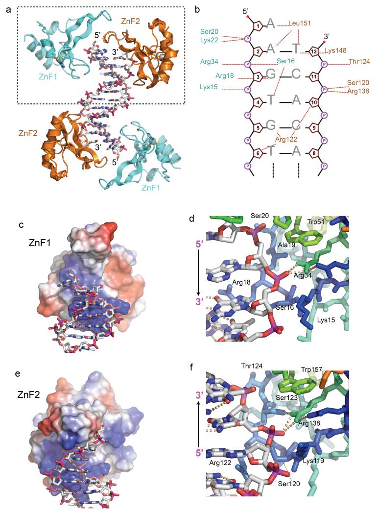
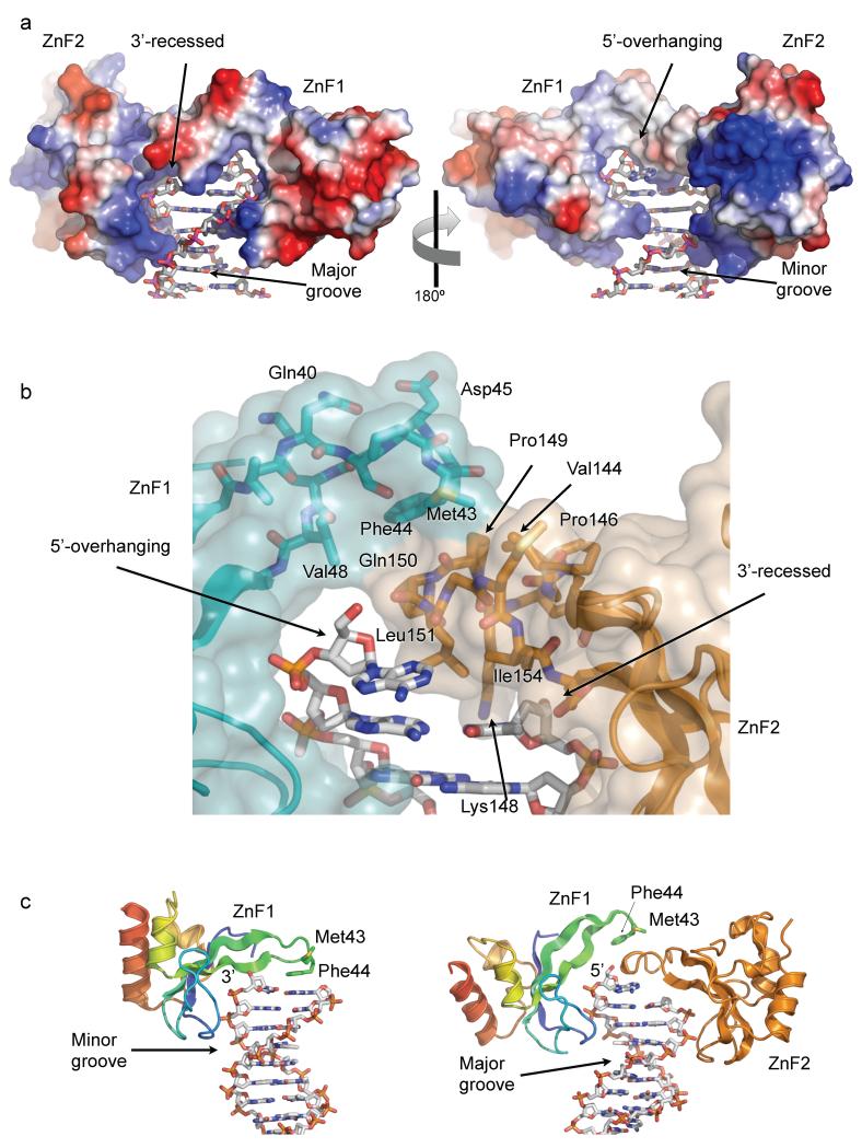
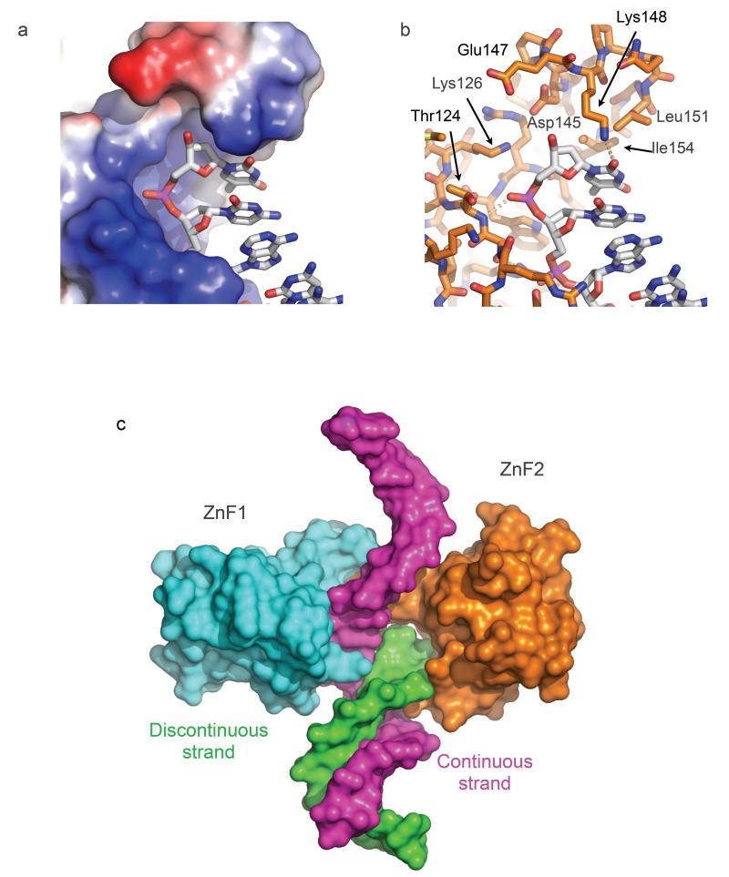
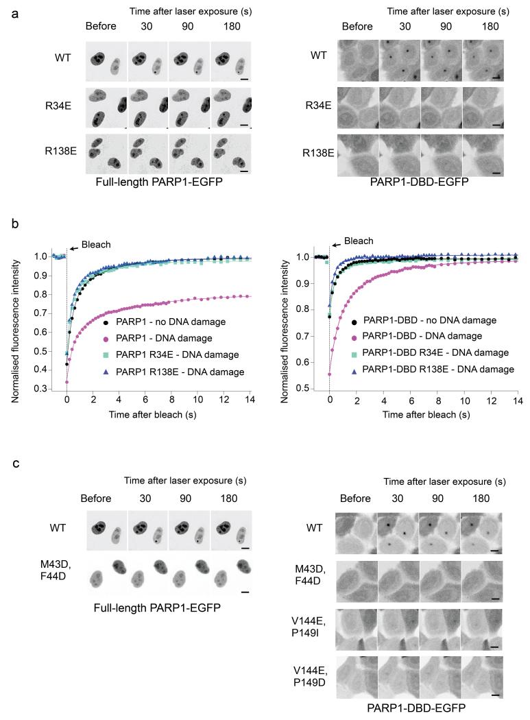
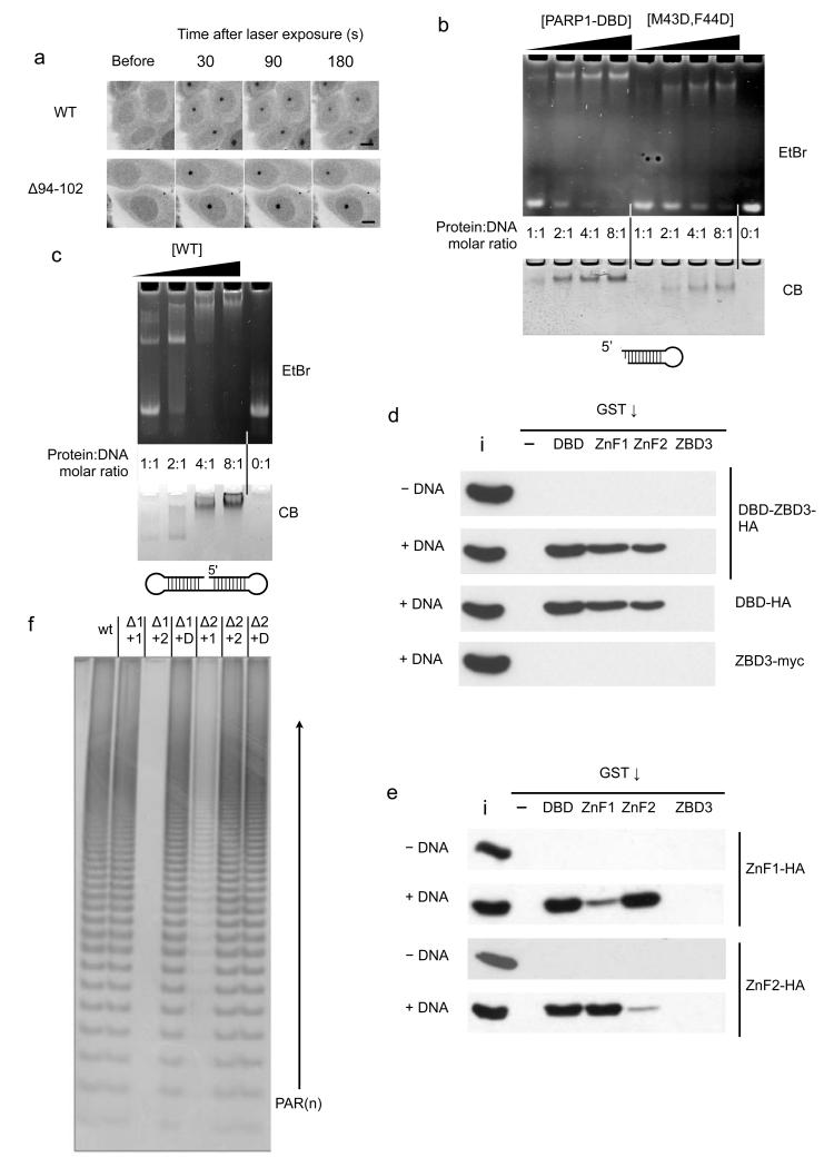
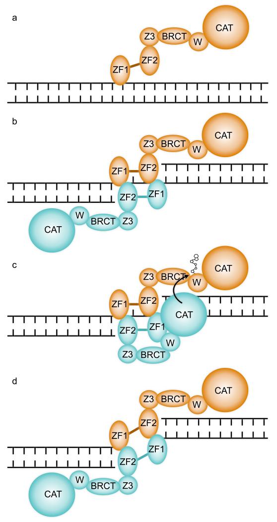
Poly(ADP-ribose) polymerase 1 (PARP1) is a primary DNA damage sensor whose (ADP-ribose) polymerase activity is acutely regulated by interaction with DNA breaks. Upon activation at sites of DNA damage, PARP1 modifies itself and other proteins by covalent addition of long, branched polymers of ADP-ribose, which in turn recruit downstream DNA repair and chromatin remodeling factors. PARP1 recognizes DNA damage through its N-terminal DNA-binding domain (DBD), which consists of a tandem repeat of an unusual zinc-finger (ZnF) domain. We have determined the crystal structure of the human PARP1-DBD bound to a DNA break. Along with functional analysis of PARP1 recruitment to sites of DNA damage in vivo, the structure reveals a dimeric assembly whereby ZnF1 and ZnF2 domains from separate PARP1 molecules form a strand-break recognition module that helps activate PARP1 by facilitating its dimerization and consequent trans-automodification.


Figures

FIGURE 1
FIGURE 1. PARP1-DNA interactions
a) Structure of the PARP1 DNA-binding domain (DBD) bound to a DNA duplex with a 5’-overhang. Each end of the non-self-complementary duplex is bound by a complex of the PARP1 ZnF1 and ZnF2 domains. b) Schematic of the interactions of PARP1 DBD with the DNA duplex within the dashed box in a). Dotted lines indicate interactions with phosphate (circled P), sugar (pentagon) or base (letters). c) ZnF1 DNA-interacting surface coloured by electrostatic potential (+ve – blue to −ve – red) interacts with the sugar-phosphate backbone of the overhanging strand and the major groove. d) Details of ZnF1-DNA interactions, centred on the polar interaction of Arg34 and a DNA phosphate group. e) As c) but for ZnF2, which interacts with the sugar-phosphate backbone of the recessed strand and the minor groove. f) Details of ZnF2-DNA interactions, centred on the polar interaction of Arg138 and a DNA phosphate group. The conserved DNA interacting groups in the ZnF1 and ZnF2 domains, are able to interact with a DNA strand in either polarity.
FIGURE 2
FIGURE 2. DNA end-binding by the ZnF1-ZnF2 complex
a) Electrostatic-colored (blue – basic; red –acidic) molecular surface of the DNA-end binding structure formed by PARP1 ZnF1 and ZnF2 domains, extending across the surface of the DNA end and interacting with both grooves of the duplex. The 3’-recessed end of one strand is on the left – the 5’-overhanging end of the other is on the right. b) Detail of the interface between the tips of the β2-3 connecting loops of ZnF1 and ZnF2, which form the bridge overlying the terminal base-pair of the duplex. Transparent molecular surface and carbon atoms are colored by domain; ZnF1 – cyan, ZnF2 – gold. c) Comparison of the interaction of isolated PARP1-ZnF1 with a blunt-end oligonucleotide (left) and PARP1-ZnF1 in the context of the PARP1-DBD construct used in this work (right). In the two different structures PARP1-ZnF1 uses essentially the same residues to interact with the sugar-phosphate backbone in a 3’-5’ orientation via the minor groove (ZnF1 only) or in a 5’-3’ orientation via the major groove (ZnF1-ZnF2 complex).
FIGURE 3
FIGURE 3. DNA break detection by PARP1 ZnF2
a) The β2-3 loop of PARP1 ZnF2 projects over the basic DNA-binding groove and blocks continuation of the DNA strand, thereby conferring binding specificity to DNA ends. b) Detail of the end-binding structure of ZnF2. Leu151 and Ile154 provide a hydrophobic wedge that overlies the terminal base on the recessed strand. The polar pocket formed by the side chains of Lys126, Asp145, Glu147 and Arg156 would be capable of accommodating a range of end chemistries, including 3’-PO4, 5’-OH and 5’- PO4, as well as the 3’-OH in the present crystal structure. c) Model of gap recognition by the combined PARP1 ZnF1 and ZnF2 structure. Specific recognition of the discontinuity in one strand (green) is provided by the β2-3 loop of ZnF2 which completely blocks the end of the DNA-binding groove, while the other strand (magenta) can continue comfortably through the open DNA-binding groove of ZnF1 with only a small kink introduced.
FIGURE 4
FIGURE 4. DNA damage focus formation by PARP1 and its mobility
a) Recruitment of C-terminal EGFP fusions with full-length PARP1 (left) or PARP1-DBD (right) to damage foci following laser damage. Fluorescent foci are evident 30 seconds post-irradiation in the nuclei of cells expressing the wild-type full-length PARP1 (only the bottom, right nucleus was laser-damaged) or PARP1-DBD (all nuclei damaged) constructs and diminish in intensity over subsequent minutes. No significant foci are formed by either full-length or DBD constructs harbouring mutations designed to disrupt DNA binding by ZnF1 (R34E) or by ZnF2 (R138E), showing a clear requirement for both zinc-finger domains in DNA damage recognition. Scale bar indicates 10 μm. b) FRAP analysis shows that an intact PARP1-DBD is required for PARP1 retention at damaged DNA. In the absence of DNA damage, wild-type PARP1 full-length and PARP1-DBD are not immobilized on DNA. In contrast, under DNA damage conditions, wild-type PARP1 constructs are immobilized, while disruption of DNA binding by either ZnF1 or ZnF2 abolishes retention. c) Recruitment of full-length PARP1 (left) or PARP1-DBD (right) to damage foci is abolished by polar mutations of Met 43 and Phe 44 in the ZnF1 domain, or of Val 144 and Pro 149 in the ZnF2 domain (not determined for full-length PARP1), which together form the hydrophobic protein-protein interface between ZnF1 and ZnF2. Scale bar indicates 10 μm.
FIGURE 5
FIGURE 5. Intermolecular dimerisation of PARP1 Znf1 and ZnF2 domain
a) A nine residue deletion in the linker connecting ZnF1 and ZnF2 has no effect on the ability of the PARP1 - DBD to localise to damage foci. Scale bar indicates 10 μm. b) Wild-type PARP1-DBD (WT) forms a strong interaction with the DNA in an electrophoretic mobility assay (EMSA) that is only saturated at a 2:1 protein:DNA ratio. DNA binding of the M43D, F44D PARP1-DBD mutant is substantially impaired. c) EMSA of a ‘dumbbell’ oligonucleotide shows some complex formation at 1:1 protein-DNA ratio, but a 2:1 ratio is required to achieve saturation. d) Pull-down assay showing that the DBD and isolated DNA-binding zinc-finger domains, but not the third zinc-binding domain (ZBD3), are able to co-precipitate constructs incorporating the DBD in a DNA dependent manner. ‘i’ = 20% input, ‘-‘ = empty beads. e) Pull-down assay showing that either of the isolated tagged DNA-binding zinc-finger domains can be efficiently co-precipitated in a DNA-dependent manner, by the GST-DBD fusion or the GST-fusion with the other zinc-finger domain, but not by the same zinc-finger domain or by GST-ZBD3. ‘i’ = 5% input, ‘-‘ = empty beads. f) Catalytic activity of PARP1 constructs deleted for either ZnF1 (Δ1) or ZnF2 (Δ2) domain can be complemented by presentation of the deleted domain in trans (+1, +2, or D = +1 and 2), consistent with the requirement for both DNA-binding zinc-finger domains to form an intermolecular interaction for PARP1 activation at sites of DNA damage.
FIGURE 6
FIGURE 6. A Mechanism for DNA Damage Dependent trans-automodification by PARP1
a) PARP1 is constitutively associated with chromatin in the absence of DNA damage - and may associate with undamaged DNA, via the generally weak interaction of ZnF1 with the sugar-phosphate backbone. ZF1 and ZF2 – first and second zinc-finger domains; Z3 – zinc-binding domain; BRCT – BRCA1 C-terminal homology domain; W – WGR domain; CAT – catalytic domain. b) A discontinuity in the DNA backbone permits additional binding of ZnF2 and formation of a functional break-recognition module via dimerization with a second PARP1 molecule. An inter-molecular ZnF1-ZnF2 complex could form at both margins if the single-strand gap is sufficiently large. c) The close proximity of two PARP1 molecules bound cooperatively at a site of DNA damage, facilitates access of the catalytic domain of one to the poly-(ADP-ribose) acceptor sites on the other to achieve the well-described trans-automodification that initiates PARP1 signalling. Formation of a productive trans-autoribosylation complex is likely to involve conformational changes and multiple inter- and intra-molecular interactions of the other domains of PARP1, which are yet to be elucidated. d) Formation of two break recognition modules formed by inter-molecular dimerization of ZnF1 and ZnF2 domains from two PARP1 molecules, explains the ability of PARP1 to maintain synapsis of DNA double-strand breaks and thereby facilitate a Ku-independent end-joining reaction -.

Comment in

  • PARP pairs up to PARsylate.
    Coquelle N, Glover JN. Coquelle N, et al. Nat Struct Mol Biol. 2012 Jul 5;19(7):660-1. doi: 10.1038/nsmb.2306. Nat Struct Mol Biol. 2012. PMID: 22767236 No abstract available.

Similar articles

Cited by

References

    1. Pion E, et al. DNA-induced dimerization of poly(ADP-ribose) polymerase-1 triggers its activation. Biochemistry. 2005;44:14670–81. - PubMed
    1. Kirsten E, Kun E, Mendeleyev J, Ordahl CP. Activity assays for poly-ADP ribose polymerase. Methods Mol Biol. 2004;287:137–49. - PubMed
    1. Huang K, et al. Analysis of nucleotide sequence-dependent DNA binding of poly(ADP-ribose) polymerase in a purified system. Biochemistry. 2004;43:217–23. - PubMed
    1. Shall S, de Murcia G. Poly(ADP-ribose) polymerase-1: what have we learned from the deficient mouse model? Mutation Research-Dna Repair. 2000;460:1–15. - PubMed
    1. Ame JC, Spenlehauer C, de Murcia G. The PARP superfamily. Bioessays. 2004;26:882–93. - PubMed

Publication types

Associated data