. 2012 Jun 10;19(7):677-84.
doi: 10.1038/nsmb.2317.

Structural dynamics of the aminoacylation and proofreading functional cycle of bacterial leucyl-tRNA synthetase

Affiliations

Structural dynamics of the aminoacylation and proofreading functional cycle of bacterial leucyl-tRNA synthetase

Andrés Palencia et al. Nat Struct Mol Biol. .

Abstract

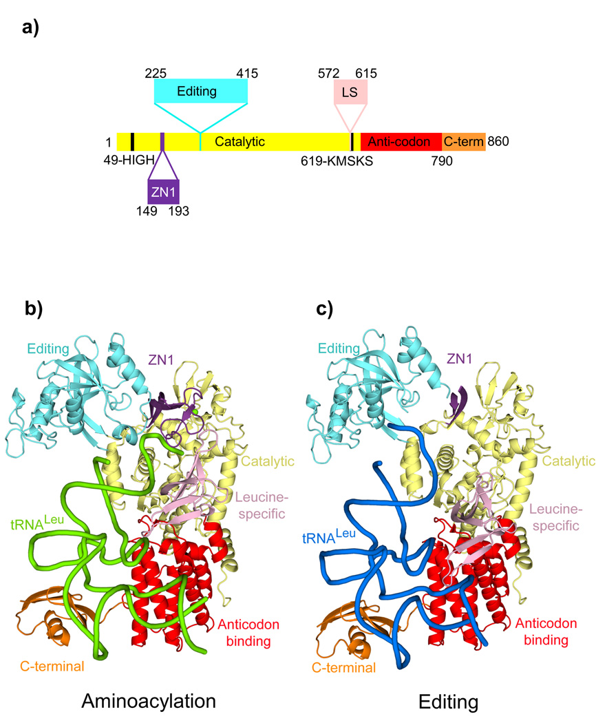
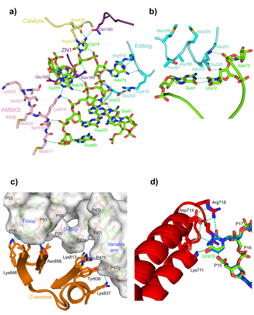
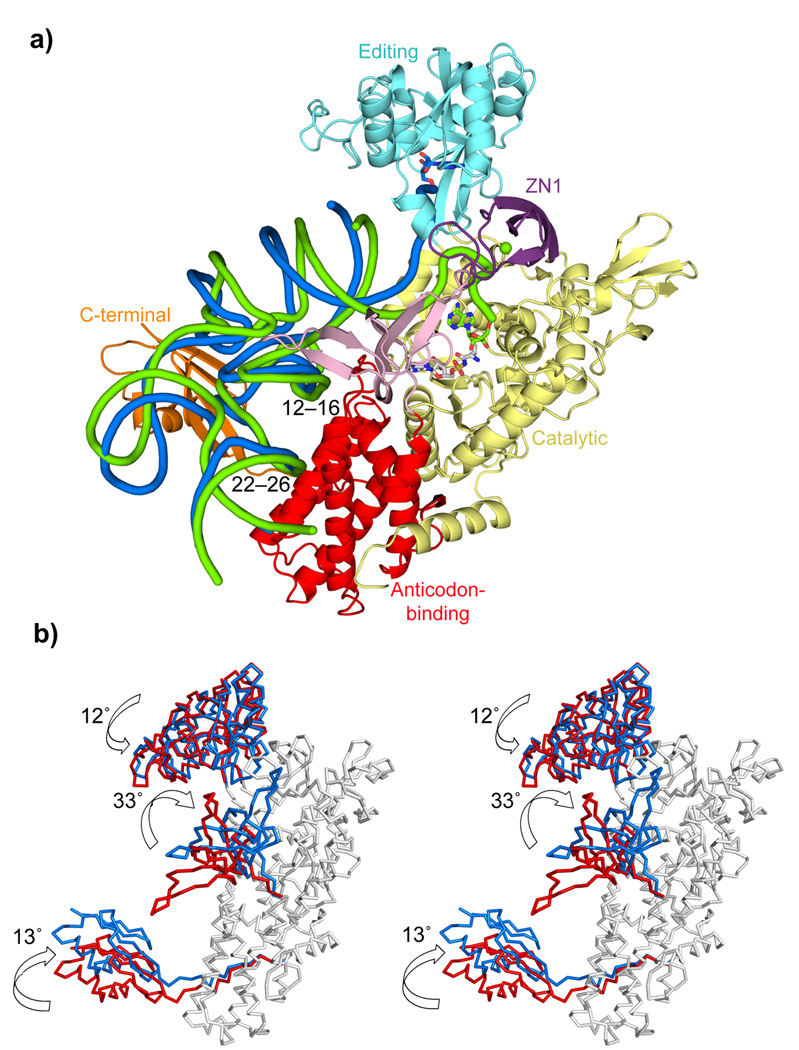
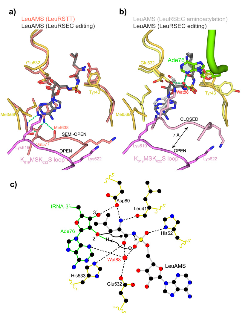
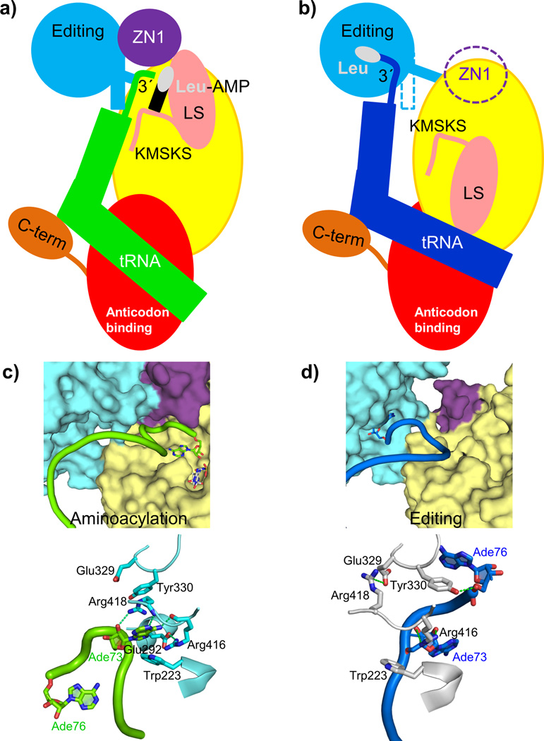
Leucyl-tRNA synthetase (LeuRS) produces error-free leucyl-tRNA(Leu) by coordinating translocation of the 3' end of (mis-)charged tRNAs from its synthetic site to a separate proofreading site for editing. Here we report cocrystal structures of the Escherichia coli LeuRS-tRNA(Leu) complex in the aminoacylation or editing conformations, showing that translocation involves correlated rotations of four flexibly linked LeuRS domains. This pivots the tRNA to guide its charged 3' end from the closed aminoacylation state to the editing site. The editing domain unexpectedly stabilizes the tRNA during aminoacylation, and a large rotation of the leucine-specific domain positions the conserved KMSKS loop to bind the 3' end of the tRNA, promoting catalysis. Our results give new insight into the structural dynamics of a molecular machine that is essential for accurate protein synthesis.


Figures

Figure 1
Figure 1. Structures of the E. coli LeuRS-tRNALeu complex in the aminoacylation and editing states
a. The domain structure of LeuRSEC. Residue numbers indicate domain boundaries. The color code used throughout this paper for the various domains is catalytic (yellow), zinc (ZN1) (purple) with the zinc ion in green, editing (cyan), leucine-specific (pink), anticodon-binding (red) and C-terminal (orange). b. Aminoacylation conformation with the tRNA in green. c. Editing conformation with the tRNA in blue. In this state, the ZN1 domain is partially disordered.
Figure 2
Figure 2. LeuRS-tRNALeu interactions in the aminoacylation complex
a. Several domains (color coded as in Fig. 1) of LeuRS are involved in binding and stabilizing the conformation of nucleotides 69 to 76 of the 3′ end of tRNALeu (green). The base of Gua71 is omitted for clarity. b. The α-helix 291–298 of the editing domain stacks on the G1-U72 base pair and contacts the backbone of Ade73. c. Interactions of the C-terminal domain with the T-loop, D-loop and long-variable arm of the tRNA (surface representation) are conserved in the editing and aminoacylation states (overlaid). d. A network of interactions from the anti-codon binding domain, conserved between the two states (overlaid), specifically recognizes the base of Ura16 (see also Supplementary Figs. 4a,b).
Figure 3
Figure 3. Comparison between the aminoacylation and the editing configurations
a. Global comparison of the two states. The LeuRSEC-tRNALeu aminoacylation complex (tRNA green tube) is shown together with the tRNA in the editing conformation (tRNA blue tube) after superimposition of the catalytic and the anticodon-binding domains of the two complexes. b. Stereo diagram showing rotations of the flexibly linked domains between the editing (red) and aminoacylation states (blue), after superposition of the body of the enzyme (grey).
Figure 4
Figure 4. tRNA 3′ end binding induces full closure of the KMSKS loop
a. Comparison between the synthetic sites of the LeuRSEC-LeuAMS-tRNALeu complex with the tRNA in the editing configuration (catalytic domain yellow, fully open KMSKS loop purple, LeuAMS dark grey) and the LeuRSTT-LeuAMS complex (PDB 1H3N) with the KMSKS loop in the semi-open conformation (all chains and LeuAMS salmon). Hydrogen bonds between the semi-open KMSKS loop conformation and the adenine base of the LeuAMS are indicated by green dotted lines but are absent in the open conformation. b. Comparison between the synthetic sites of the LeuRS-LeuAMS-tRNALeu complex in the aminoacylation configuration (catalytic domain pale yellow, closed KMSKS loop light purple, LeuAMS light grey, tRNA green) and editing configuration (as in (a)). In the aminoacylation state, the presence of Ade76 necessitates a side-chain flip of Tyr43 and a water molecule (Wat88, red sphere) is coordinated by Ade76 N3, the re-orientated Glu532 carboxylate, a LeuAMS sulphate oxygen and the carbonyl-oxygen of the substrate leucine as indicated by green dotted lines (see text and (c)). c. Schematic diagram of the LeuRSEC active site indicating the presumed substrate assisted mechanism of aminoacylation (see text).
Figure 5
Figure 5. Dynamics of the functional cycle of bacterial LeuRS
a, b. Schematic diagram of the structural changes between the aminoacylation and proof-reading conformations of the LeuRSEC-tRNALeu complex (see text). Color code for each domain is as in Figure 1, leucine is represented as a white oval and AMP as a black rectangle. c, d. Dynamic rearrangements in the interface between the editing, ZN1 and catalytic domain that allow translocation of the tRNA 3′ end from the synthetic site (c) to the editing site for proof-reading (d). Some elements of the catalytic domain have been removed for clarity. Top panels show the cavities of the catalytic and editing sites in surface representation, and bottom panels the residues important for the translocation of the tRNA (rotated 180 ° respect to the top panels for clarity).

References

    1. Karkhanis VA, Mascarenhas AP, Martinis SA. Amino acid toxicities of Escherichia coli that are prevented by leucyl-tRNA synthetase amino acid editing. J Bacteriol. 2007;189:8765–8768. - PMC - PubMed
    1. Lincecum TL, Jr, et al. Structural and mechanistic basis of pre- and posttransfer editing by leucyl-tRNA synthetase. Mol Cell. 2003;11:951–963. - PubMed
    1. Tukalo M, Yaremchuk A, Fukunaga R, Yokoyama S, Cusack S. The crystal structure of leucyl-tRNA synthetase complexed with tRNALeu in the post-transfer-editing conformation. Nat Struct Mol Biol. 2005;12:923–930. - PubMed
    1. Hagiwara Y, Field MJ, Nureki O, Tateno M. Editing Mechanism of Aminoacyl-tRNA Synthetases Operates by a Hybrid Ribozyme/Protein Catalyst. J Am Chem Soc - PubMed
    1. Rock FL, et al. An antifungal agent inhibits an aminoacyl-tRNA synthetase by trapping tRNA in the editing site. Science. 2007;316:1759–1761. - PubMed

Publication types

Substances

LinkOut - more resources