Crystallization and preliminary X-ray analysis of the reductase component of p-hydroxyphenylacetate 3-hydroxylase from Acinetobacter baumannii
- PMID: 22684080
- PMCID: PMC3370920
- DOI: 10.1107/S1744309112016909
Crystallization and preliminary X-ray analysis of the reductase component of p-hydroxyphenylacetate 3-hydroxylase from Acinetobacter baumannii
Abstract
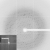
p-Hydroxyphenylacetate 3-hydroxylase (HPAH) from Acinetobacter baumannii catalyzes the hydroxylation of p-hydroxyphenylacetate (HPA) at the ortho position to yield 3,4-dihydroxyphenylacetate (DHPA). HPAH from A. baumannii is a two-component flavoprotein consisting of a smaller reductase (C(1)) component and a larger oxygenase (C(2)) component. The C(1) component supplies a reduced flavin in its free form to the C(2) counterpart for hydroxylation. In addition, HPA can bind to C(1) and enhance the flavin-reduction rate without becoming hydroxylated. The recombinant C(1) component was purified and crystallized using the microbatch method at 295 K. X-ray diffraction data were collected to 2.3 Å resolution using synchrotron radiation on the BL13B1 beamline at NSRRC, Taiwan. The crystal belonged to the orthorhombic space group P2(1)2(1)2(1), with unit-cell parameters a = 47.78, b = 59.92, c = 211.85 Å, and contained two molecules of C(1) per asymmetric unit.
Figures


Similar articles
-
4-Hydroxyphenylacetate 3-Hydroxylase (4HPA3H): A Vigorous Monooxygenase for Versatile O-Hydroxylation Applications in the Biosynthesis of Phenolic Derivatives.Int J Mol Sci. 2024 Jan 19;25(2):1222. doi: 10.3390/ijms25021222. Int J Mol Sci. 2024. PMID: 38279222 Free PMC article. Review.
-
The reductase of p-hydroxyphenylacetate 3-hydroxylase from Acinetobacter baumannii requires p-hydroxyphenylacetate for effective catalysis.Biochemistry. 2005 Aug 2;44(30):10434-42. doi: 10.1021/bi050615e. Biochemistry. 2005. PMID: 16042421
-
Kinetic mechanisms of the oxygenase from a two-component enzyme, p-hydroxyphenylacetate 3-hydroxylase from Acinetobacter baumannii.J Biol Chem. 2006 Jun 23;281(25):17044-17053. doi: 10.1074/jbc.M512385200. Epub 2006 Apr 20. J Biol Chem. 2006. PMID: 16627482
-
Crystal structure of the flavin reductase of Acinetobacter baumannii p-hydroxyphenylacetate 3-hydroxylase (HPAH) and identification of amino acid residues underlying its regulation by aromatic ligands.Arch Biochem Biophys. 2018 Sep 1;653:24-38. doi: 10.1016/j.abb.2018.06.010. Epub 2018 Jun 22. Arch Biochem Biophys. 2018. PMID: 29940152
-
Monooxygenation of aromatic compounds by flavin-dependent monooxygenases.Protein Sci. 2019 Jan;28(1):8-29. doi: 10.1002/pro.3525. Protein Sci. 2019. PMID: 30311986 Free PMC article. Review.
Cited by
-
Mechanistic insights into allosteric regulation of the reductase component of p-hydroxyphenylacetate 3-hydroxylase by p-hydroxyphenylacetate: a model for effector-controlled activity of redox enzymes.RSC Chem Biol. 2024 Dec 5;6(1):81-93. doi: 10.1039/d4cb00213j. eCollection 2025 Jan 2. RSC Chem Biol. 2024. PMID: 39649338 Free PMC article.
-
Crystals on the cover 2013.Acta Crystallogr Sect F Struct Biol Cryst Commun. 2013 Jan 1;69(Pt 1):1. doi: 10.1107/S1744309112051950. Epub 2012 Dec 31. Acta Crystallogr Sect F Struct Biol Cryst Commun. 2013. PMID: 23295475 Free PMC article.
-
Advances in 4-Hydroxyphenylacetate-3-hydroxylase Monooxygenase.Molecules. 2023 Sep 19;28(18):6699. doi: 10.3390/molecules28186699. Molecules. 2023. PMID: 37764475 Free PMC article. Review.
-
The C-terminal domain of 4-hydroxyphenylacetate 3-hydroxylase from Acinetobacter baumannii is an autoinhibitory domain.J Biol Chem. 2012 Jul 27;287(31):26213-22. doi: 10.1074/jbc.M112.354472. Epub 2012 Jun 3. J Biol Chem. 2012. PMID: 22661720 Free PMC article.
-
4-Hydroxyphenylacetate 3-Hydroxylase (4HPA3H): A Vigorous Monooxygenase for Versatile O-Hydroxylation Applications in the Biosynthesis of Phenolic Derivatives.Int J Mol Sci. 2024 Jan 19;25(2):1222. doi: 10.3390/ijms25021222. Int J Mol Sci. 2024. PMID: 38279222 Free PMC article. Review.
References
-
- Arunachalam, U., Massey, V. & Miller, S. M. (1994). J. Biol. Chem. 269, 150–155. - PubMed
-
- Arunachalam, U., Massey, V. & Vaidyanathan, C. S. (1992). J. Biol. Chem. 267, 25848–25855. - PubMed
-
- Chaiyen, P., Suadee, C. & Wilairat, P. (2001). Eur. J. Biochem. 268, 5550–5561. - PubMed
-
- Chayen, N. E., Shaw Stewart, P. D. & Blow, D. M. (1992). J. Cryst. Growth, 122, 176–180.
Publication types
MeSH terms
Substances
LinkOut - more resources
Full Text Sources
Miscellaneous

