

Definition of a fluorescence in-situ hybridization score identifies high- and low-level FGFR1 amplification types in squamous cell lung cancer
- PMID: 22684217
- PMCID: PMC4089812
- DOI: 10.1038/modpathol.2012.102
Definition of a fluorescence in-situ hybridization score identifies high- and low-level FGFR1 amplification types in squamous cell lung cancer
Abstract
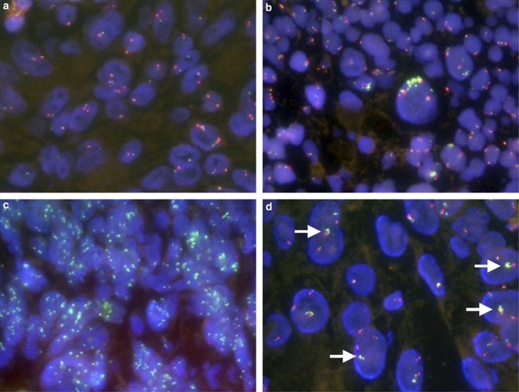
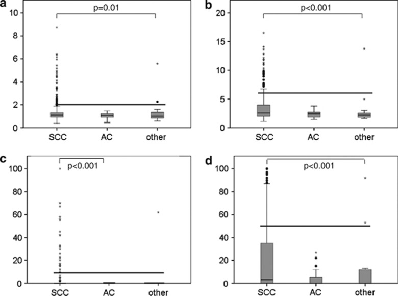
We recently reported fibroblast growth factor receptor-type 1 (FGFR1) amplification to be associated with therapeutically tractable FGFR1 dependency in squamous cell lung cancer. This makes FGFR1 a novel target for directed therapy in these tumors. To reproducibly identify patients for clinical studies, we developed a standardized reading and evaluation strategy for FGFR1 fluorescence in-situ hybridization (FISH) and propose evaluation criteria, describe different patterns of low- and high-level amplifications and report on the prevalence of FGFR1 amplifications in pulmonary carcinomas. A total of 420 lung cancer patients including 307 squamous carcinomas, 100 adenocarcinomas of the lung and 13 carcinomas of other types were analyzed for FGFR1 amplification using a dual color FISH. We found heterogeneous and different patterns of gene copy numbers. FGFR1 amplifications were observed in 20% of pulmonary squamous carcinomas but not in adenocarcinomas. High-level amplification (as defined by an FGFR1/centromer 8 (CEN8) ratio ≥2.0, or average number of FGFR1 signals per tumor cell nucleus ≥6, or the percentage of tumor cells containing ≥15 FGFR1 signals or large clusters ≥10%) was detected at a frequency of 16% and low-level amplification (as defined by ≥5 FGFR1 signals in ≥50% of tumor cells) at a frequency of 4%. We conclude that FGFR1 amplification is one of the most frequent therapeutically tractable genetic lesions in pulmonary carcinomas. Standardized reporting of FGFR1 amplification in squamous carcinomas of the lung will become increasingly important to correlate therapeutic responses with FGFR1 inhibitors in clinical studies. Thus, our reading and evaluation strategy might serve as a basis for identifying patients for ongoing and upcoming clinical trials.
Figures
References
-
- Mok TS, Wu YL, Thongprasert S, et al. Gefitinib or carboplatin-paclitaxel in pulmonary adenocarcinoma. N Engl J Med. 2009;361:947–957. - PubMed
-
- Soda M, Choi YL, Enomoto M, et al. Identification of the transforming EML4-ALK fusion gene in non-small-cell lung cancer. Nature. 2007;448:561–566. - PubMed
-
- Khuder SA. Effect of cigarette smoking on major histological types of lung cancer: a meta-analysis. Lung Cancer. 2001;31:139–148. - PubMed
Publication types
MeSH terms
Substances
LinkOut - more resources
Full Text Sources
Other Literature Sources
Medical
Miscellaneous
