. 2012 Dec;61(12):2273-82.
doi: 10.1007/s00262-012-1276-7. Epub 2012 Jun 10.

Mast cells impair the development of protective anti-tumor immunity

Affiliations

Mast cells impair the development of protective anti-tumor immunity

Anna Wasiuk et al. Cancer Immunol Immunother. 2012 Dec.

Abstract

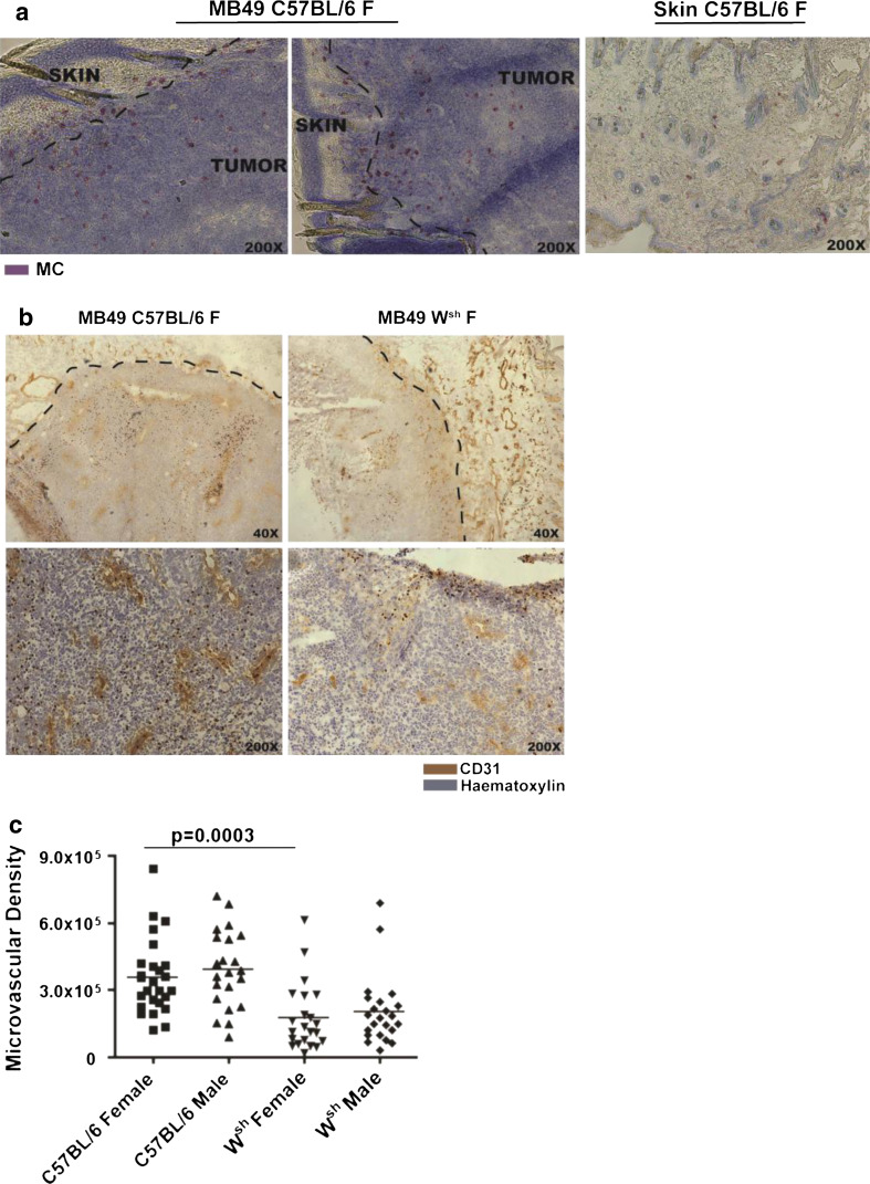
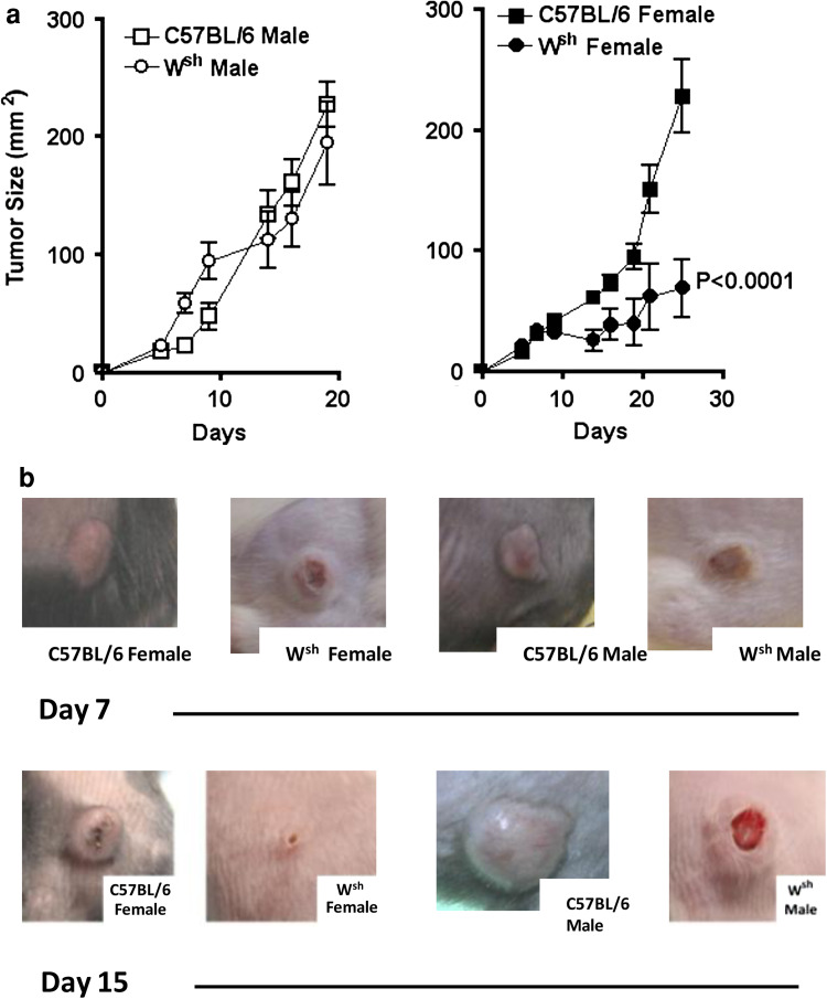
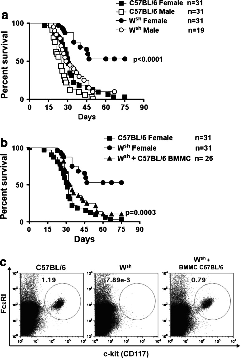
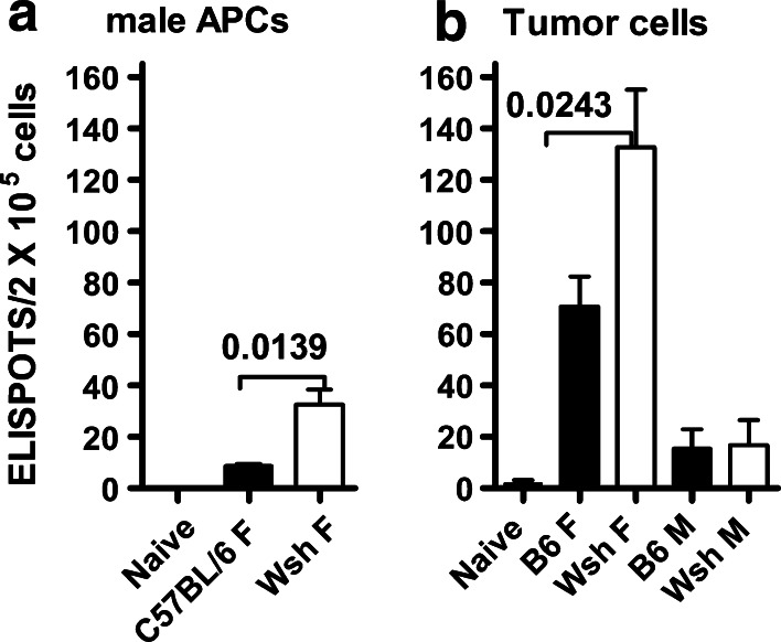
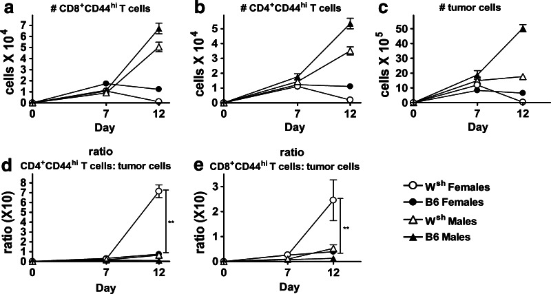
Mast cells have emerged as critical intermediaries in the regulation of peripheral tolerance. Their presence in many precancerous lesions and tumors is associated with a poor prognosis, suggesting mast cells may promote an immunosuppressive tumor microenvironment and impede the development of protective anti-tumor immunity. The studies presented herein investigate how mast cells influence tumor-specific T cell responses. Male MB49 tumor cells, expressing HY antigens, induce anti-tumor IFN-γ(+) T cell responses in female mice. However, normal female mice cannot control progressive MB49 tumor growth. In contrast, mast cell-deficient c-Kit(Wsh) (W(sh)) female mice controlled tumor growth and exhibited enhanced survival. The role of mast cells in curtailing the development of protective immunity was shown by increased mortality in mast cell-reconstituted W(sh) mice with tumors. Confirmation of enhanced immunity in female W(sh) mice was provided by (1) higher frequency of tumor-specific IFN-γ(+) CD8(+) T cells in tumor-draining lymph nodes compared with WT females and (2) significantly increased ratios of intratumoral CD4(+) and CD8(+) T effector cells relative to tumor cells in W(sh) mice compared to WT. These studies are the first to reveal that mast cells impair both regional adaptive immune responses and responses within the tumor microenvironment to diminish protective anti-tumor immunity.


Conflict of interest statement

The authors declare no competing financial interests.

Figures

Fig. 1
Fig. 1
Mast cells accumulate around MB49 tumors and contribute to increased microvascular density in tumors of WT mice. (a) Toluidine blue staining of MB49 tumor sections show mast cells surrounding the tumor. Two different sections from MB49 tumors (day 12) in WT female mice are shown. One representative normal skin section stained with Toluidine blue is shown. b Representative images of CD31 staining on tumor sections (day 12) from C57BL/6 female mice and Wsh female mice. The dotted lines on the top images show the boundary between tumor and normal tissues. c Quantification of immunohistochemical staining for CD31 on sections of C57BL/6 female and male and Wsh female and male i.d. tumor sections (n = 4 per group). Microvascular density was calculated by dividing number of pixels staining with CD31 over number of pixels in total skin section examined
Fig. 2
Fig. 2
Mast cell-deficient Wsh female mice show superior regression of MB49 tumors. a MB49 tumor growth curves in C57BL/6 female and male mice and the genetically mast cell-deficient Wsh male and female mice. Mice were inoculated intradermally (i.d.) with 2.5 × 105 cells per mouse and perpendicular diameters of tumors were measured; shown are the mean and SEM for 6–8 mice per group. The graph shown is representative of 10 experiments. Tumor growth in female B6 compared with female Wsh mice was significantly different (2-way ANOVA). b Top panels are images of representative day 7 tumors. Bottom panels are images of representative day 15 tumors in WT males and female and Wsh males and females
Fig. 3
Fig. 3
Enhanced survival of Wsh females with systemic MB49 tumors was abolished by their reconstitution with bone-marrow-derived mast cells. a MB49 tumor was injected systemically through the tail vein, and mice were monitored for survival. Survival curve is a composite of four independent experiments. b Systemic reconstitution of Wsh female mice with C57BL/6 bone-marrow-derived mast cells (BMMC). Cells were inoculated i.v., i.d., intraperitoneally, and subcutaneously to ensure mast cell reconstitution in all anatomical locations. Survival graphs show data from three independent experiments. c FACS plots showing representative samples from C57BL/6, Wsh, and Wsh reconstituted with WT bone-marrow mast cells. Cells are from an intraperitoneal lavage stained for FcεRI and CD117 (c-kit), to identify mast cells
Fig. 4
Fig. 4
Enhanced resistance of Wsh female mice to tumors is T cell mediated and can be adoptively transferred into mast cell-sufficient mice. Groups of mice were given MB49 tumors i.v. and monitored for survival. a and b Enhanced survival of Wsh female mice with systemic MB49 tumors was abolished by antibody-mediated depletion of CD8+ (α-CD8 Wsh F) or CD4+ T cells (α-CD4 Wsh F). Survival curves of Wsh mice depleted of CD4+ T cells and CD8+ T cells were significantly different from survival of intact Wsh mice *p < 0.04. c and d MB49 tumor was injected into groups of mice by i.d. route. Growth rates (perpendicular diameters) of i.d. MB49 tumor are shown with each point representing the mean and SEM of 6–8 mice. c Growth rate of MB49 tumor in Wsh females and B6 female and male controls shown for comparison to those in d. d Adoptive transfer of combined CD4+ and CD8+ T cells isolated from spleen and dLNs of Wsh female mice that had previously completely rejected i.d. MB49 (immune T cells), or from naïve Wsh female mice into naïve C57BL/6 female mice. Growth of MB49 tumor in recipients of immune T cells was significantly reduced compared with growth rate of MB49 tumors in naïve Wsh mice (p < 0.001). Data show composite results from 3 independent experiments. p values were calculated by the 2-way ANOVA
Fig. 5
Fig. 5
Increased IFN-γ responses in tumor dLNs of Wsh female tumor-bearing mice. The frequencies of tumor-responsive CD8+ T cells were measured in an ELISPOT assay. Tumor-draining inguinal lymph nodes from four mice were pooled and CD8+ T cells were enriched to 90–95 % purity and plated into triplicate wells for each condition. Enriched CD8+ T cells were plated at 2 × 105 cells per well with a T-depleted male spleens as antigen-presenting cells (APCs) or b irradiated MB49 tumor cells. Bars show the mean and SEM of ELISPOTS per 2 × 105 input CD8+ T cells with 3–10 points per group measured in four independent experiments. On the X-axis, the following abbreviations were used: B6 F, WT females; Wsh F, Wsh females; B6M, WT males; Wsh M, Wsh males. Significance was determined by unpaired t test
Fig. 6
Fig. 6
Wsh female mice have significantly increased ratios of intratumoral CD4+ and CD8+ T effector cells relative to tumor cells compared with other groups. Mice were given MB49 tumors by i.d. route. Tumors were excised and analyzed by flow cytometry on days 7 and 12. Absolute numbers of a CD4+CD44hi T cells b CD8+CD44hi T cells c and tumor cells in tumors from WT and Wsh female and male mice. d Ratios of intratumoral CD4+CD44hi T cells per total numbers of tumor cells are shown for each group. Wsh female compared with WT females p = 0.0024**. e Ratios of intratumoral CD8+CD44hi T cells per total tumor cells are shown for each group. Ratios for Wsh females compared with WT females, p = 0.006**. Points represent the mean and SEM of 8–9 mice/group on day 7 and 10–16 mice/group on day 12, data points were obtained in two independent replicate experiments. The Mann–Whitney test was used to determine significance

References

    1. Keibel A, Singh V, Sharma MC. Inflammation, microenvironment, and the immune system in cancer progression. Curr Pharm Des. 2009;15(17):1949–1955. doi: 10.2174/138161209788453167. - DOI - PubMed
    1. Coussens LM, Raymond WW, Bergers G, et al. Inflammatory cells up-regualate angiogenesis during squamous epithelial carcinogenesis. Genes Dev. 1999;13:1382–1397. doi: 10.1101/gad.13.11.1382. - DOI - PMC - PubMed
    1. Gounaris E, Erdman S, Restaino C, et al. Mast cells are an essential hematopoietic component for polyp development. Proc Natl Acad Sci USA. 2007;104:19977–19982. doi: 10.1073/pnas.0704620104. - DOI - PMC - PubMed
    1. Soucek L, Lawlor ER, Soto D, Shchors K, Swigart LB, Evan GI. Mast cells are required for angiogenesis and macroscopic expansion of Myc-induced pancreatic islet tumors. Nat Med. 2007;13(10):1211–1218. doi: 10.1038/nm1649. - DOI - PubMed
    1. Yang FC, Ingram DA, Chen S, Zhu Y, Yuan J, Li X, Yang X, Knowles S, Horn W, Li Y, Zhang S, Yang Y, Vakili ST, Yu M, Burns D, Robertson K, Hutchins G, Parada LF, Clapp DW. Nf1-dependent tumors require a microenvironment containing Nf1 ± and c-kit-dependent bone marrow. Cell. 2008;135(3):437–448. doi: 10.1016/j.cell.2008.08.041. - DOI - PMC - PubMed

Publication types