

Brain volume findings in 6-month-old infants at high familial risk for autism
- PMID: 22684595
- PMCID: PMC3744332
- DOI: 10.1176/appi.ajp.2012.11091425
Brain volume findings in 6-month-old infants at high familial risk for autism
Abstract
Objective: Individuals with autism as young as 2 years have been observed to have larger brains than healthy comparison subjects. Studies using head circumference suggest that brain enlargement is a postnatal event that occurs around the latter part of the first year. To the authors' knowledge, no previous brain imaging studies have systematically examined the period prior to age 2. In this study they used magnetic resonance imaging (MRI) to measure brain volume in 6-month-olds at high familial risk for autism.
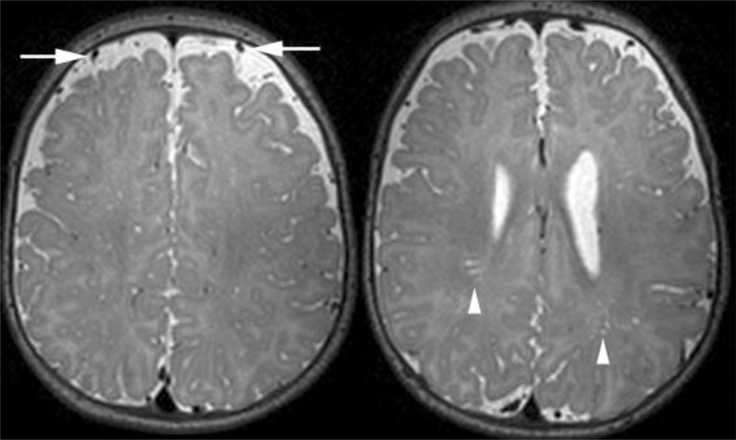
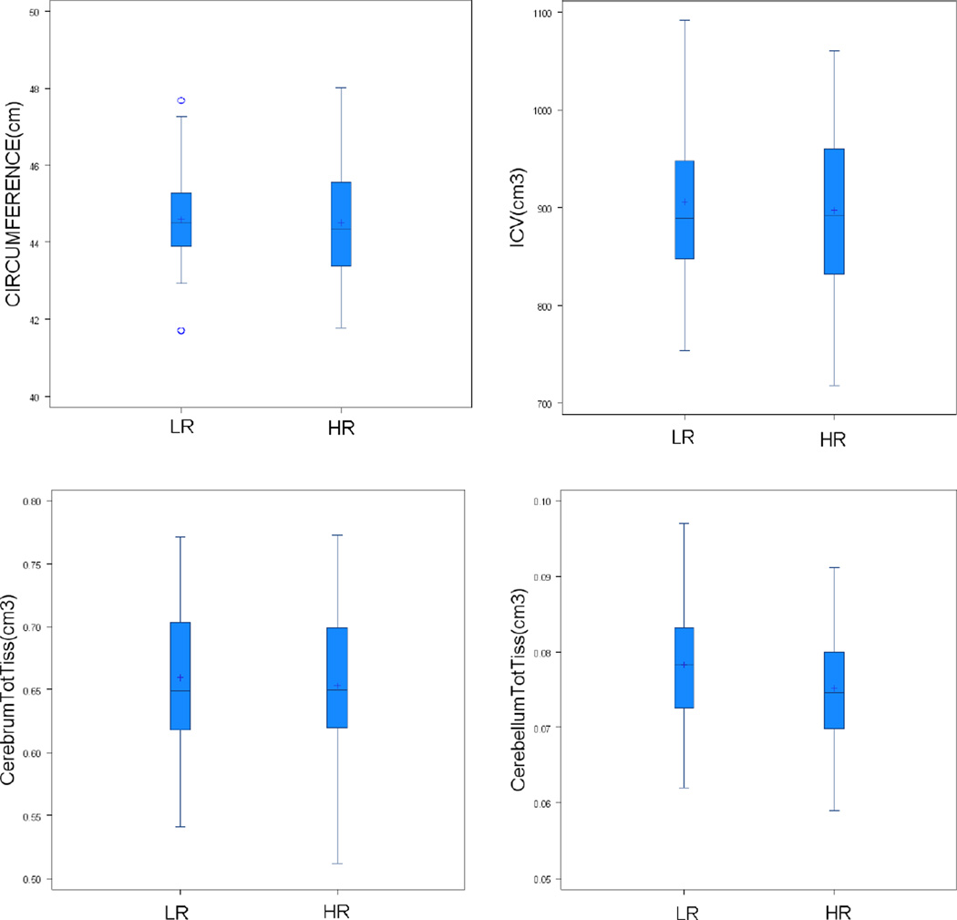
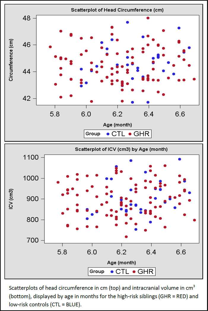
Method: The Infant Brain Imaging Study (IBIS) is a longitudinal imaging study of infants at high risk for autism. This cross-sectional analysis compared brain volumes at 6 months of age in high-risk infants (N=98) and infants without family members with autism (N=36). MRI scans were also examined for radiologic abnormalities
Results: No group differences were observed for intracranial, cerebrum, cerebellum, or lateral ventricle volume or for head circumference.
Conclusions: The authors did not observe significant group differences for head circumference, brain volume, or abnormalities in radiologic findings from a group of 6-month-old infants at high risk for autism. The authors are unable to conclude that these abnormalities are not present in infants who later go on to receive a diagnosis of autism; rather, abnormalities were not detected in a large group at high familial risk. Future longitudinal studies of the IBIS study group will examine whether brain volume differs in infants who go on to develop autism.
Figures

Comment in
-
Promise for finding brain biomarkers among infants at high familial risk for developing autism spectrum disorders.Am J Psychiatry. 2012 Jun;169(6):551-3. doi: 10.1176/appi.ajp.2012.12030397. Am J Psychiatry. 2012. PMID: 22684588 No abstract available.
References
-
- Piven J, Nehme E, Simon J, Barta P, Pearlson G, Folstein SE. Magnetic resonance imaging in autism: Measurement of the cerebellum, pons, and fourth ventricle. Biol Psychiatry. 1992;31:491–504. - PubMed
-
- Piven J, Ardnt S, Bailey J, Andreasen N. Regional brain enlargement in autism: A magnetic resonance imaging study. J Am Acad Child Adol Psychiatry. 1996;35:530–536. - PubMed
-
- Courchesne E, Karns CM, Davis HR, Ziccardi R, Carper RA, Tigue ZD, Chisum HJ, Moses P, Pierce K, Lord C, Lincoln AJ, Pizzo S, Schreibman L, Haas RH, Akshoomoff NA, Courchesne RY. Unusual brain growth patterns in early life in patients with autistic disorder: An MRI study. Neurology. 2001;57:245–254. - PubMed
-
- Sparks BF, Friedman SD, Shaw DW, Aylward EH, Echelard D, Artru AA, Maravilla KR, Giedd JN, Munson J, Dawson G, Dager SR. Brain structural abnormalities in young children with autism spectrum disorder. Neurology. 2002;59(2):184–192. - PubMed
-
- Bailey A, Le Couteur A, Gottesman I, Bolton P, Simonoff E, Yuzda E, Rutter M. Autism as a strongly genetic disorder: Evidence from a British twin study. Psychol Med. 1995;25:63–77. - PubMed
Publication types
MeSH terms
Grants and funding
LinkOut - more resources
Full Text Sources
Other Literature Sources
