. 2012 Sep;40(16):8059-71.
doi: 10.1093/nar/gks504. Epub 2012 Jun 8.

Control of alternative splicing by forskolin through hnRNP K during neuronal differentiation

Affiliations

Control of alternative splicing by forskolin through hnRNP K during neuronal differentiation

Wenguang Cao et al. Nucleic Acids Res. 2012 Sep.

Abstract

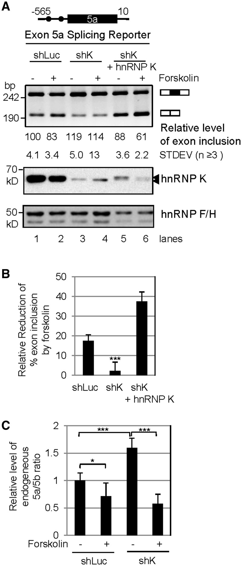
The molecular basis of cell signal-regulated alternative splicing at the 3' splice site remains largely unknown. We isolated a protein kinase A-responsive ribonucleic acid (RNA) element from a 3' splice site of the synaptosomal-associated protein 25 (Snap25) gene for forskolin-inhibited splicing during neuronal differentiation of rat pheochromocytoma PC12 cells. The element binds specifically to heterogeneous nuclear ribonucleo protein (hnRNP) K in a phosphatase-sensitive way, which directly competes with the U2 auxiliary factor U2AF65, an essential component of early spliceosomes. Transcripts with similarly localized hnRNP K target motifs upstream of alternative exons are enriched in genes often associated with neurological diseases. We show that such motifs upstream of the Runx1 exon 6 also bind hnRNP K, and importantly, hnRNP K is required for forskolin-induced repression of the exon. Interestingly, this exon encodes the peptide domain that determines the switch of the transcriptional repressor/activator activity of Runx1, a change known to be critical in specifying neuron lineages. Consistent with an important role of the target genes in neurons, knocking down hnRNP K severely disrupts forskolin-induced neurite growth. Thus, through hnRNP K, the neuronal differentiation stimulus forskolin targets a critical 3' splice site component of the splicing machinery to control alternative splicing of crucial genes. This also provides a regulated direct competitor of U2AF65 for cell signal control of 3' splice site usage.


Figures

Figure 1.
Figure 1.
Isolation of a KARRE from the upstream 3′ splice site of the exon 5a of Snap25 gene based on forskolin reduction of 5a/5b ratios in PC12 cells during neuronal differentiation. (A) Forskolin-induced neuronal differentiation of PC12 cells. Shown are representative images of PC12 cells without or with forskolin addition for 18 h. (B) Diagram of the alternative splicing of the exons 5a and 5b of the Snap25 gene. Arrows: sites of restriction enzymes NdeI (5a) and AvaII (5b) used to digest the PCR products. Arrowheads: PCR primers. (C) RT-PCR products from untreated (−) and treated PC12 cells. ETOH: ethanol, vehicle for forskolin and Dex (dexamethasone); H89: a PKA-specific inhibitor; NC: PCR negative control; M: molecular size marker. (D) Diagram of the deletion/replacement mutants of Snap25 splicing reporters and their exon inclusion levels in the presence of PKAm or active PKA. Asterisks: indicating P value levels in one tail, paired Student’s t-test (***P < 0.001, *P < 0.05, n ≥ 3). (E) An agarose gel showing the PKA response of the heterologous S175NK reporter containing the 17 nt Snap25a element upstream of a stronger exon (175NK, about 75% inclusion with PKAm coexpression).
Figure 2.
Figure 2.
Identification of hnRNP K and U2AF65 as specific factors binding to the KARRE. (A) Alignment of the KARRE-containing upstream 3 splice sites of Snap25 exon 5a from different species. The TCCCT, as well as a TCCT, (with heavy bars above) is mostly conserved within the polypyrimidine tract (boxed) among vertebrates. The 17 nt mouse KARRE sequence is above the dotted heavy line. (B) Sequence of the Biotin-RNA wild type and mutant probes used to pull down 3 splice site-binding proteins, with the hnRNP K target consensus motifs underlined. (C) A Coomassie-stained SDS-PAGE gel of proteins pulled down from HeLa nuclear extracts using the probes in B. 1: nucleolin, 2: hnRNP K, 3: PTB, 4: YB-1, as identified in mass spectrometry with significance against random events. (D) Human hnRNP K protein sequence, with the tryptic peptide hits in MALDI-TOF mass spectrometry highlighted and underlined. (E) A Western blot of U2AF65 proteins similarly pulled down as in C.
Figure 3.
Figure 3.
Regulation of hnRNP K binding to the KARRE by forskolin and its direct competition with U2AF65. (A) Western blot of hnRNP K proteins pulled down from PC12 nuclear extracts using the biotin-RNA probes in Figure 2B. Et: vehicle ethanol, Fsk: forskolin (10 µM). (B) UV cross-linking immunoprecipitation of hnRNP K in forskolin-treated PC12 nuclear extracts with the 3′ splice site of exon 5a and its sensitivity to pretreatment by PPase. The wild type RNA probe sequence is the same as in Figure 2B except that it is without biotin. The upper panel is a phosphorimage and the lower a Western blot for the hnRNP K protein in the same SDS-PAGE gel. (C) (Upper panel) a phosphorimage of recombinant His-hnRNP K incubated with active or heat-inactivated PKA in the presence of [32P-γ]ATP in in vitro kinase assay. Lower panel is a Western bot image of the same gel showing equal loading of His-hnRNP K. (D) HnRNP K interacts with the endogenous Snap25 pre-mRNA transcript. Above the gel is a diagram of the PCR target pre-mRNA region of Snap25, with thin lines as introns, boxes as exons and arrowheads as locations of PCR primers. The agarose gel shows the RT-PCR products from RNA samples isolated from the nuclear extracts of forskolin-treated PC12 cells, or from immunoprecipitates using anti-hnRNP K (anti-K) or protein G beads. Each RNA sample was treated with DNase I and one of them also with RNase (A + T1) as indicated. a: a band insensitive to either DNase or RNase treatment, probably nonspecific product from the PCR primers. (E) Western blots of hnRNP K and U2AF65 proteins pulled down from HeLa nuclear extracts with increasing amount of His-hnRNP K added, using the wild type biotin-RNA probe in Figure 2B. The blot was first probed with anti-hnRNP K antibody, stripped with SDS buffer and then reprobed with anti-U2AF65. (F) UV cross-linking of hnRNP K and U2AF65 to the 3′ splice site of exon 5a. The hnRNP K consensus motifs (underlined) and its C to G mutations (italicized) are shown above the denaturing PAGE gel. b: a protein band enhanced by the mutation, likely preferring the G tracts in the mutant, at similar size as hnRNP F/H.
Figure 4.
Figure 4.
Effect of lentiviral vector-mediated expression of shRNA against hnRNP K (shK) or rescue with an exogenous HA-tagged hnRNP K on forskolin-regulated exon 5a inclusion. (A). Shown are the exon 5a insert (top), an agarose gel of the spliced products (middle) and Western blots of hnRNP K and loading control hnRNP F/H. The intron lengths of Snap25 are indicated above the insert. Dots: hnRNP K consensus motifs. shLuc: control shRNA, against luciferase. The two arrowheads point to the positions of the endogenous (lower) and exogenous (upper) hnRNP K proteins. (B) A bar graph of the relative forskolin-reduction (average ± SD, n = 3) of exon 5a inclusion of the splicing reporter in A, which is calculated as the net reduction of percent exon 5a inclusion relative to its level without forskolin (same as in the followings). (C) A bar graph of endogenous 5a/5b ratio levels in PC12 cells transduced with shK relative to that in the mock- or shLuc-transduced cells without (−) or with (+) forskolin treatment (average ± SD, n ≥ 3). *P < 0.05, ***P < 0.001.
Figure 5.
Figure 5.
Functional categories of transcripts containing similar hnRNP K target motifs within U2AF65 binding sites and an example for the essential role of hnRNP K in forskolin-regulated splicing of an endogenous neuronal gene. (A) Percentage of the 46 hnRNP K target transcripts/genes in each significant category of biological functions and diseases. These transcripts, containing similar hnRNP K target motifs within U2AF65 binding sites, were identified from the alternative splicing database ASAPII. The categories are as follows: 1, cell morphology; 2, cell cycle; 3, neurological diseases; 4, drug metabolism; 5, endocrine system; 6, inflammatory response and 7, lipid metabolism. (B) Diagram of the upstream 3′ splice site sequences (upper), splicing pattern (middle) and protein domains (lower) of the vertebrate Runx1 (also called acute myeloid leukemia (AML)-1B, GenBank accession #: NM_001754) gene. The relative position of the splice site, exon, encoded peptide and its location in the protein domains are linked with dotted lines. The black dot denotes the 3′ splice site with TCCCT motifs. The 192 nt exon 6-encoded peptide (red) contains the critical sequence (underlined) required for binding by the Sin3A transcriptional corepressor. Runt: Runt homology DNA-binding domain (brown), AD: activation domain (green). Relative locations of the AD domain and Sin3A binding site/activity are based on published data by Lutterbach, with the expected variant transcriptional activities in brackets at the bottom. (C) Sequences of wild type or mutant RunX1 RNA probe for UV cross-linking experiments. Note that all cytidines (underlined) in the hnRNP K target motifs are mutated to guanines. (D) HnRNP K and U2AF65 binding to the upstream 3′ splice site of RunX1 exon 6 in UV cross-linking with HeLa nuclear extracts, carried out similarly as in Figure 3F. On the right (lanes 6 and 7) is a higher contrast image of the IP samples in lanes 4 and 5. a: protein bands also abolished by the mutations, at sizes similar to PTB doublets. b: a protein only in the mutant sample, likely preferring the G tracts, at a size similar to hnRNP F/H. (E) An agarose gel of the Runx1 transcripts in PC12 cells with or without expressing the shRNA against hnRNP K (upper) and a bar graph of the relative reduction of exon 6 inclusion by forskolin (lower, average ± SD, n ≥ 3). **P < 0.01, compared with controls. Arrowheads in B and C demarcate intron–exon junctions.
Figure 6.
Figure 6.
Effect of knocking down hnRNP K on forskolin-induced neurite growth. Shown are bright field images of PC12 cells expressing shRNA against luciferase (shLuc, control) or hnRNP K (shK) and treated with forskolin (10 µM) for 18 h as indicated. To the right of the images is a bar graph of the levels of neurite densities measured in the two groups (***P < 0.005). Images representative of three samples treated separately.
Figure 7.
Figure 7.
Diagram of our proposed model for the observed forskolin effect on exon skipping through direct hnRNP K competition with U2AF65 at the 3′ splice site during neuronal differentiation of PC12 cells. Briefly, on stimulation of PC12 cells with the external stimulus forskolin and activation of the PKA or other pathways, hnRNP K binding to the KARRE motifs at the upstream 3′ splice sites of the exons is enhanced inside the nucleus (gray oval), competing with U2AF65 binding (detailed in the enlarged box with Snap25 and Runx1 3′ splice sites as examples). The hnRNP K target consensus KARRE motifs are in red and underlined. For Snap25, hnRNP K likely acts together with an unknown factor (blue oval with a question mark) for the forskolin repression of exon 5a. For Runx1, hnRNP K alone is sufficient to mediate the forskolin repression. The two hnRNP K proteins above the polypyrimidine tract of Runx1 are to reflect the multiple binding motifs available for the protein to compete with U2AF65. The regulation leads to exon skipping and neurite growth in the presence of the resulting splice variants (such as reduced Snap25 a/b ratio or increased Runx1-E16). Without hnRNP K and this regulation (upper right corner), the splicing switches/changes of a group of neuronal splice variants (Supplementary Tables SI and SII), including Snap25a/b and the Runx1 + E6, are altered contributing to disrupted neurite growth. Black dot: potential branch point of Runx1. The upstream branch point of Snap25 exon 5a is further upstream according to our experiment data.

References

    1. Graveley BR. Alternative splicing: increasing diversity in the proteomic world. Trends Genet. 2001;17:100–107. - PubMed
    1. Maniatis T, Tasic B. Alternative pre-mRNA splicing and proteome expansion in metazoans. Nature. 2002;418:236–243. - PubMed
    1. Black DL. Mechanisms of alternative pre-messenger rna splicing. Annu. Rev. Biochem. 2003;72:291–336. - PubMed
    1. Wang ET, Sandberg R, Luo S, Khrebtukova I, Zhang L, Mayr C, Kingsmore SF, Schroth GP, Burge CB. Alternative isoform regulation in human tissue transcriptomes. Nature. 2008;456:470–476. - PMC - PubMed
    1. Tazi J, Bakkour N, Stamm S. Alternative splicing and disease. Biochim. Biophys. Acta. 2009;1792:14–26. - PMC - PubMed

Publication types

MeSH terms

Substances