. 2012 Aug 15;520(12):2730-41.
doi: 10.1002/cne.23078.

Spatiotemporal distribution of vasoactive intestinal polypeptide receptor 2 in mouse suprachiasmatic nucleus

Affiliations

Spatiotemporal distribution of vasoactive intestinal polypeptide receptor 2 in mouse suprachiasmatic nucleus

Sungwon An et al. J Comp Neurol. .

Abstract

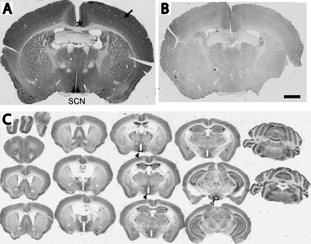
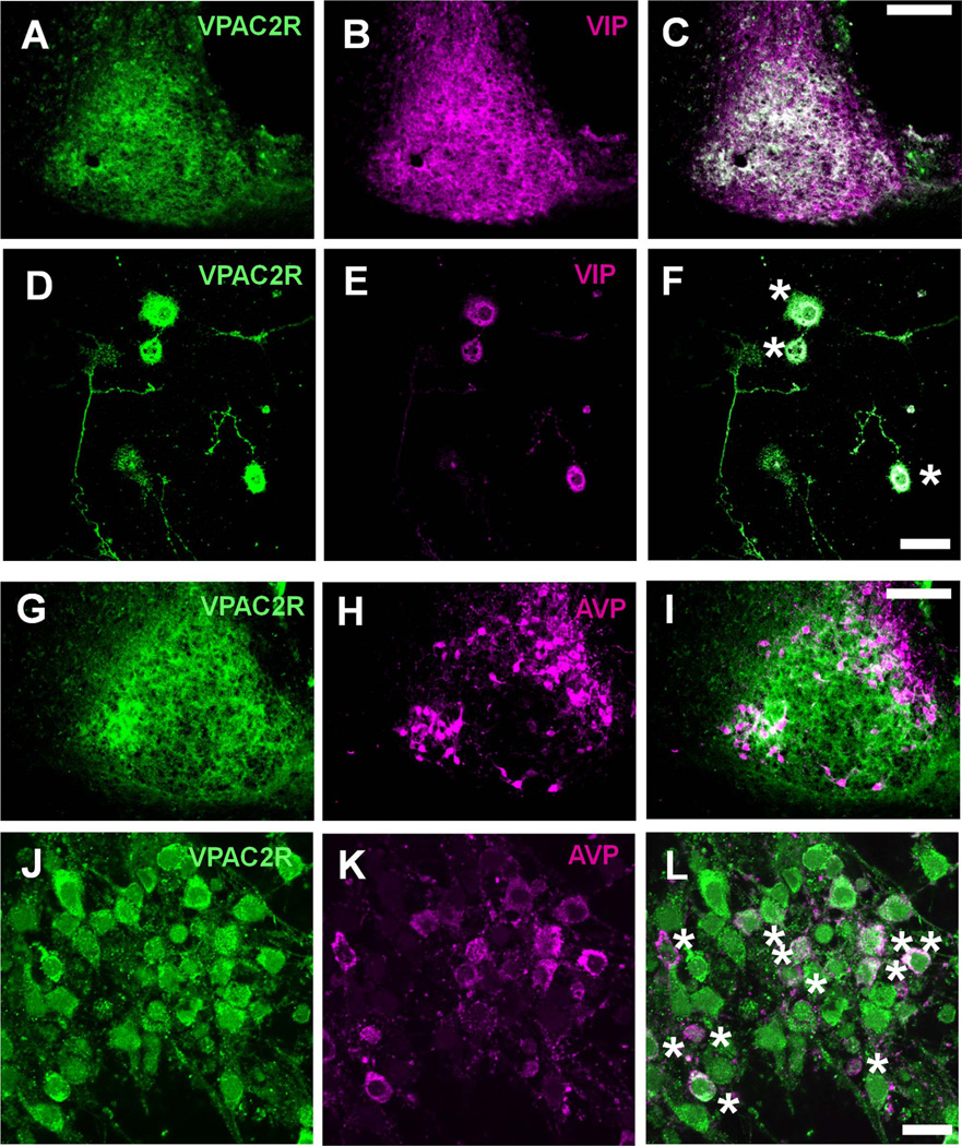
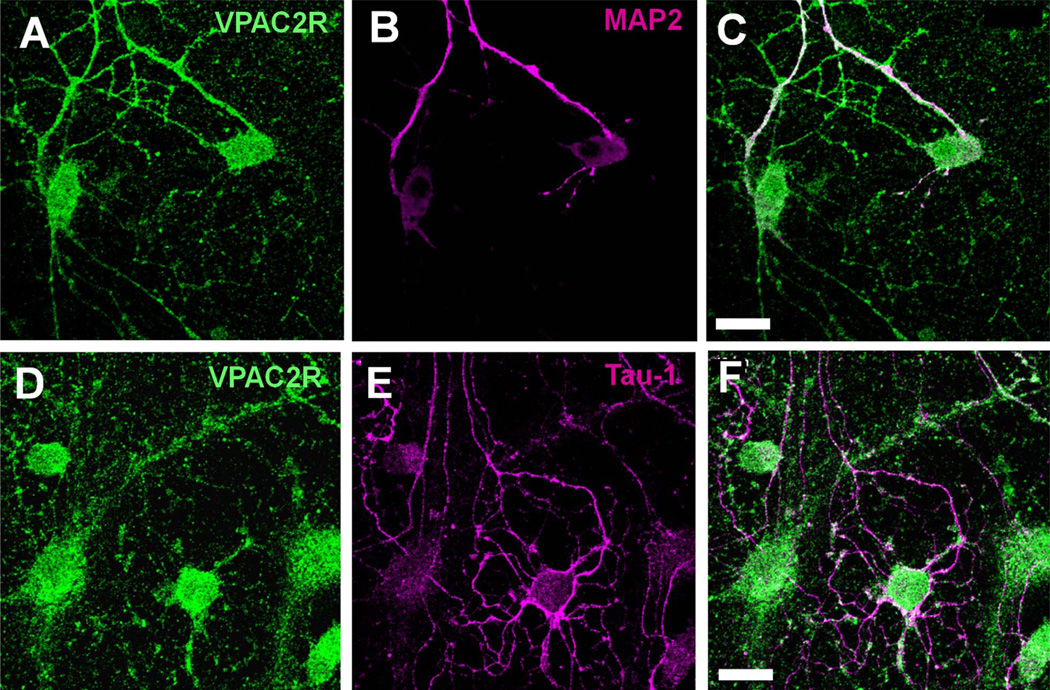
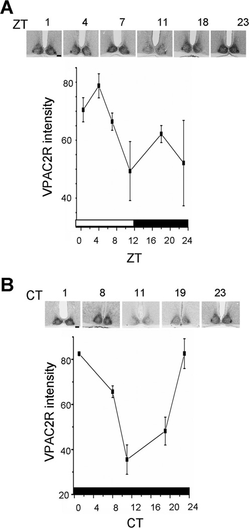
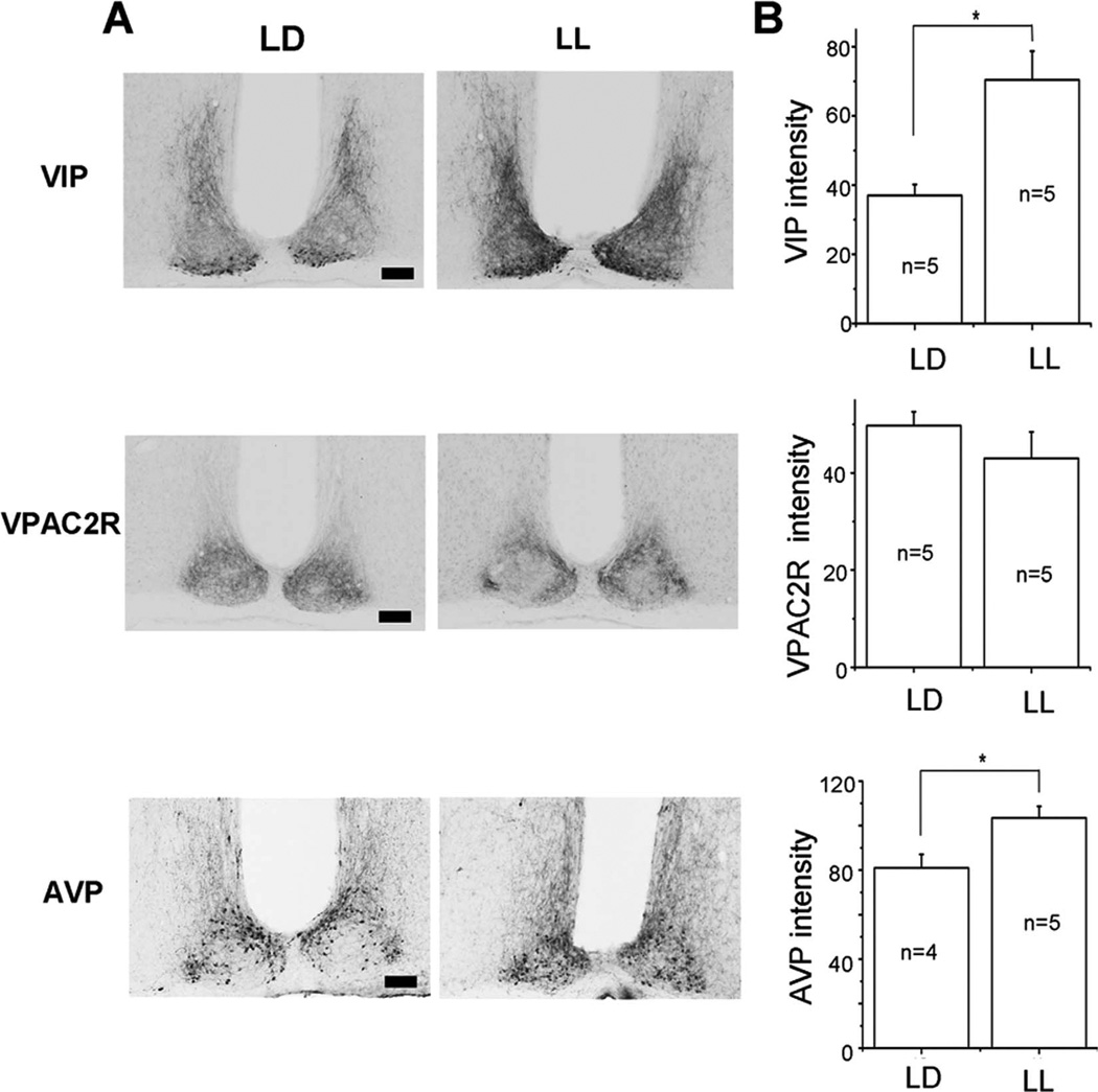
Vasoactive intestinal polypeptide (VIP) signaling is critical for circadian rhythms. For example, the expression of VIP and its main receptor, VPAC2R, is necessary for maintaining synchronous daily rhythms among neurons in the suprachiasmatic nucleus (SCN), a master circadian pacemaker in animals. Where and when VPAC2R protein is expressed in the SCN and other brain areas has not been examined. Using immunohistochemistry, we characterized a new antibody and found that VPAC2R was highly enriched in the SCN and detectable at low levels in many brain areas. Within the SCN, VPAC2R was circadian, peaking in the subjective morning, and abundantly expressed from the rostral to caudal margins with more in the dorsomedial than ventrolateral area. VPAC2R was found in nearly all SCN cells including neurons expressing either VIP or vasopressin (AVP). SCN neurons mainly expressed VPAC2R in their somata and dendrites, not axons. Finally, constant light increased VIP and AVP expression, but not VPAC2R. We conclude that the circadian clock, not the ambient light level, regulates VPAC2R protein localization. These results are consistent with VPAC2R playing a role in VIP signaling at all times of day, broadly throughout the brain and in all SCN cells.


Figures

Figure 1
Figure 1
VPAC2R was strongly expressed in the suprachiasmatic nucleus (SCN). A: A representative coronal section of a wild-type mouse brain showing intense immunostaining in the SCN, moderate to weak staining in the neocortex (arrow) and no staining in axonal projections like the corpus callosum (asterisk). B: A representative brain section from a Vipr2−/− mouse showing little or no VPAC2R immunoreactivity. Sections in A and B were imaged, processed and displayed using identical protocols. C: Serial coronal sections of a mouse brain immunolabeled for VPAC2R. These sections were processed to reveal the intense staining in the SCN (closed triangles) and moderate staining in the lateral posterior arcuate nucleus (open triangles). Less intense labeling is seen broadly throughout the brain including the hippocampus, neocortex, and cerebellum. Scale bar = 1 mm in B (applies to A,B)
Figure 2
Figure 2
Broad VPAC2R expression in the SCN. VPAC2R immunoreactivity was present throughout the rostral (top left) to caudal extent (bottom right) of the SCN. Note that VPAC2R labeling tended to be more intense in the dorsomedial than ventrolateral SCN. Scale bar = 1 mm.
Figure 3
Figure 3
VPAC2R expression overlapped with VIP and AVP expression in the SCN. A–L: Representative images of SCN cells co-stained for VPAC2R and VIP (A–F) or VPAC2R and AVP (G–L). Images show either the left SCN of a coronal brain section (A–C, G–I) or SCN cells 1 week after being plated at low density in vitro (D–F, J–L). The composite images show the extensive overlap (yellow) between VPAC2R (green) and VIP (red), and VPAC2R and AVP (red) expression (C,F,I,L). Dissociated SCN neurons expressing both VPAC2R and either neuropeptide were marked with an asterisk (F and L). Scale bar = 100 µm in C (applies to A–C) and I (applies to G–I); 20 µm in F (applies to D–F) and L (applies to J–L).
Figure 4
Figure 4
VPAC2R co-localized with dendritic markers but not with axonal markers in the SCN. A–F: Representative images of dissociated SCN neurons stained for VPAC2R (A,D) and MAP2, a dendritic marker protein (B), or Tau-1, an axonal marker protein (E). Note that VPAC2R (green) staining colocalized with MAP2 (red) along processes (C) but not with Tau-1 (red) staining (F). Scale bar = 20 µm in C (applies to A–C) and F (applies to D–F).
Figure 5
Figure 5
VPAC2R expression oscillated in a light–dark cycle and in constant darkness. Data points represent the mean ± SEM of three brains at each time point. A: In LD, VPAC2R expression was rhythmic, with a peak after dawn (ZT1) and trough around dusk (ZT11). B: In DD, the pattern was similar, with peak VPAC2R expression around subjective dawn (CT1) and trough near subjective dusk (CT11). Insets show sections of the SCN from representative mice at each time point. The bars at the bottom of each plot show the times of lights on (white) and off (black) experienced by the mice. Scale bar = 100 µm in A,B.
Figure 6
Figure 6
Constant light increased VIP and AVP, but not VPAC2R expression in the SCN. A: Representative micrographs illustrate VIP-immunoreactive cell bodies in the ventral SCN and their dense projections into the dorsomedial SCN in light–dark (LD; left) and constant light (LL; right). Note the darker projections and cell bodies in LL. Similarly, AVP immunoreactivity increased, whereas VPAC2R labeling did not change in LL B: Mean ± SEM of the staining intensity in the SCN above background for VIP, VPAC2R, and AVP from mice maintained in LD or LL. Asterisks indicate significance, at P < 0.05, and n indicates the number of mice used. Scale bar = 100 µm in A.

References

    1. Abrahamson EE, Moore RY. Suprachiasmatic nucleus in the mouse: retinal innervation, intrinsic organization and efferent projections. Brain Res. 2001;916:172–191. - PubMed
    1. An S, Irwin RP, Allen CN, Tsai CA, Herzog ED. Vasoactive intestinal polypeptide requires parallel changes in adenylate cyclase and phospholipase C to entrain circadian rhythms to a predictable phase. J Neurophysiol. 2011;105:2289–2296. - PMC - PubMed
    1. Aton SJ, Herzog ED. Come together, right…now: synchronization of rhythms in a mammalian circadian clock. Neuron. 2005;48:531–534. - PMC - PubMed
    1. Aton SJ, Colwell CS, Harmar AJ, Waschek J, Herzog ED. Vasoactive intestinal polypeptide mediates circadian rhythmicity and synchrony in mammalian clock neurons. Nat Neurosci. 2005;8:476–483. - PMC - PubMed
    1. Ben-Barak Y, Russell JT, Whitnall MH, Ozato K, Gainer H. Neurophysin in the hypothalamo-neurohypophysial system. I. Production and characterization of monoclonal antibodies. J Neurosci. 1985;5:81–97. - PMC - PubMed

Publication types

MeSH terms

Substances

LinkOut - more resources