. 2012 Jun 26;109(26):10376-81.
doi: 10.1073/pnas.1200174109. Epub 2012 Jun 8.

Anoctamin 1 (Tmem16A) Ca2+-activated chloride channel stoichiometrically interacts with an ezrin-radixin-moesin network

Affiliations

Anoctamin 1 (Tmem16A) Ca2+-activated chloride channel stoichiometrically interacts with an ezrin-radixin-moesin network

Patricia Perez-Cornejo et al. Proc Natl Acad Sci U S A. .

Abstract

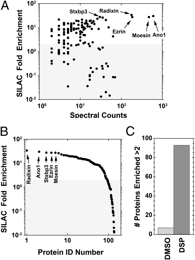
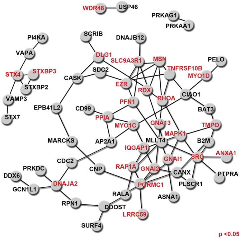
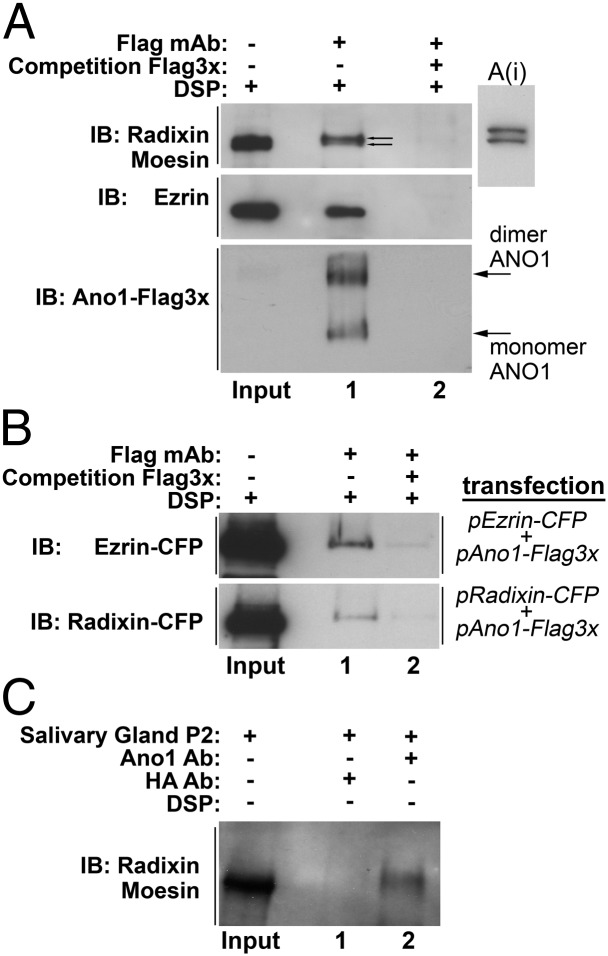
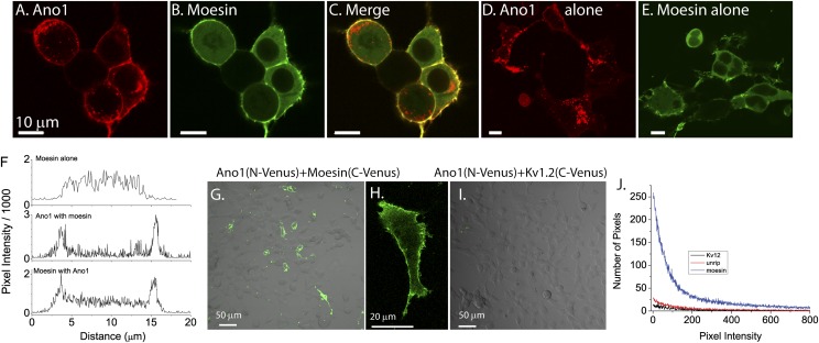
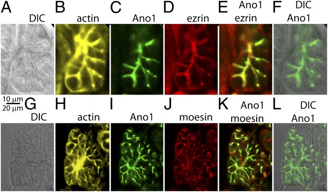
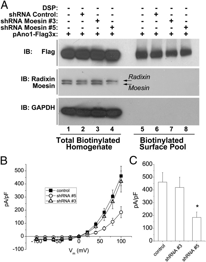
The newly discovered Ca(2+)-activated Cl(-) channel (CaCC), Anoctamin 1 (Ano1 or TMEM16A), has been implicated in vital physiological functions including epithelial fluid secretion, gut motility, and smooth muscle tone. Overexpression of Ano1 in HEK cells or Xenopus oocytes is sufficient to generate Ca(2+)-activated Cl(-) currents, but the details of channel composition and the regulatory factors that control channel biology are incompletely understood. We used a highly sensitive quantitative SILAC proteomics approach to obtain insights into stoichiometric protein networks associated with the Ano1 channel. These studies provide a comprehensive footprint of putative Ano1 regulatory networks. We find that Ano1 associates with the signaling/scaffolding proteins ezrin, radixin, moesin, and RhoA, which link the plasma membrane to the cytoskeleton with very high stoichiometry. Ano1, ezrin, and moesin/radixin colocalize apically in salivary gland epithelial cells, and overexpression of moesin and Ano1 in HEK cells alters the subcellular localization of both proteins. Moreover, interfering RNA for moesin modifies Ano1 current without affecting its surface expression level. Another network associated with Ano1 includes the SNARE and SM proteins VAMP3, syntaxins 2 and -4, and syntaxin-binding proteins munc18b and munc18c, which are integral to translocation of vesicles to the plasma membrane. A number of other regulatory proteins, including GTPases, Ca(2+)-binding proteins, kinases, and lipid-interacting proteins are enriched in the Ano1 complex. These data provide stoichiometrically prioritized information about mechanisms regulating Ano1 function and trafficking to polarized domains of the plasma membrane.


Conflict of interest statement

The authors declare no conflict of interest.

Figures

Fig. 1.
Fig. 1.
Immunoaffinity purification of Ano1 supramolecular complexes from Ano1–FLAG3× cell line. (A) Silver-stained SDS/PAGE gel. (B) Immunoblot. Ano1 migrates as ∼130-kDa monomer and a ∼260-kDa dimer (25). HEK cells were cross-linked with DSP. Triton X-100 soluble lysates were incubated with anti-FLAG magnetic beads. Input: total lysate from Ano1–FLAG3× HEK cells. Lanes 1–4: Bound proteins were eluted with SDS/PAGE sample buffer. Lane 1: No antibody control, beads lacking antibody. Lane 2: Irrelevant antibody control, beads coated with SV2 antibody. Lane 3: beads coated with anti-FLAG antibody. Note the band corresponding to Ano1 at ∼130 kDa. Lane 4: Excess antigen control, beads coated with anti-FLAG antibody in the presence of 340 μM FLAG3× peptide. Lane 5: Proteins eluted from anti-FLAG coated beads (as in lane 3) with wash buffer A. Lane 6: Proteins eluted from anti-FLAG coated beads (as in lane 3) with 340 μM FLAG3× peptide.
Fig. 2.
Fig. 2.
Summary of SILAC experiment. Untransfected HEK cells were incubated in isotopically “light” (R0K0) DMEM. HEK cells stably transfected with Ano1–FLAG3× were incubated in isotopically “heavy” (R10K8) medium. Cells were substoichiometrically cross-linked using DSP. Lysates were immunoprecipitated using magnetic beads decorated with FLAG antibody. Ano1 supramolecular complexes were eluted with FLAG3× peptide. Samples were combined at a 1:1 ratio and analyzed by nano-LS MS/MS. Peptides enriched more than twofold with the R10K8 amino acids were considered as potential Ano1 interactors. The list of proteins was refined by curation against a list of peptides that nonspecifically bind to the immunomagnetic beads. The Venn diagram shows the number of peptides identified in the experiment. A total of 509 proteins were identified, 93 of which were enriched more than twofold in R10K8 and did not bind nonspecifically to immunomagnetic beads.
Fig. 3.
Fig. 3.
Fold-enrichment and spectral counts of peptides identified in SILAC experiment. (A) Fold enrichment of heavy:light of the top 209 proteins vs. the spectral counts. (B) Data in A replotted as fold enrichment vs. protein rank. The top five enriched proteins are labeled. (C) Number of proteins enriched more than twofold from cells treated with vehicle (DMSO) or DSP.
Fig. 4.
Fig. 4.
DAPPLE analysis of interactions among the proteins in the Ano1 interactome. Proteins from Dataset S1 were submitted for analysis using parameters: iterations = 10,000, common interactor binding degree cutoff = 6, iteration = on. Red labels are nodes that achieved a P value <0.05, indicating the probability that the connections at these nodes were observed by chance.
Fig. 5.
Fig. 5.
Coimmunoprecipitation of ezrin, radixin, and moesin with Ano1. (A) Ano1–FLAG3× stable cell line was cross-linked and Ano1 complexes were purified by immunoaffinity chromatography (lane 1). Control sample (lane 2) was applied to the anti-FLAG affinity column in the presence of excess FLAG3× peptide. Eluted proteins were run on Western blot and probed with antibodies against moesin (67.8 kDa) and radixin (67.8 kDa), ezrin, or FLAG (Ano1). Insert A(i) shows a better separation of radixin and moesin than in Fig. 8A. (B) HEK cells transiently coexpressing Ano1–FLAG3× and radixin–CFP or moesin–cyan fluorescent protein were cross-linked and Ano1 complexes purified and probed as in A. (C) Moesin/radixin were immunoprecipiated from salivary gland P2 Triton X-100 soluble fraction (Fig. S5) with a polyclonal antibody against Ano1 (lane 2) or an irrelevant antibody against hemaglutinin (control, lane 1).
Fig. 6.
Fig. 6.
Colocalization of moesin and Ano1 in HEK cells. (AC) HEK cells were transfected with Ano1–mCherry (red, A) and moesin–EGFP (green, B). (C) A and B superimposed. (D and E) Cells transfected with Ano1–mCherry alone (D) or moesin–EGFP alone (E). (F) Profiles of transcellular fluorescence in cells expressing only moesin–EGFP or moesin–EGFP plus Ano1–mCherry. Profiles were obtained by drawing a line across the cell avoiding the nucleus where moesin is excluded. Profiles are representative of >10 cells selected at random. (GJ) Bimolecular fluorescence complementation (BiFC). (G) Cells were cotransfected with Ano1 tagged with Venus(1–155) and Moesin tagged with Venus(156–239). (H) High power showing membrane localization of the BiFC fluorescence. (I) Control for BiFC. Cells were transfected with Ano1–Venus(1–155) and Kv1.2–Venus(156–239).(J) Quantification of BiFC. Histograms of pixel intensity for fields transfected with Ano1–Venus(1–155) plus moesin–Venus(156–239), the potassium channel Kv1.2–Venus(156–239), or the survival of motor neurons (SMN) complex subunit unrip–Venus(156–239).
Fig. 7.
Fig. 7.
Localization of Ano1, ezrin, moesin, and actin in salivary gland. Coloring is as follows: moesin and ezrin are red, Ano1 is green, and actin (stained with Alexa 647 phalloidin) is yellow. (AF). Colocalization of Ano1, ezrin, and actin. (GL). Colocalization of Ano1, moesin, and actin. (A and G) DIC images. (B and H) Actin. (C and I) Ano1. (D) Ezrin. (E) Ano1 and ezrin overlay. (J) Moesin. (K) Ano1 and moesin overlay. (F and L) Ano1 and DIC overlay.
Fig. 8.
Fig. 8.
Effect of knockdown of moesin on Ano1 currents. (A) Western blot of total cell extracts (lanes 1–4) and surface biotinylated proteins (lanes 5–8). Cells were transfected with Ano1–FLAG3× and with empty shRNA vector or two different shRNA constructs (nos. 3 and 5). Cells transfected with shRNA vectors were selected with puromycin. (Upper) Ano1 expression detected with Ano1 antibody. (Lower) Radixin and moesin expression. Control shRNA: cells were transfected with the empty shRNA vector and selected with puromycin as with the other shRNA constructs. shRNA 5 almost completely eliminated moesin expression. (B) IV curves of whole-cell patch clamped Ano1 currents in a cell line stably expressing Ano1 tagged with EGFP on the C terminus treated with control vector, shRNA 3 or shRNA 5. Intracellular (pipet) Ca2+ concentration was 600 nM. (C) Average current densities at +100 mV. *P < 0.05.

References

    1. Duran C, Thompson CH, Xiao Q, Hartzell HC. Chloride channels: Often enigmatic, rarely predictable. Annu Rev Physiol. 2010;72:95–121. - PMC - PubMed
    1. Hartzell C, Putzier I, Arreola J. Calcium-activated chloride channels. Annu Rev Physiol. 2005;67:719–758. - PubMed
    1. Yang YD, et al. TMEM16A confers receptor-activated calcium-dependent chloride conductance. Nature. 2008;455:1210–1215. - PubMed
    1. Schroeder BC, Cheng T, Jan YN, Jan LY. Expression cloning of TMEM16A as a calcium-activated chloride channel subunit. Cell. 2008;134:1019–1029. - PMC - PubMed
    1. Caputo A, et al. TMEM16A, a membrane protein associated with calcium-dependent chloride channel activity. Science. 2008;322:590–594. - PubMed

Publication types