. 2012 Jun 26;109(26):E1695-704.
doi: 10.1073/pnas.1201516109. Epub 2012 Jun 8.

Nanoparticle-based therapy in an in vivo microRNA-155 (miR-155)-dependent mouse model of lymphoma

Affiliations

Nanoparticle-based therapy in an in vivo microRNA-155 (miR-155)-dependent mouse model of lymphoma

Imran A Babar et al. Proc Natl Acad Sci U S A. .

Abstract

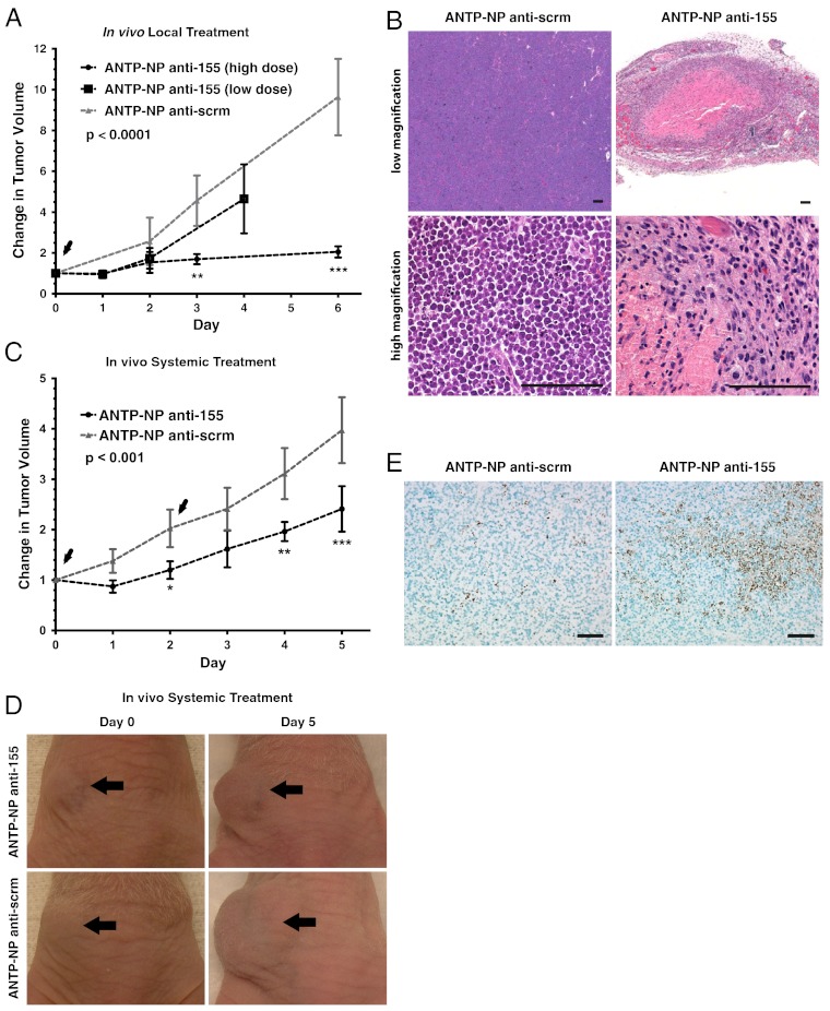
MicroRNA-155 (miR-155) is an oncogenic microRNA that regulates several pathways involved in cell division and immunoregulation. It is overexpressed in numerous cancers, is often correlated with poor prognosis, and is thus a key target for future therapies. In this work we show that overexpression of miR-155 in lymphoid tissues results in disseminated lymphoma characterized by a clonal, transplantable pre-B-cell population of neoplastic lymphocytes. Withdrawal of miR-155 in mice with established disease results in rapid regression of lymphadenopathy, in part because of apoptosis of the malignant lymphocytes, demonstrating that these tumors are dependent on miR-155 expression. We show that systemic delivery of antisense peptide nucleic acids encapsulated in unique polymer nanoparticles inhibits miR-155 and slows the growth of these "addicted" pre-B-cell tumors in vivo, suggesting a promising therapeutic option for lymphoma/leukemia.


Conflict of interest statement

The authors declare no conflict of interest.

Figures

Fig. 1.
Fig. 1.
Controlled expression of miR-155 in mir-155LSLtTA mice. (A) Schematic of controlled gene expression in mir-155LSLtTA mice. miR-155 expression is prevented until exposure to Cre recombinase, which excises a stop cassette allowing transcription to proceed. Transcription can subsequently be suppressed by exposure to doxycycline. Modified from (36). (B) Recombination and stop cassette removal detected by PCR in the spleen, thymus, and bone marrow of a 2-wk-old NesCre8; mir-155LSLtTA mouse (n = 20). (C) miR-155 levels in the spleens of wild-type mice and NesCre8; mir-155LSLtTA mice with and without doxycycline. Graph represents averages of three mice per group ± SD. (D) Percentage of mice that showed signs of lymphadenopathy and/or paresis (median time 115 d); p was calculated with a log-rank (Mantel-Cox) test (n = 20).
Fig. 2.
Fig. 2.
mir-155LSLtTA mice develop disseminated lymphoma. (A) NesCre8; mir-155LSLtTA mouse with ruffled fur and massively enlarged peripheral and axillary lymph nodes (arrowheads) (n = 20). (B) Gross anatomical level of enlarged spleen (arrow) and lymph nodes (arrowheads) (n = 20). (C, D) Representative H&E sections from wild-type mice reveal distinct periarteriolar lymphocyte sheaths (PALS) in spleen and normal cortex and medullary in thymus; (n = 20). (E, F) Representative H&E sections from NesCre8; mir-155LSLtTA mouse spleen and thymus. Spleen and thymic parenchyma are effaced and replaced by a monotonous population of lymphoblasts(n = 20). (GJ) Lymphocyte infiltration into liver, submandibular lymph nodes (SMLN), meninges of the brain (I, asterisk), paravertebral lymph nodes (J, arrows), and meninges of the spinal cord (J, arrowhead). Scale bars, 500 μ (n = 20). (K) Lymphadenopathy is observed in nude mice when injected subcutaneously with NesCre8; mir-155LSLtTA spleen cells (n = 5). (L) Representative flow cytometry plot reveals that NesCre8; mir-155LSLtTA spleens (p = 0.0006) and thymuses (p = 0.0002) are highly enriched with a B220+, IgM population of cells (Upper Left) (n = 3). The following channels were used for gating: FL1 channel to detect FITC-conjugated antibodies, FL2 to detect PE-conjugated antibodies, FL3 to detect APC-conjugated antibodies, and FL4 to detect PI.
Fig. 3.
Fig. 3.
Doxycycline-induced withdrawal of miR-155 leads to rapid tumor regression. (A) Regression of enlarged lymph nodes in NesCre8; mir-155LSLtTA mice exposed to doxycycline (n = 10). (B) H&E spleens from NesCre8; mir-155LSLtTA mice without doxycycline or on doxycycline for 48 h or 2 wk. In recovered mice, spleen architecture is restored similar to wild-type mice (n = 10). (C) Nude mice injected subcutaneously with lymphoma spleen cells develop aggressive tumors. Tumors regress rapidly when exposed to doxycycline (n = 3 mice per group). (D) Change in tumor volume, V = (length × width2)/2 (n = 3 mice per group, represented as mean ± SD); t test was used for statistical analysis. (E) Kaplan-Meier survival curve. After developing conspicuous signs of disease, mice were fed with doxycycline food for recovery or normal food (n = 10 mice per group).
Fig. 4.
Fig. 4.
Tumor regression cccurs in part via apoptosis. (A) TUNEL staining of tumors from nude mice fed with doxycycline for 48 h or without doxycycline (n = 3). (B. C) Nude tumors from mice fed with or without doxycycline for 48 h and analyzed for annexin-V binding and/or CD43 staining via flow cytometry (n = 3). (D) Western blot of spleens from wild-type and NesCre8; mir-155LSLtTA mice treated with doxycycline for 72 h with or without doxycycline (n = 3). (E) AlamarBlue assay to detect relative viability of cells with and without doxycycline (n = 3, data represented as mean ± SD).
Fig. 5.
Fig. 5.
Nanoparticle delivery of anti-miR-155 PNAs inhibits miR-155 in vitro. (A) miR-155 dual luciferase sensor demonstrates feasibility of nanoparticle-mediated anti-miR delivery to Toledo B cells (n = 3, data represented as mean ± SD); t test was used for statistical analysis relative to ANTP-NP anti-155 group. (B) Dose response of cultured Toledo B cells to anti-miR treatment with (siSHIP1) and without knockdown of SHIP1. After 48 h nanoparticle incubation, cell viability was measured using CellTiter-Blue (Promega) (n = 3, data represented as mean ± SD). Two-way ANOVA was used for statistical analysis relative to ANTP-NP anti-155-treated SHIP1 knockdown group. (C) miR-155 levels in miR-155-addicted pre-B cells treated with anti-miRs (n = 3, data represented as mean ± SD); t test was used for statistical analysis relative to ANTP-NP anti-scrm group; (D) Western blot of pre-B cells treated with anti-miRs demonstrates SHIP1 response to miR-155 inhibition. Shown is a representative blot of n = 3 independent experiments per group. (E) Dose response of cultured pre-B cells to anti-miR treatment. Cell viability measured using CellTiter-Blue (Promega) (n = 3, data represented as mean ± SD). Two-way ANOVA was used for statistical analysis.
Fig. 6.
Fig. 6.
Nanoparticle-based inhibition of miR-155 induces therapeutic effects. (A) Fold change in tumor growth in response to locally administered anti-miRs. Arrowheads, nanoparticle injection (n = 3 mice for control group, n = 4 mice for high dose treatment group, n = 3 for low dose treatment group); on day 1, one high dose anti-miR-155-treated mouse was removed for early time point analysis. Data represented as mean ± SD. Statistics were done using two-way ANOVA. (B) H&E analysis of locally treated tumors. Scale bar, 100 µm for both high and low magnification images. Representative images shown for n = 3 tumors. (C) Fold change in tumor growth in response to systemically administered anti-miRs. Arrowheads indicate nanoparticle injection (n = 3 mice per group, data represented as mean ± SD). Two-way ANOVA was used for statistical analysis. (D) Representative tumor images before and after systemic treatment, n = 3 mice per group. In accordance with proper animal care regulations, mice were euthanized when tumors reached a volume of 2,000 mm3. (E) TUNEL staining of systemically treated tumors. Scale bar, 50 µm. Representative images shown for n = 3 tumors.
Fig. P1.
Fig. P1.
A schematic of the oncomiR-addiction cancer model to evaluate cell-penetrating NP as an anti-miR delivery technology. (1) Transgenic mice that express high levels of miR-155 develop disseminated lymphoma comprising neoplastic lymphocytes that are dependent on miR-155 overexpression. (2) These cells can establish aggressive subcutaneous flank tumors that are acutely sensitive to miR-155 withdrawal. (3) Coating NPs with ANTP and polyethylene glycol allows them to overcome in vivo delivery barriers. (4) Once inside target tumor cells, ANTP-NPs release encapsulated anti-miR-155 molecules that (5) bind to and inhibit miR-155 in order to attenuate the oncogenic functions of the oncomiR.

Similar articles

Cited by

References

    1. Bartel DP. MicroRNAs: Genomics, biogenesis, mechanism, and function. Cell. 2004;116:281–297. - PubMed
    1. Babar IA, Slack FJ, Weidhaas JB. miRNA modulation of the cellular stress response. Future Oncol. 2008;4:289–298. - PubMed
    1. Alvarez-Garcia I, Miska EA. MicroRNA functions in animal development and human disease. Development. 2005;132:4653–4662. - PubMed
    1. Bushati N, Cohen SM. microRNA functions. Annu Rev Cell Dev Biol. 2007;23:175–205. - PubMed
    1. Chen X. MicroRNA biogenesis and function in plants. FEBS Lett. 2005;579:5923–5931. - PMC - PubMed

Publication types