. 2012 Jun 26;109(26):E1715-23.
doi: 10.1073/pnas.1205918109. Epub 2012 Jun 8.

Conservation of complex knotting and slipknotting patterns in proteins

Affiliations

Conservation of complex knotting and slipknotting patterns in proteins

Joanna I Sułkowska et al. Proc Natl Acad Sci U S A. .

Abstract

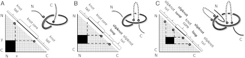
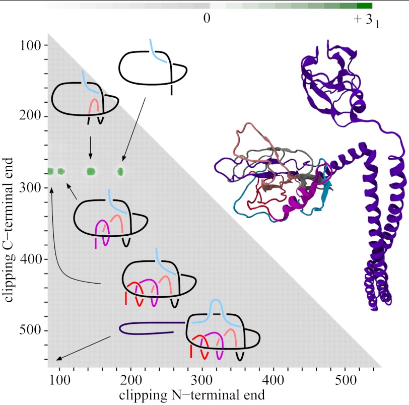
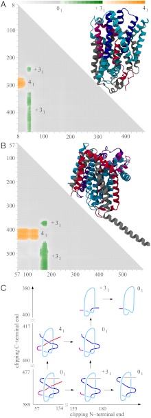
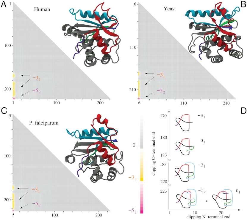
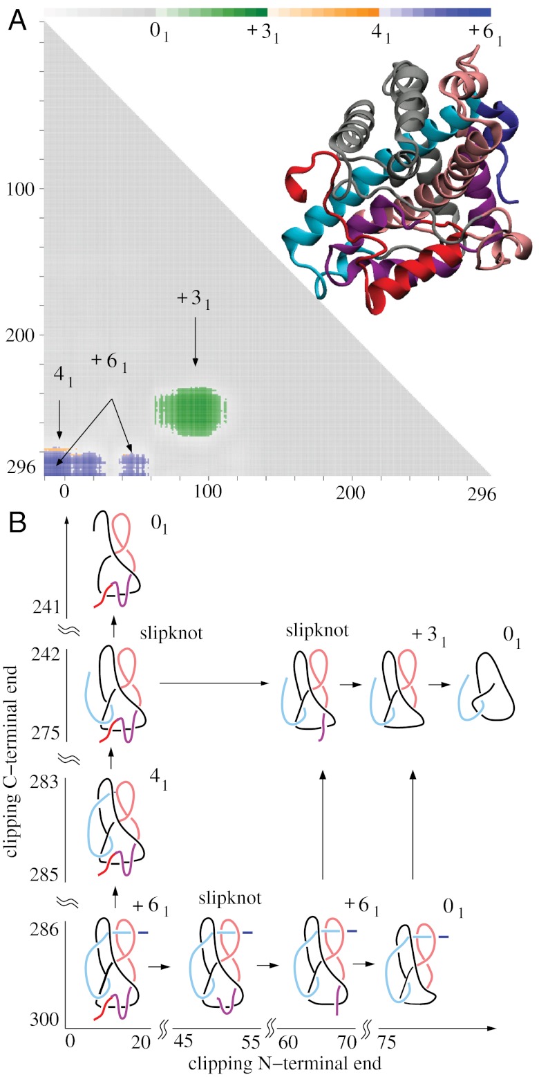
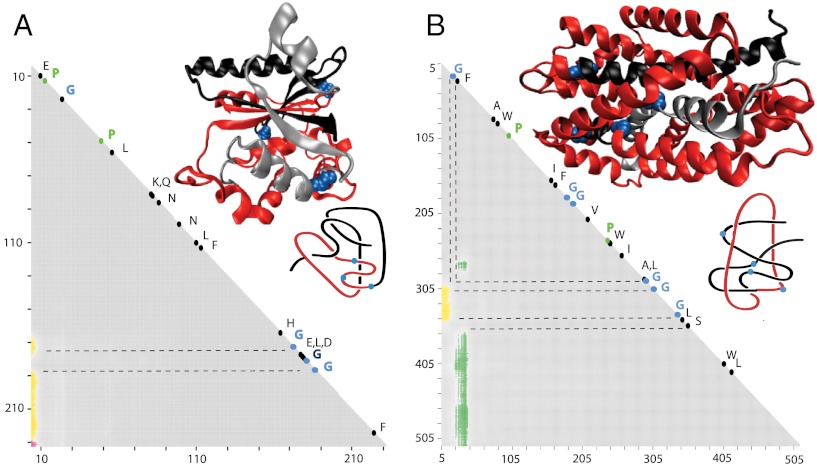
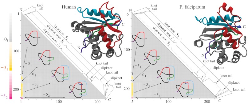
While analyzing all available protein structures for the presence of knots and slipknots, we detected a strict conservation of complex knotting patterns within and between several protein families despite their large sequence divergence. Because protein folding pathways leading to knotted native protein structures are slower and less efficient than those leading to unknotted proteins with similar size and sequence, the strict conservation of the knotting patterns indicates an important physiological role of knots and slipknots in these proteins. Although little is known about the functional role of knots, recent studies have demonstrated a protein-stabilizing ability of knots and slipknots. Some of the conserved knotting patterns occur in proteins forming transmembrane channels where the slipknot loop seems to strap together the transmembrane helices forming the channel.


Conflict of interest statement

The authors declare no conflict of interest.

Figures

Fig. 1.
Fig. 1.
Matrix presentations of protein knotting. Each entry in the matrix indicates the knot type formed by one continuous subchain by its shading (Fig. 1) or color (Figs. 2–5). In each case, the subchain starts with the N-terminal amino acid at position x and ends with the C-terminal amino acid at position y, indicated on the horizontal and vertical axes, respectively. Equivalently, this subchain can be interpreted as a part of the diagonal, delimited by the corresponding coordinates x and y, where the entire diagonal corresponds to the entire polypeptide chain. (A) A case of a knotted protein. Notice that the entry in the lower left-hand corner, which corresponds to the entire protein, is shaded; this indicates that the corresponding chain or subchain is knotted. The knot core is defined as the shortest subchain that still forms a knot (see the thickened part of the protein in the sketch above). The two remaining parts of the chain form knot tails, and their length is conveniently represented along the diagonal. Subchains that do not include at least short bits of both knot tails do not form knots and therefore the matrix entries corresponding to these subchains are not shaded. (B and C) Cases of protein slipknots. Notice that in the case of slipknots the entire protein is unknotted (the element in the lower corner is white) but as one (B) or both termini (C) are trimmed to some extent, the remaining fragment forms a trefoil knot, denoted 31 in Table 1 and SI Appendix, Table S2, and the corresponding matrix entries are therefore darkened in the matrix. Schematic drawings of the polypeptide chains forming trefoil knots and slipknots illustrate which parts of the polypeptide chains constitute the knot cores (thickened), the knot and slipknot tails (solid line), and the slipknot loops (dashed line).
Fig. 5.
Fig. 5.
The structure and knotting of Colicin E3, zigzag motif (S31313131). The molecular structure and schematic drawing of Colicin E3 show that the entire protein is unknotted. However, the matrix representation reveals slipknots producing four distinct 31 territories on the matrix. Schematic drawings of the protein structure explain how this row of slipknots is created upon polypeptide chain clipping.
Fig. 4.
Fig. 4.
LeuT(Aa) and BetP proteins conserve the same knotting notations K314131 and similar knotting fingerprints despite large sequence divergence. (A and B) The molecular structure and matrix presentation of LeuT(Aa) and BetP knotting. Notice that the matrices have a similar pattern of knots and slipknots. The protein LeuT(Aa) matrix resembles the BetP matrix with 60 aa removed. (C) The schematic drawing reflects the overall structure of the LeuTAa and BetP proteins and shows how clipping the C terminus and/or N terminus (termini) from the entire polypeptide chains (with global unknotted knotting) leads to the formation of subchains that form 41 and 31 knots.
Fig. 3.
Fig. 3.
Molecular structures and matrix presentation for ubiquitin C-terminal hydrolases from (A) human, (B) yeast, and (C) P. falciparum plasmodium cells form the same knotting motif, K523131. Notice that in all three cases the proteins form 52 knots with nearly the same sizes and positions with respect to a linear map of their polypeptide chains. In all cases, the 52 knot is unknotted by removing a few amino acids from the N terminus, whereas removing a similar number of amino acids from its C terminus transforms the remaining portion of the protein into a 31 knot. (D) Schematic drawings reflecting the overall structure of ubiquitin C-terminal hydrolases explain how the 52 knot is converted into an unknot or 31 knot depending which end is trimmed.
Fig. 2.
Fig. 2.
The DehI protein forms the Stevedore’s knot as a whole but some of its subchains form 41 and 31 knots or form Stevedore’s and trefoil slipknots. (A) Matrix presentation of the DehI knotting. The color scale shows the dominant knot type formed by a given subchain and the frequency (shown via the color opacity) of its formation. The color bar above a matrix presents the corresponding frequencies at 10%, 20%, …, 100%. Notice the narrow territory of 41 knots above the territory of the Stevedore’s knot. (B) Schematic drawing explaining how the progressive clipping of the C and/or N terminus from the entire polypeptide chain with Stevedore’s knot leads to subchains forming 41 and 31 knots or Stevedore’s and trefoil slipknots.
Fig. 6.
Fig. 6.
Sequence conservation in related proteins with complex knotting fingerprints. (A) Ubiquitin C-terminal hydrolase (knot notation K523131). (B) Five families of proteins with knotting notation S314131 (see Table 1). The positions of the protein regions where varying the length of the analyzed subchain leads to a passage from a knot to the unknot and then again to a knot are indicated with dashed lines. These regions show strong sequence conservation with a high proportion of conserved glycines. The placement of these glycines can be seen in the molecular structures of representative proteins belonging to the analyzed groups (Yuh1 and LeuT for A and B, respectively) and also on the schematic drawings.
Fig. P1.
Fig. P1.
The conservation of complex knotting pattern in ubiquitin C-terminal hydrolases from H. sapiens and P. falciparum. The matrix presentation of protein knotting shows the knot type formed by subchains delimited by the corresponding amino acid residue positions indicated on the horizontal and vertical axis. Each of the complete protein chains forms a knot with five crossings, known as the 52 knot. Clipping a few amino acid residues from the N terminus unknots each of the proteins. Progressive truncation from the C terminus results first in a fragment that is unknotted when considered as a whole, but its shorter subchains can form 31 knots. To understand this matrix presentation of protein knotting, the entire polypeptide chain, unfolded for this purpose, is presented along the diagonal of the matrices. The corresponding regions of knots and slipknots are indicated. The strict conservation of the knotting pattern is obvious despite the fact that these two proteins share only 28% sequence identity.

References

    1. Mansfield ML. Are there knots in proteins? Nat Struct Biol. 1994;1:213–214. - PubMed
    1. King NP, Yeates EO, Yeates TO. Identification of rare slipknots in proteins and their implications for stability and folding. J Mol Biol. 2007;373:153–166. - PubMed
    1. Lua RC, Grosberg AY. Statistics of knots, geometry of conformations, and evolution of proteins. PLoS Comput Biol. 2006;2:e45. - PMC - PubMed
    1. Virnau P, Mirny LA, Kardar M. Intricate knots in proteins: Function and evolution. PLoS Comput Biol. 2006;2:e122. - PMC - PubMed
    1. Sulkowska JI, Sulkowski P, Onuchic JN. Dodging the crisis of folding proteins with knots. Proc Natl Acad Sci USA. 2009;106:3119–3124. - PMC - PubMed

Publication types

LinkOut - more resources