Review
. 2012 Sep;194(18):4802-9.
doi: 10.1128/JB.00576-12. Epub 2012 Jun 8.

The MscS and MscL families of mechanosensitive channels act as microbial emergency release valves

Affiliations
Review

The MscS and MscL families of mechanosensitive channels act as microbial emergency release valves

Ian R Booth et al. J Bacteriol. 2012 Sep.

Abstract

Single-celled organisms must survive exposure to environmental extremes. Perhaps one of the most variable and potentially life-threatening changes that can occur is that of a rapid and acute decrease in external osmolarity. This easily translates into several atmospheres of additional pressure that can build up within the cell. Without a protective mechanism against such pressures, the cell will lyse. Hence, most microbes appear to possess members of one or both families of bacterial mechanosensitive channels, MscS and MscL, which can act as biological emergency release valves that allow cytoplasmic solutes to be jettisoned rapidly from the cell. While this is undoubtedly a function of these proteins, the discovery of the presence of MscS homologues in plant organelles and MscL in fungus and mycoplasma genomes may complicate this simplistic interpretation of the physiology underlying these proteins. Here we compare and contrast these two mechanosensitive channel families, discuss their potential physiological roles, and review some of the most relevant data that underlie the current models for their structure and function.


Figures

Fig 1
Fig 1
Hydrophobicity plots (window = 19) showing MscS-Ec (blue) and representatives of the largest (MscK-Ec; green) and intermediate-sized (YnaI-Ec; yellow) members of the MscS family. In addition, MscS-Cg, which is an MscS homologue from Corynebacterium glutamicum that is similar to MscS along its length from the amino terminus to the base of the β domain, is shown. Thereafter, the sequences diverge, and the MscS-Cg protein has a long, essentially cytoplasmic extension (note, however, that this domain contains a potential transmembrane span located 300 amino acids after the end of β domain). The hydrophobicity plot scale is ±4.5 units, and any segment averaged over 19 residues that has a value above 1.8 at any point in the window has the potential to form a transmembrane span (44). A scale bar representing 100 amino acids is shown, and all the homologues are on the same scale.
Fig 2
Fig 2
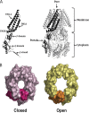
(A) Structure of the closed form of MscS-Ec (2oau) (8, 72). (B) Depiction of TM3A helix movements associated with forming an open state (reprinted from reference 80). (A) A single subunit of the homoheptamer is colored black and is shown integrated into (right) and separate from (left) the MscS structure. Each distinct structural element is labeled. (B) The closed (left) MscS pore viewed from the periplasmic face. Residues T93 to N117 only are shown in space-filled mode (pink), with a single subunit indicated in crimson. The leucine rings form visible blocks to ion conduction by creating a hydrophobic constriction that, despite appearing to be open, is constrained in its ability to pass water and ions (see the text). An open structure, obtained by crystallization of a mutant, A106V MscS-Ec, is shown (right; yellow) with a single subunit highlighted (orange). The closed pore has a diameter of ∼4.8 Å, and the open pore has a diameter of ∼13 Å (80). The radial movement of the TM3A helices, accompanied by a small rotation, completely removes the seal residues (L105 and L109) from the center of the channel, leaving a large open pore.
Fig 3
Fig 3
Current models of the MscL channel. The top panels depict the crystal structure obtained for MscL-Tb, as observed across the plane of the membrane, with the approximate locations of the membrane shown as horizontal lines (left) or from the periplasmic side of the membrane (right); note the black arrow in the latter near the green subunit which describes the corkscrew movement of TM1 that is predicted to occur early in the gating process. In the top center, a single subunit is shown in isolation to better show the domains: S1 is the N-terminal amphipathic α helix that lies along the cytoplasmic membrane, TM1 and TM2 are the transmembrane domains, Peri Loop is the periplasmic loop connecting them, and Cyto Helix is the cytoplasmic helical bundle. The bottom panels show a model for the positions of the transmembrane domains in the open structure, as derived from EPR and other studies and as indicated in the text. Note the tilting of these domains within the membrane.

Similar articles

Cited by

References

    1. Akitake B, Anishkin A, Sukharev S. 2005. The “dashpot” mechanism of stretch-dependent gating in MscS. J. Gen. Physiol. 125:143–154 - PMC - PubMed
    1. Anishkin A, et al. 2003. On the conformation of the COOH-terminal domain of the large mechanosensitive channel MscL. J. Gen. Physiol. 121:227–244 - PMC - PubMed
    1. Anishkin A, Sukharev S. 2009. State-stabilizing interactions in bacterial mechanosensitive channel gating and adaptation. J. Biol. Chem. 284:19153–19157 - PMC - PubMed
    1. Anishkin A, Sukharev S. 2004. Water dynamics and dewetting transitions in the small mechanosensitive channel MscS. Biophys. J. 86:2883–2895 - PMC - PubMed
    1. Balleza D, Gomez-Lagunas F. 2009. Conserved motifs in mechanosensitive channels MscL and MscS. Eur. Biophys. J. 38:1013–1027 - PubMed

Publication types

LinkOut - more resources