. 2012 Jul 27;287(31):26302-11.
doi: 10.1074/jbc.M112.342113. Epub 2012 Jun 8.

Elevation of highly up-regulated in liver cancer (HULC) by hepatitis B virus X protein promotes hepatoma cell proliferation via down-regulating p18

Affiliations

Elevation of highly up-regulated in liver cancer (HULC) by hepatitis B virus X protein promotes hepatoma cell proliferation via down-regulating p18

Yumei Du et al. J Biol Chem. .

Abstract

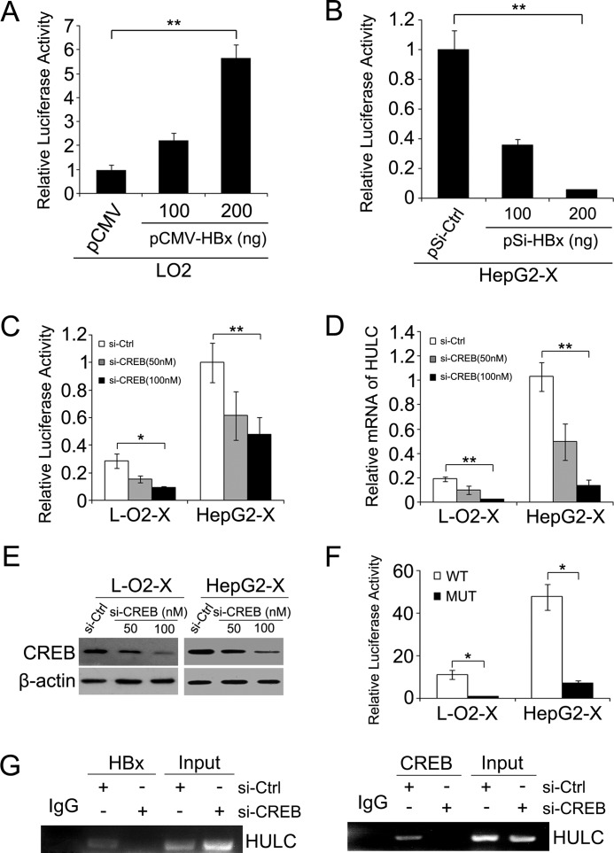
Long noncoding RNAs (lncRNAs) play crucial roles in human cancers. It has been reported that lncRNA highly up-regulated in liver cancer (HULC) is dramatically up-regulated in hepatocellular carcinoma (HCC). Hepatitis B virus X protein (HBx) contributes importantly to the development of HCC. However, the function of HULC in HCC mediated by HBx remains unclear. Here, we report that HULC is involved in HBx-mediated hepatocarcinogenesis. We found that the expression levels of HULC were positively correlated with those of HBx in clinical HCC tissues. Moreover, we revealed that HBx up-regulated HULC in human immortalized normal liver L-O2 cells and hepatoma HepG2 cells. Luciferase reporter gene assay and chromatin immunoprecipitation (ChIP) assay showed that HBx activated the HULC promoter via cAMP-responsive element-binding protein. We further demonstrated that HULC promoted cell proliferation by methyl thiazolyl tetrazolium, 5-ethynyl-2'-deoxyuridine, colony formation assay, and tumorigenicity assay. Next, we hypothesized that HULC might function through regulating a tumor suppressor gene p18 located near HULC in the same chromosome. We found that the mRNA levels of p18 were inversely correlated with those of HULC in the above clinical HCC specimens. Then, we validated that HULC down-regulated p18, which was involved in the HULC-enhanced cell proliferation in vitro and in vivo. Furthermore, we observed that knockdown of HULC could abolish the HBx-enhanced cell proliferation through up-regulating p18. Thus, we conclude that the up-regulated HULC by HBx promotes proliferation of hepatoma cells through suppressing p18. This finding provides new insight into the roles of lncRNAs in HBx-related hepatocarcinogenesis.


Figures

FIGURE 1.
FIGURE 1.
HBx up-regulates HULC in liver cells and hepatoma cells. A, real-time PCR analysis of HULC expression in 21 paired HCC tissues and their corresponding nontumorous tissues. Data were analyzed using 2−ΔΔCt. Statistical significance of HULC expression difference between HCC and normal liver was determined by Wilcoxon signed-rank test (***, p < 0.001, versus nontumorous tissues). B, correlation between the relative mRNA levels of HBx and HULC in 33 HCC tissue specimens. Statistical analysis was performed using Pearson's correlation coefficient (r = 0.8173, p < 0.0001, Pearson's correlation). C, correlation between HBx and HULC expression in the 19 HBx-positive nontumorous liver tissues. Statistical analysis was performed using Pearson's correlation coefficient (r = 0.7925, p < 0.0001, Pearson's correlation). The data presented were from three independent experiments. D, the relative expression levels of HULC were quantified by real-time PCR in L-O2 and HepG2 cells, as well as their corresponding HBx stably transfected cells (L-O2-X and HepG2-X), respectively. E, the relative mRNA levels of HULC were measured by real-time PCR in L-O2 cells transfected with pCMV-HBx plasmid (upper portion). Western blot analysis was performed to determine the efficiency of overexpressing HBx (lower portion). F, the relative expression levels of HULC were detected by real-time PCR in HepG2-X transfected with pSi-HBx plasmid (upper portion). Western blot analysis was performed to measure the efficiency of silencing HBx (lower portion). Data are reported as mean ± S.D. for three independent experiments (*, p < 0.05, **, p < 0.01, Student's t test).
FIGURE 2.
FIGURE 2.
HBx activates the HULC promoter via CREB. A, the promoter activities of HULC were examined by luciferase reporter gene assay in L-O2 cells overexpressing HBx. B, the promoter activities of HULC were measured by luciferase reporter gene assay in HepG2-X cells silencing HBx. Renilla luciferase vector was used as an internal control. Normalized luciferase activity in negative control (pCMV or pSi-Ctrl) transfected cells was set to 1. C, luciferase activity of HULC promoter was determined by luciferase reporter gene assay after silencing CREB. D, the relative mRNA levels of HULC were determined by real-time PCR in L-O2-X/HepG2-X transfected with si-CREB. E, Western blot analysis was performed to determine the expression level of CREB. F, the effect of HBx on the promoter activity of HULC was detected by luciferase reporter gene assays when the CREB binding site in the promoter region of HULC was deleted. MUT, mutant. G, the interaction between HBx and the promoter region of HULC was examined by ChIP analysis. Data are reported as mean ± S.D. for three independent experiments (*, p < 0.05, **, p < 0.01, Student's t test).
FIGURE 3.
FIGURE 3.
HULC promotes proliferation of hepatoma cells in vitro. After silencing or overexpressing HULC, cell proliferation was determined by (A–D) MTT assay, (E) EdU incorporation assay, and (F) colony formation assay. Data are reported as mean ± S.D. for three independent experiments. Statistically significant differences are indicated: *, p < 0.05, **, p < 0.01, ***, p < 0.001, Student's t test. OD Value, optical density value.
FIGURE 4.
FIGURE 4.
HULC promotes proliferation of hepatoma cells in vivo. A, the growth curves of tumors derived from HepG2-X cells transfected with 100 nm si-HULC or si-Ctrl (**, p < 0.01, versus control, Student's t test). B, the average weight of tumors (**, p < 0.01, versus control, Student's t test). C, the images of tumors from nude mice. D, the relative mRNA levels of HULC in the tumor tissues from mice were detected by real-time PCR. Statistically significant differences are indicated: ***, p < 0.001, Student's t test. Data are reported as mean ± S.D. for three independent experiments. E, the expression levels of Ki-67 were measured by immunohistochemistry assay (IHC) in the tumor tissues from mice. Results are representative of three independent experiments.
FIGURE 5.
FIGURE 5.
HULC down-regulates p18 in hepatoma cells. A, a diagram of genes located around HULC. B, relative mRNA levels of p18 in 21 paired HCC tissues and their corresponding nontumorous tissues were detected by real-time PCR analysis. C, correlation between the relative mRNA levels of HULC and p18 in 33 HCC tissue specimens (r = −0.5229, p = 0.0018, Pearson's correlation). D, the mRNA and protein levels of p18 were assessed by RT-PCR and Western blot in HepG2 cells transfected with si-HULC, respectively. E, the promoter activity of p18 was determined by luciferase reporter gene assay in L-O2 cells overexpressing HULC (left). The promoter activity of p18 was measured by luciferase reporter gene assay in HepG2 cells silencing HULC (right). Normalized luciferase activity in negative control (pcDNA3.1 or si-Ctrl) transfected cells was set to 1. F, the protein levels of p18 in tumors from mice shown in Fig. 4C were examined by Western blot analysis. Data are reported as mean ± S.D. for three independent experiments (**, p < 0.01, ***, p < 0.001, Student's t test).
FIGURE 6.
FIGURE 6.
HULC promotes proliferation of hepatoma cells through down-regulating p18 in vitro and in vivo. A, the proliferation of L-O2 cells treated with si-p18 was measured by MTT assay, and the protein levels of p18 were confirmed by Western blot analysis. OD Value, optical density value. B, cell proliferation of HepG2 cells co-transfected with si-HULC and si-p18 was determined by MTT assay. C, the average weight of tumors derived from HepG2 cells transfected with 100 nm si-HULC or/and si-p18. D, the growth curves of tumors. E, the image of tumors from nude mice. F, immunohistochemical staining for Ki-67 and BrdU in the tumor tissues from mice. Data are reported as mean ± S.D. for three independent experiments (**, p < 0.01, ***, p < 0.001, Student's t test).
FIGURE 7.
FIGURE 7.
Decrease of HULC abolishes HBx-enhanced cell proliferation through up-regulating p18. A, the protein levels of p18 were determined by Western blot in L-O2/HepG2, L-O2-X/HepG2-X, and L-O2-X/HepG2-X treated by pSi-HBx or si-HULC, respectively. B, cell proliferation was detected by MTT in HepG2, HepG2-X, and HepG2-X treated by pSi-HBx or si-HULC. Data are reported as mean ± S.D. for three independent experiments (**, p < 0.05, ***, p < 0.01, Student's t test). OD Value, optical density value.

References

    1. El-Serag H. B., Rudolph K. L. (2007) Hepatocellular carcinoma: epidemiology and molecular carcinogenesis. Gastroenterology 132, 2557–2576 - PubMed
    1. Liu Q., Chen J., Liu L., Zhang J., Wang D., Ma L., He Y., Liu Y., Liu Z., Wu J. (2011) The X protein of hepatitis B virus inhibits apoptosis in hepatoma cells through enhancing the methionine adenosyltransferase 2A gene expression and reducing S-adenosylmethionine production. J. Biol. Chem. 286, 17168–17180 - PMC - PubMed
    1. Kim C. M., Koike K., Saito I., Miyamura T., Jay G. (1991) HBx gene of hepatitis B virus induces liver cancer in transgenic mice. Nature 351, 317–320 - PubMed
    1. Lee S., Tarn C., Wang W. H., Chen S., Hullinger R. L., Andrisani O. M. (2002) Hepatitis B virus X protein differentially regulates cell cycle progression in X-transforming versus nontransforming hepatocyte (AML12) cell lines. J. Biol. Chem. 277, 8730–8740 - PubMed
    1. Wang W. H., Hullinger R. L., Andrisani O. M. (2008) Hepatitis B virus X protein via the p38MAPK pathway induces E2F1 release and ATR kinase activation mediating p53 apoptosis. J. Biol. Chem. 283, 25455–25467 - PMC - PubMed

Publication types

MeSH terms

Substances