. 2012 Jul 27;287(31):26029-37.
doi: 10.1074/jbc.M112.372672. Epub 2012 Jun 8.

Elongation factor 1A is the target of growth inhibition in yeast caused by Legionella pneumophila glucosyltransferase Lgt1

Affiliations

Elongation factor 1A is the target of growth inhibition in yeast caused by Legionella pneumophila glucosyltransferase Lgt1

Yury Belyi et al. J Biol Chem. .

Abstract

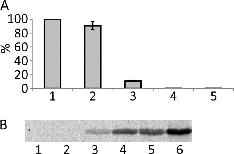
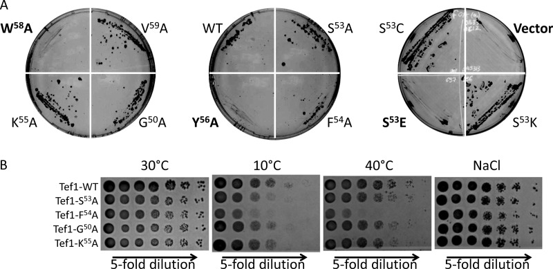
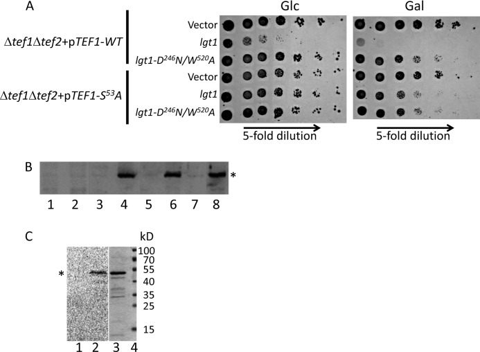
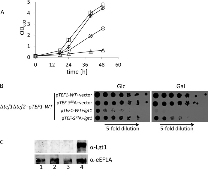
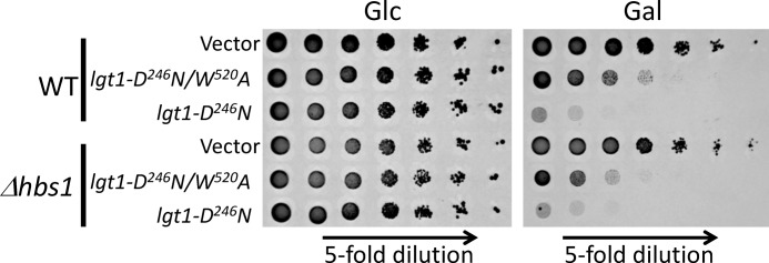
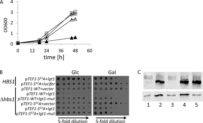
Legionella is a pathogenic Gram-negative bacterium that can multiply inside of eukaryotic cells. It translocates numerous bacterial effector proteins into target cells to transform host phagocytes into a niche for replication. One effector of Legionella pneumophila is the glucosyltransferase Lgt1, which modifies serine 53 in mammalian elongation factor 1A (eEF1A), resulting in inhibition of protein synthesis and cell death. Here, we demonstrate that similar to mammalian cells, Lgt1 was severely toxic when produced in yeast and effectively inhibited in vitro protein synthesis. Saccharomyces cerevisiae strains, which were deleted of endogenous eEF1A but harbored a mutant eEF1A not glucosylated by Lgt1, were resistant toward the bacterial effector. In contrast, deletion of Hbs1, which is also an in vitro substrate of the glucosyltransferase, did not influence the toxic effects of Lgt1. Serial mutagenesis in yeast showed that Phe(54), Tyr(56) and Trp(58), located immediately downstream of serine 53 of eEF1A, are essential for the function of the elongation factor. Replacement of serine 53 by glutamic acid, mimicking phosphorylation, produced a non-functional eEF1A, which failed to support growth of S. cerevisiae. Our data indicate that Lgt1-induced lethal effect in yeast depends solely on eEF1A. The region of eEF1A encompassing serine 53 plays a critical role in functioning of the elongation factor.


Figures

FIGURE 1.
FIGURE 1.
Toxic effects of Lgt1 in wild type yeast. A, 14C-glucosylation of GST-tagged target decapeptide 50GKGSFKYAWV59 of eEF1A by wild type Lgt1 (circles), Lgt1 with D246N substitution (diamonds), Lgt1 with D246N/W520A substitutions (squares), and Lgt1 with D246N/W520A/N293A substitutions (triangles). The amount of the 14C-glucosylated eEF1A-derived peptide at indicated time periods is given. Left and right panels represent graphs with different Y-scales to accommodate different glucosylation activities of wild type Lgt1 and its mutated forms. Reaction with the wild type Lgt1 is not shown on the right panel. B, spot-test assay of Lgt1-accomplished yeast toxicity. S. cerevisiae MH272α was transformed with the vector pESC-Ura or pESC-Ura-based plasmids, coding for different variants of Lgt1 (wild type and mutated), titrated 5-fold and spotted onto supplemented SD agar (with glucose, Glc) or SGal agar (with galactose, Gal). Variants of proteins, coded by the plasmids, are indicated on the left. C, Western blotting analysis of Lgt1 production in yeast, transformed with vector pESC-Ura or pESC-Ura-based plasmids, coding for different variants of Lgt1 (wild type and mutated). S. cerevisiae were cultivated in liquid SD. (i.e. with glucose, lanes 1–5) or SGal (i.e. with galactose, lanes 6–10). Lanes 1 and 6, pESC-Ura; lanes 2 and 7, lgt1-WT; lanes 3 and 8, lgt1-D246N; lanes 4 and 9, lgt1-D246N/W520A; lanes 5 and 10, lgt1-D246N/W520A/N293A. Nitrocellulose membranes were probed with anti-Lgt1 serum overnight at 4 °C and anti-mouse horseradish peroxidase conjugate for 1 h at 22 °C. The position of reacting protein bands is marked by an asterisk.
FIGURE 2.
FIGURE 2.
Influence of Lgt1 upon protein synthesis in S. cerevisiae translation extracts. A, synthesis of luciferase in yeast translation extracts in the absence (bars 1 and 5) or presence of 9 nm (bar 2), 90 nm (bar 3), and 280 nm (bar 4) Lgt1. Bar 5, experiment without luciferase-coding mRNA (i.e. no translation). Each bar represents means of two experiments ± S.D. Data are shown as percentage of maximal translation without added Lgt1. B, modification of eEF1A in yeast translational extracts by Lgt1. The reaction conditions were exactly the same as for in vitro translation but with 10 μm UDP-[14C]glucose instead of unlabeled UDP-glucose. Subsequently, the reaction mix was subjected to SDS-PAGE and autoradiography. Lanes 1 and 2, 9 nm Lgt1; lanes 3 and 4, 90 nm Lgt1; lanes 5 and 6, 280 nm Lgt1. Lanes 1, 3, and 5, incubation time of 10 min; lane 2, 4, and 6, incubation time of 60 min.
FIGURE 3.
FIGURE 3.
Growth phenotypes of yeast strains expressing wild type (WT) and mutated TEF1. A, Δtef1Δtef2 yeast strain expressing TEF1 on a URA3-containing plasmid p561 (pYE-TEF1) was transformed with different variants of TEF1 cloned into pRS313. Strains harboring both plasmids were cultivated on 5-FOA-containing plates for 1 week at 30 °C. Only strains that had lost the URA3-containing plasmid and in addition harbored a functional variant of TEF1 as an insert in pRS313 vector were able to form colonies. Constructions resulting in non-functional Tef1 are shown in boldface type. The experiment was repeated twice with identical results. Representative plates are shown. B, spot-test assay of growth phenotypes of selected TEF1 mutants grown on YPD at 30, 10, or 40 °C and in the presence of 0.9 m NaCl. TEF1 mutants, encoded on pRS313-based plasmids, are shown on the left.
FIGURE 4.
FIGURE 4.
Toxic effect of Lgt1 in S. cerevisiae variants expressing wild type and mutated TEF1 genes. A, spot-test assay of growth phenotypes of S. cerevisiae containing plasmid-born wild type Tef1 and Tef1-S53A as the only eEF1A present in yeast cells. Yeast variants were transformed with pESC-Ura or pESC-Ura-based plasmids coding for wild type lgt1 and lgt1-D246A/W520A. Strains were analyzed as described under “Experimental Procedures” on SD (left panel, glucose, Glc) and SGal (right panel, galactose, Gal). The transformed variants of lgt1 and types of Tef1 in recipient yeast strains are indicated on the left. B, Western blotting, demonstrating Lgt1 production in S. cerevisiae strains transformed by pESC-Ura-based plasmids coding for wild type lgt1 and lgt1-D246A/W520A. Yeast cultures were grown in SD liquid medium (i.e. with glucose, lanes 1, 3, 5, and 7) or SGal (i.e. with galactose, lanes 2, 4, 6, and 8). Lanes 1 and 2, lgt1-WT in S. cerevisiae expressing wild type TEF1; lanes 3 and 4, lgt1-D246A/W520A in S. cerevisiae expressing wild type TEF1; lanes 5 and 6, lgt1 in S. cerevisiae expressing TEF1-S53A; lanes 7 and 8, lgt1-D246A/W520A in S. cerevisiae expressing Tef1-S53A. Western blotting was probed with anti-Lgt1 serum. The position of the glucosyltransferase is labeled with an asterisk. C, In vitro glucosylation of yeast extract prepared from S. cerevisiae containing Tef1-S53A and transformed with wild type lgt1. After cultivation in SGal, yeast cells were collected, disrupted by glass beads, and tested for Lgt1-dependent glucosylation in the 14C-glucosylation assay without (lane 1) and with (lane 2) addition of purified His-tagged Tef1. Coomassie-stained purified His-tagged Tef1 (lane 3) and molecular mass markers (lane 4) are shown. Molecular masses of used markers are shown on the right. The position of a protein band representing glucosylated Tef1 is labeled with an asterisk.
FIGURE 5.
FIGURE 5.
Rescue of toxic effects of Lgt1 in the S. cerevisiaetef1Δtef2+pRS-TEF1-WT) strain by pYC-TEF1-S53A. A, growth of yeast strains in liquid SGal. S. cerevisiaetef1Δtef2+pRS-TEF1-WT) co-transformed by pESC-Ura and pYC-TEF-WT (squares), by pESC-Ura and pYC-TEF1-S53A (diamonds), by pESC-lgt1 and pYC-TEF1-WT (triangles), or by pESC-lgt1 and pYC-TEF1-S53A (circles). B, spot-test assay of strains as in A on SD (left panel, glucose, Glc) or SGal (right panel, galactose, Gal). C, Lgt1 production in S. cerevisiae strains grown in SGal. S. cerevisiaetef1Δtef2+pRS-TEF1-WT) co-transformed by pESC-Ura and pYC-TEF-WT (lane 1), by pESC-Ura and pYC-TEF1-S53A (lane 2), by pESC-lgt1 and pYC-TEF1-WT (lane 3), and by pESC-lgt1 and pYC-TEF1-S53A (lane 4). Western blots were probed with anti-Lgt1 serum (upper panel) or anti-eEF1A antibody (lower panel).
FIGURE 6.
FIGURE 6.
Toxicity of Lgt1 in wild type and Δhbs1 strains. Wild type and Δhbs1 S. cerevisiae, containing pESC-Ura or lgt1-D246N or lgt1-D246N/W520A within pESC-Ura-based plasmids, were analyzed on SD (left panel, glucose, Glc) or SGal (right panel, galactose, Gal). Description of strains is indicated on the left.
FIGURE 7.
FIGURE 7.
Toxicity of Lgt1 in Δhbs1, expressing exclusively plasmid-born TEF1-WT or TEF1-S53A. Growth of S. cerevisiaetef1Δtef2Δhbs1+pRS-TEF1-S53A) harboring pESC-Ura, pESC-Ura-coded lgt1, or pESC-Ura-coded lgt1-D246N/W520A/N293A was compared with growth of the corresponding strain expressing wild type HBS1. A, growth of engineered yeast strains in liquid SGal. Δtef1Δtef2Δhbs1+pRS-TEF1+lgt1 (closed triangles), Δtef1Δtef2Δhbs1+pRS-TEF1-S53A+pESC-Ura (circles), Δtef1Δtef2+pRS-TEF1-S53A+lgt1 (squares) and Δtef1Δtef2Δhbs1+pRS-TEF1-S53A+lgt1 (open triangles). B, growth phenotypes of yeast strains on SD and SGal plates. A description of the strains is indicated on the left. lgt1-mut indicates lgt1-D246N/W520A/N293A. A strain expressing firefly luciferase (lucifer) served as an additional control. C, expression of lgt1 in SGal by engineered S. cerevisiae strains: Δtef1Δtef2Δhbs1+pRS-TEF1+lgt1 (lane 1), Δtef1Δtef2Δhbs1+pRS-TEF1+lgt1-D246N/W520A/N293A (lane 2), Δtef1Δtef2Δhbs1+pRS-TEF1-S53A+pESC-Ura (lane 3), Δtef1Δtef2Δhbs1+pRS-TEF1-S53A+lgt1 (lane 4), and Δtef1Δtef2Δhbs1+pRS-TEF1-S53A+lgt1-D246N/W520A/N293A (lane 5). Western blots were probed with anti-Lgt1 serum (upper panel) or anti-eEF1A antibody (lower panel).

Similar articles

Cited by

References

    1. McDade J. E., Shepard C. C., Fraser D. W., Tsai T. R., Redus M. A., Dowdle W. R. (1977) Legionnaires' disease: Isolation of a bacterium and demonstration of its role in other respiratory disease. N. Engl. J. Med. 297, 1197–1203 - PubMed
    1. Fraser D. W., Tsai T. R., Orenstein W., Parkin W. E., Beecham H. J., Sharrar R. G., Harris J., Mallison G. F., Martin S. M., McDade J. E., Shepard C. C., Brachman P. S. (1977) Legionnaires' disease: Description of an epidemic of pneumonia. N. Engl. J. Med. 297, 1189–1197 - PubMed
    1. Jules M., Buchrieser C. (2007) Legionella pneumophila adaptation to intracellular life and the host response: Clues from genomics and transcriptomics. FEBS Lett. 581, 2829–2838 - PubMed
    1. Ensminger A. W., Isberg R. R. (2009) Legionella pneumophila Dot/Icm translocated substrates: A sum of parts. Curr. Opin. Microbiol. 12, 67–73 - PMC - PubMed
    1. Hubber A., Roy C. R. (2010) Modulation of host cell function by Legionella pneumophila type IV effectors. Annu. Rev. Cell Dev. Biol. 26, 261–283 - PubMed

Publication types

MeSH terms