. 2012 Jul 27;287(31):26038-51.
doi: 10.1074/jbc.M112.345611. Epub 2012 Jun 8.

Biological significance of nuclear localization of mitogen-activated protein kinase Pmk1 in fission yeast

Affiliations

Biological significance of nuclear localization of mitogen-activated protein kinase Pmk1 in fission yeast

Laura Sánchez-Mir et al. J Biol Chem. .

Abstract

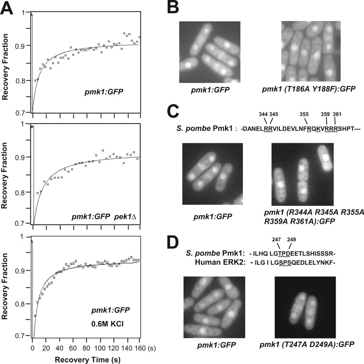
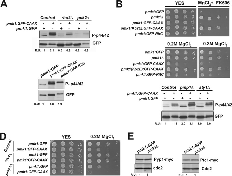
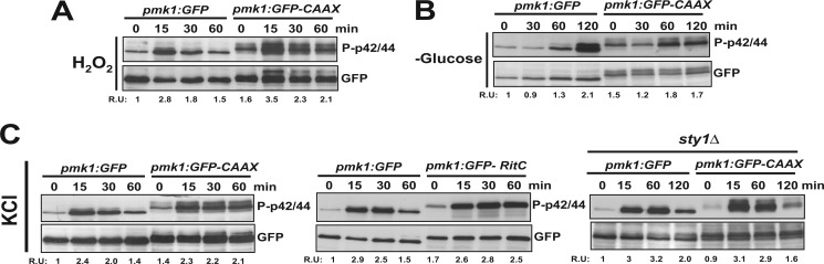
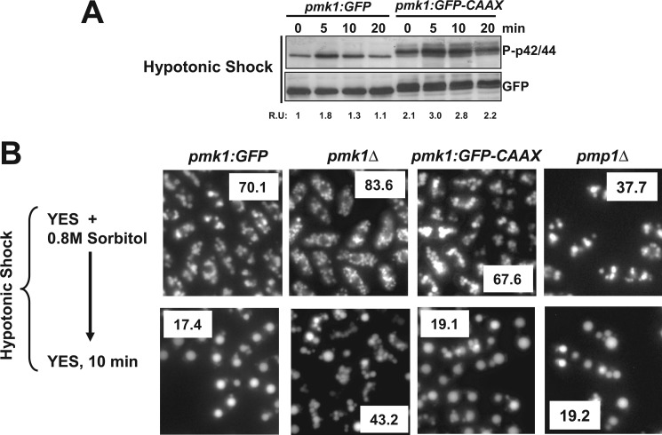
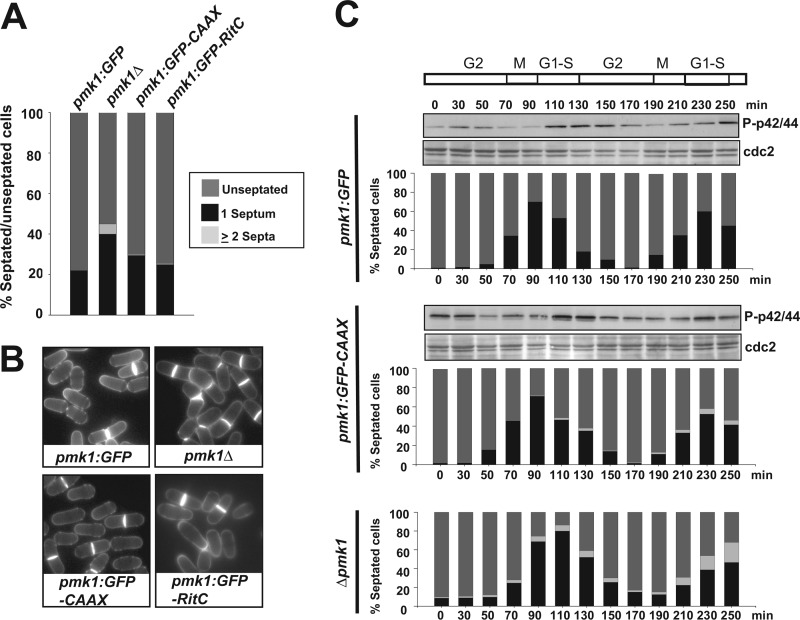
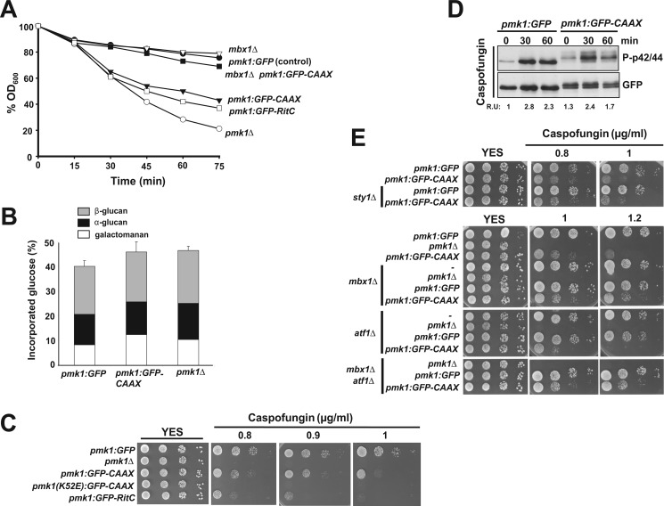
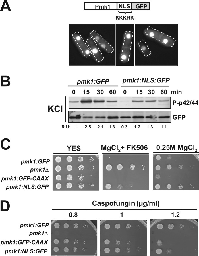
Mitogen-activated protein kinase (MAPK) signaling pathways play a fundamental role in the response of eukaryotic cells to environmental changes. Also, much evidence shows that the stimulus-dependent nuclear targeting of this class of regulatory kinases is crucial for adequate regulation of distinct cellular events. In the fission yeast Schizosaccharomyces pombe, the cell integrity MAPK pathway, whose central element is the MAPK Pmk1, regulates multiple processes such as cell wall integrity, vacuole fusion, cytokinesis, and ionic homeostasis. In non-stressed cells Pmk1 is constitutively localized in both cytoplasm and nucleus, and its localization pattern appears unaffected by its activation status or in response to stress, thus questioning the biological significance of the presence of this MAPK into the nucleus. We have addressed this issue by characterizing mutants expressing Pmk1 versions excluded from the cell nucleus and anchored to the plasma membrane in different genetic backgrounds. Although nuclear Pmk1 partially regulates cell wall integrity at a transcriptional level, membrane-tethered Pmk1 performs many of the biological functions assigned to wild type MAPK like regulation of chloride homeostasis, vacuole fusion, and cellular separation. However, we found that down-regulation of nuclear Pmk1 by MAPK phosphatases induced by the stress activated protein kinase pathway is important for the fine modulation of extranuclear Pmk1 activity. These results highlight the importance of the control of MAPK activity at subcellular level.


Figures

FIGURE 1.
FIGURE 1.
Active and inactive forms of Pmk1 localize into the nucleus in S. pombe. A, nuclear recovery of Pmk1-GFP was measured by fluorescence recovery after photobleaching in strains LS111 (control; upper panel), MI306 (pek1Δ, Pmk1-GFP; middle panel), and in LS111 cells subjected to a saline stress with 0.6 m KCl for 20 min (lower panel). B, shown is fluorescence microscopy of growing cells from strains LS111 (Pmk1-GFP; control) and LS112 (Pmk1(T186A/Y188F)-GFP). C, basic residues (underlined) that define a putative NLS in Pmk1 amino acid sequence are shown. Fluorescence microscopy of growing cells from strains LS111 (control) and LS114 (Pmk1 (R344A/R345A/R355A/R359A/R361A)-GFP) are shown. D, the SPS motif present in ERKs is partially conserved in Pmk1. Fluorescence microscopy of growing cells from strains LS111 (control) and LS113 (Pmk1(T247A/D249A)-GFP).
FIGURE 2.
FIGURE 2.
Construction of Pmk1 versions excluded from the nucleus. A, shown is a scheme representing a version of Pmk1 fused at its C terminus to GFP and followed by the addition of the nine terminal residues of Rho2 GTPase. The amino acid sequence SSTKCCIIS incorporates the cysteine residue (underlined) that mediates Pmk1 targeting to the plasma membrane. B, fluorescence microscopy of growing cells of strain LS115 expressing the Pmk1-GFP-CAAX fusion and stained with calcofluor white is shown. C, shown is fluorescence microscopy of fixed growing cells of strain LS115 and stained with DAPI. D, Western blot analysis of fractionated cell extracts from strains LS111 (Pmk1-GFP) and LS115 is shown. P1, pellet fractions. S2 and P2 correspond to supernatant and membrane fractions, respectively, obtained from P1 samples treated with 7 m urea (see “Experimental Procedures”). E, strains LS134 (pmk1Δ, Pyp2–13myc), LS135 (Pmk1-GFP, Pyp2–13myc) (control), and LS136 (Pmk1-GFP-CAAX Pyp2–13myc), were grown to mid log-phase and treated with 0.9 m potassium chloride. pyp2+ mRNA levels (upper panel) were detected by Northern blot analysis after hybridization with 32P-labeled probes for pyp2+ and leu1+ (loading control) genes. Pyp2 protein levels (lower panel) were detected by immunoblotting with anti-c-myc antibody. Anti-Cdc2 antibody was used as the loading control. F, shown is a scheme representing a version of Pmk1 fused at its C terminus to GFP and followed by the addition of the C-terminal plasma membrane-targeting domain of the mammalian non-CAAX GTPase Rit (RitC). G, shown is fluorescence microscopy of growing cells of strain LS137 expressing the Pmk1-GFP-RitC fusion.
FIGURE 3.
FIGURE 3.
Effect of nuclear exclusion of Pmk1 on chloride homeostasis. A, upper panel, cells from strains LS111 (Pmk1-GFP) (control), LS115 (Pmk1-GFP-CAAX), LS119 (rho2Δ, Pmk1-GFP), LS120 (rho2Δ, Pmk1-GFP-CAAX), LS121 (pck2Δ, Pmk1-GFP), and LS122 (pck2Δ, Pmk1-GFP-CAAX) were grown in YES medium to mid-log phase. Basal Pmk1 activity was detected by immunoblotting of cell extracts with anti-(phosho-p42/44) antibody, and total Pmk1 was detected with anti-GFP antibody as loading control (R.U. indicates relative units). Lower panel, cells from strains LS111 (Pmk1-GFP) (control), LS115 (Pmk1-GFP-CAAX), and LS137 (Pmk1-GFP-RitC) were grown in YES medium, and basal Pmk1 activity was detected and quantified as described above. B, shown are vic phenotype and chloride sensitivity assays for strains LS111 (control), MI102 (pmk1Δ), LS115, and LS116 (Pmk1(K52E)-GFP-CAAX; kinase dead). After growth in YES medium, 104, 103, 102, or 10 cells were spotted onto YES plates supplemented with 0.2 m MgCl2 plus 0.5 μg/ml FK506 (vic) or with 0.2–0.3 M MgCl2 alone and incubated for 3 days at 28 °C before being photographed. C, basal Pmk1 activity in growing cells from strains LS111 (control), LS115, LS117 (pmp1Δ, Pmk1-GFP), LS118 (pmp1Δ, Pmk1-GFP-CAAX), LS132 (sty1Δ, Pmk1-GFP), and LS133 (sty1Δ, Pmk1-GFP-CAAX) was detected as described above. D, chloride sensitivity assay for the strains described in C is shown. E, strains LS138 (Pmk1-GFP, Pyp1–13myc) and LS139 (pmk1Δ, Pyp1–13myc) (left panel), and LS140 (Pmk1-GFP, Ptc1–13myc) and LS141 (pmk1Δ, Ptc1–13myc) (right panel) were grown to mid log-phase, and either Pyp1 or Ptc1 protein levels were detected by immunoblotting with anti-c-myc antibody. Anti-Cdc2 antibody was used as loading control.
FIGURE 4.
FIGURE 4.
Stress-induced phosphorylation of membrane-targeted Pmk1. A, strains LS111 (Pmk1-GFP; control) and LS115 (Pmk1-GFP-CAAX) were grown in YES medium to mid log-phase and treated with 1 mm hydrogen peroxide. At timed intervals either activated or total Pmk1 were detected by immunoblotting with anti-phospho-p42/44 or anti-GFP antibodies, respectively. B, both activated and total Pmk1 were detected in strains LS111 and LS115 after glucose depletion. C, exponentially growing cells from strains LS111 and LS115 (left panel), LS111 and LS137 (Pmk1-GFP-RitC; middle panel), or LS132 (sty1Δ, Pmk1-GFP) and LS133 (sty1Δ, Pmk1-GFP-CAAX; right panel) were treated with 0.6 m potassium chloride, and both activated and total Pmk1 were detected as described above.
FIGURE 5.
FIGURE 5.
Membrane-tethered Pmk1 promotes vacuole fusion induced during hypotonic stress. A, strains LS111 (Pmk1-GFP; control) and LS115 (Pmk1-GFP-CAAX) were grown in YES medium plus 0.8 m sorbitol to mid log-phase and shifted to the same medium without polyol. At timed intervals either activated or total Pmk1 was detected by immunoblotting with anti-phospho-p42/44 or anti-GFP antibodies, respectively. B, vacuole fusion is shown. Strains LS111 (Pmk1-GFP; control), MI102 (pmk1Δ), LS115 (Pmk1-GFP-CAAX), and MI212 (pmp1Δ) were grown in YES medium plus 0.8 m sorbitol. Cells were labeled with CDCFDA and resuspended in YES medium, and vacuole size and fusion visualized by fluorescence microscopy. The average number of vacuoles per cell is shown as an insert for control and mutant strains.
FIGURE 6.
FIGURE 6.
Cell separation defect in cells expressing a Pmk1 version excluded from the nucleus. A, strains LS111 (Pmk1-GFP; Control), MI102 (pmk1Δ), LS115 (Pmk1-GFP-CAAX), and LS137 (Pmk1-GFP-RitC) were grown in YES medium plus 0.8 m sorbitol to mid log-phase, and the percentage of unseptated/septated cells in cultures was determined by fluorescence microscopy after cell staining (n≥400) with calcofluor white. B, representative cells from the strains described above as observed by fluorescence microscopy. C, cells from strains LS123 (cdc25-22, Pmk1-GFP), LS124 (cdc25-22, Pmk1-GFP-CAAX), and MI601 (cdc25-22, pmk1Δ) were grown to A600 = 0.3 at 25 °C, shifted to 37 °C for 3.5 h, and then released from the growth arrest by transfer back to 25 °C. Aliquots were taken at different time intervals, and activated Pmk1 was detected by immunoblotting with anti-phospho-p42/44 antibodies. Anti-Cdc2 antibody was employed as loading control. Percentage of unseptated, septated, and multiseptated cells at each time point is shown as described in A.
FIGURE 7.
FIGURE 7.
Cell wall integrity is partially compromised in cells expressing a nuclear-excluded version of Pmk1. A, strains LS111 (Pmk1-GFP; control), MI102 (pmk1Δ), LS115 (Pmk1-GFP-CAAX), LS137 (Pmp1-GFP-RitC), LS127 (mbx1Δ, Pmk1-GFP), and LS128 (mbx1Δ, Pmk1-GFP-CAAX) were grown in YES medium (A600 = 0.5) and assayed for β-glucanase sensitivity. Cell lysis was monitored by measuring decay in A600 for different incubation periods. Results shown are the mean value of three independent experiments. B, cell wall composition of strains LS111 (control), MI102, and LS115 is shown. Strains were grown in YES medium at 32 °C, and [14C]glucose was added 6 h before harvesting. Values shown are the mean of three independent experiments with duplicate samples, and error bars represent S.D. for total carbohydrate values. C, caspofungin sensitivity assays for strains LS111 (Pmk1-GFP; control), MI102 (pmk1Δ), LS115 (Pmk1-GFP-CAAX), LS116 (Pmk1(K52E)-GFP-CAAX; kinase dead), and LS137 (Pmk1-GFP-RitC) are shown. After growth in YES medium, 104, 103, 102, or 10 cells were spotted onto YES plates supplemented with 0.8, 1.0, or 1.2 μg/ml caspofungin and incubated for 3 days at 28 °C before being photographed. D, strains LS111 (Pmk1-GFP; control) and LS115 (Pmk1-GFP-CAAX) were grown in YES medium to mid log-phase and treated with 1 μg/ml caspofungin. At timed intervals either activated or total Pmk1 was detected by immunoblotting with anti-phospho-p42/44 or anti-GFP antibodies, respectively. E, shown are caspofungin sensitivity assays for strains LS111, LS115, LS132 (sty1Δ, Pmk1-GFP), L133 (sty1Δ, Pmk1-GFP-CAAX), MI102, LS108 (mbx1Δ), LS109 (mbx1Δ pmk1Δ), LS127 (mbx1Δ, Pmk1-GFP), LS128 (mbx1Δ, Pmk1-GFP-CAAX), LS106 (atf1Δ), LS107 (atf1Δ pmk1Δ), LS125 (atf1Δ, Pmk1-GFP), LS126 (atf1Δ, Pmk1-GFP-CAAX), LS110 (mbx1Δ atf1Δ pmk1Δ), LS129 (mbx1Δ atf1Δ, Pmk1-GFP), and LS130 (mbx1Δ atf1Δ, Pmk1-GFP-CAAX).
FIGURE 8.
FIGURE 8.
Biological relevance of increased nuclear localization of Pmk1. A, shown is a scheme representing a version of Pmk1 fused at its C terminus to the SV40 nuclear localization signal followed by GFP epitope and growing cells of strain LS131 expressing the Pmk1-NLS-GFP fusion as seen by fluorescence microscopy. B, exponentially growing cells of strains LS111 (Pmk1-GFP, control) and LS131 (Pmk1-NLS-GFP) were treated with 0.6 m potassium chloride, and both activated and total Pmk1 were detected by immunoblotting with anti-phospho-p42/44 or anti-GFP antibodies, respectively. C, vic phenotype and chloride sensitivity assays for strains LS111 (control), MI102 (pmk1Δ), LS115, and LS131 are shown. After growth in YES medium, 104, 103, 102, or 10 cells were spotted onto YES plates supplemented with 0.2 m MgCl2 plus 0.5 μg/ml FK506 (vic), or 0.25 m MgCl2 alone and incubated for 3 days at 28 °C before being photographed. D, shown are caspofungin sensitivity assays for the strains described in C.

Similar articles

Cited by

References

    1. Kim E. K., Choi E. J. (2010) Pathological roles of MAPK signaling pathways in human diseases. Biochim. Biophys. Acta 1802, 396–405 - PubMed
    1. Qi M., Elion E. A. (2005) MAP kinase pathways. J. Cell Sci. 118, 3569–3572 - PubMed
    1. Krisak L., Strich R., Winters R. S., Hall J. P., Mallory M. J., Kreitzer D., Tuan R. S., Winter E. (1994) SMK1, a developmentally regulated MAP kinase, is required for spore wall assembly in Saccharomyces cerevisiae. Genes Dev. 8, 2151–2161 - PubMed
    1. Shiozaki K., Russell P. (1995) Cell cycle control linked to extracellular environment by MAP kinase pathway in fission yeast. Nature 378, 739–743 - PubMed
    1. Toda T., Dhut S., Superti-Furga G., Gotoh Y., Nishida E., Sugiura R., Kuno T. (1996) The fission yeast pmk1+ gene encodes a novel mitogen-activated protein kinase homolog that regulates cell integrity and functions coordinately with the protein kinase C pathway. Mol. Cell. Biol. 16, 6752–6764 - PMC - PubMed

Publication types

MeSH terms

Substances

LinkOut - more resources