. 2012 Apr 26;45(2):139-46.
doi: 10.1267/ahc.12003. Epub 2012 Apr 21.

Close association of aquaporin-2 internalization with caveolin-1

Affiliations

Close association of aquaporin-2 internalization with caveolin-1

Takeo Aoki et al. Acta Histochem Cytochem. .

Abstract

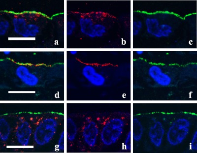
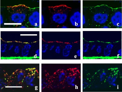
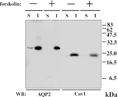
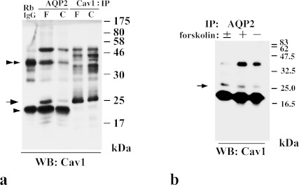
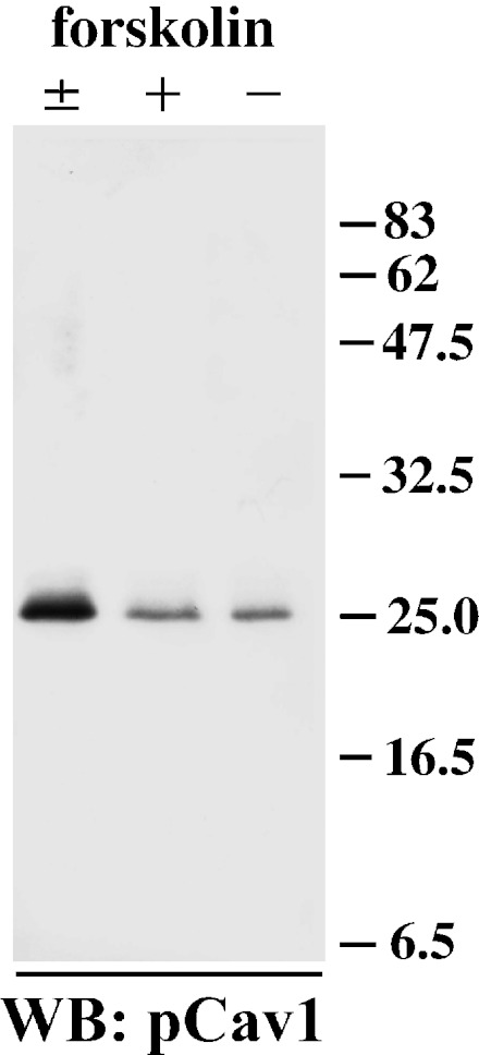
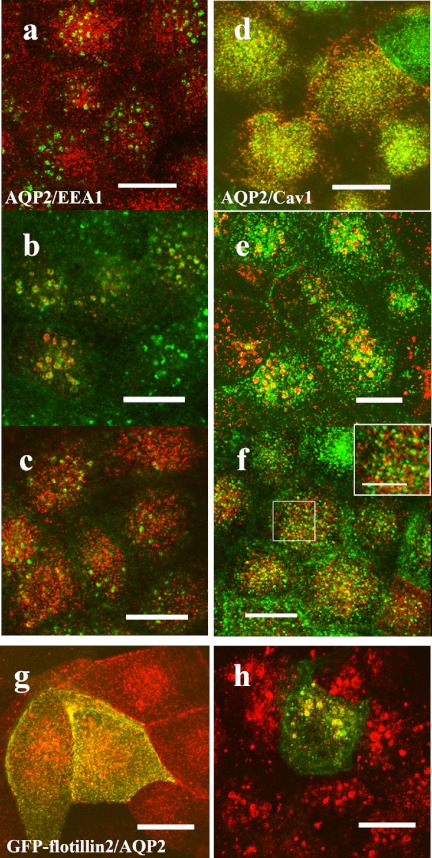
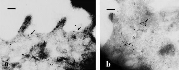
Aquaporin 2 (AQP2) is a membrane water channel protein that traffics between the intracellular membrane compartment and the plasma membrane in a vasopressin-dependent manner in the renal collecting duct cell to control the amount of water reabsorption. We examined the relation between AQP2 internalization from the plasma membrane and caveolin-1, which is a major protein in membrane microdomain caveolae, in Mardin-Darby canine kidney cells expressing human AQP2 (MDCK-hAQP2 cells). Double-immunofluorescence microscopy showed that AQP2 is colocalized with caveolin-1 in the apical plasma membrane by stimulating the intracellular signaling cascade of vasopressin with forskolin. After washing forskolin, both AQP2 and caveolin-1 were internalized to early endosomes and then separately went back to their individual compartments, which are subapical compartments and the apical membrane, respectively.Double-immunogold electron microscopy in ultrathin cryosections confirmed the colocalization of AQP2 with caveolin-1 at caveolar structures on the apical plasma membrane of forskolin-treated cells and the colocalization within the same intracellular vesicles after washing forskolin. A co-immunoprecipitation experiment showed the close interaction between AQP2 and caveolin-1 in forskolin-treated cells and in cells after washing forskolin. These results suggest that a caveolin-1-dependent and possibly caveolar-dependent pathway is a candidate for AQP2 internalization in MDCK cells.

Keywords: MDCK cells; apical membrane; aquaporin-2; caveolin-1; early endosome.


Figures

Fig. 1
Fig. 1
Trafficking of AQP2 in MDCK-hAQP2 cells. Confluent MDCK-hAQP2 cells grown on permeable supports without any treatment as a control (a–c), treated with forskolin for 30 min (d–f), or treated with forskolin for 30 min, washed, and then incubated without forskolin for 30 min (g–i) were fixed. Cells were sectioned perpendicular to the permeable support with a cryostat and double-immunofluorescently labeled for AQP2 (red) and the apical membrane marker Gp135 (green). Nuclei were stained with DAPI (blue). Merged confocal images are shown on the left. AQP2 is largely seen in the subapical cytoplasm in the control. Upon forskolin treatment, AQP2 is accumulated on the apical membrane. AQP2 is internalized and seen in the cytoplasm after washing out forskolin. Bar=10 µm.
Fig. 2
Fig. 2
Colocalization of AQP2 and caveolin-1. Confluent MDCK-hAQP2 cells grown on permeable supports without any treatment as a control (a–c), treated with forskolin for 30 min (d–f), or treated with forskolin for 30 min, washed, and then incubated without forskolin for 30 min (g–i) were fixed. Cells were sectioned perpendicular to the supports with a cryostat and double-immunofluorescently labeled for AQP2 (red) and caveolin-1 (green). Nuclei were stained with DAPI (blue). Merged confocal images are shown on the left. In the control, AQP2 is seen in the subapical cytoplasm and seems not to colocalize with caveolin-1, which is present on the apical membrane. AQP2 is largely colocalized with caveolin-1 on the apical membrane upon forskolin treatment. Some AQP2 internalized after washout is colocalized with caveolin-1. Bar=10 µm.
Fig. 3
Fig. 3
AQP2 is recovered in Triton X-100-insoluble fraction. Samples of Triton X-100-soluble (S) and -insoluble (I) fractions from MDCK-hAQP2 cell lysates with (+) or without (–) forskolin treatment for 30 min were subjected to SDS-PAGE and immunoblotting using anti-AQP2 and anti-caveolin-1 (Cav1) antibodies.
Fig. 4
Fig. 4
AQP2 co-immunoprecipitates with caveolin-1. a: In immunoprecipitates with anti-AQP2, caveolin-1 is detected as a dense band in forskolin-treated cells (F) and a faint band in control cells (C) at 22–24 kDa (arrow) with anti-caveolin-1 (Cav1). Dense bands both around 20 kDa (arrowhead) and 40 kDa (double-arrowhead) seem to be non-specific because these bands are also detected in the lane in which rabbit IgG was electrophoresed (RbIgG). b: Immunoprecipitates with anti-AQP2 from each cell lysate described as follows were subjected to immunoblotting with anti-caveolin-1 (Cav1). –: control cells, +: cells treated with forskolin for 30 min, ±: cells treated with forskolin for 30 min, washed, and then incubated without forskolin for 30 min.
Fig. 5
Fig. 5
Internalized caveolin-1 is phosphorylated. Samples of Triton X-100-insoluble fractions from MDCK-hAQP2 cell lysates were subjected to SDS-PAGE and immunoblotting using anti-phosphorylated caveolin-1 antibody. A strong band is detected in cells treated with forskolin and subsequently washed and incubated without forskolin for 30 min (±). Faint bands are detected in cells both treated (+) and untreated (–) with forskolin.
Fig. 6
Fig. 6
AQP2 is internalized to the same compartment with caveolin-1 and flotillin-2. (a–f) MDCK-hAQP2 cells were seeded on coverslips and subjected to forskolin treatment for 30 min (a, d) and subsequent washing and incubation without forskolin for 30 min (b, e) or for 2 hr (c, f). Double-immunofluorescence labeling for AQP2 and EEA1 (a–c) or for AQP2 and caveolin-1 (d–f) was carried out and their localization was observed with a laser confocal microscope. Projection images of 4 consecutive confocal images (0.4 µm intervals) are shown. AQP2 is shown in red. EEA1 and caveolin-1 are shown in green. An enlarged view of the rectangle area is shown in the inset (f). Both AQP2 and caveolin-1 are internalized in the EEA1-positive compartment 30 min after washing and then differentially localized at 2 hr. g, h: MDCK-hAQP2 cells transiently transfected with EGFP-flotillin-2 were treated with forskolin for 30 min (g) and then washed and incubated without forskolin for 30 min (h). Localization of AQP2 and flotillin-2 was observed with a laser confocal microscope. Projection images of 4 consecutive confocal images (0.4 µm intervals) are shown. AQP2 is shown in red. EGFP fluorescence is shown in green. Both AQP2 and EGFP-flotillin-2 are internalized in the same compartment. Bars=10 µm (5 µm in inset).
Fig. 7
Fig. 7
Ultrastructural localization of AQP2 and caveolin-1. MDCK-hAQP2 cells were treated with forskolin for 30 min (a), and then washed and incubated without forskolin for 30 min (b). Ultrathin cryosections were double-labeled for AQP2 (12 nm colloidal gold) and caveolin-1 (6 nm colloidal gold). In forskolin-treated cells (a), some AQP2 is colocalized with caveolin-1 at caveolar structures in the apical membrane domain (arrows). AQP2 on the microvillous membrane is not colocalized with caveolin-1. When cells were washed and incubated without forskolin for 30 min (b), AQP2 was colocalized with caveolin-1 in the intracellular vesicles (arrows). Bars=100 nm.

References

    1. Aoki T., Nomura R., Fujimoto T. Tyrosine phosphorylation of caveolin-1 in the endothelium. Exp. Cell Res. 1999;253:629–636. - PubMed
    1. Aoki T., Hagiwara H., Matsuzaki T., Suzuki T., Takata K. Internalization of caveolae and their relationship with endosomes in cultured human and mouse endothelial cells. Anat. Sci. Int. 2007;82:82–97. - PubMed
    1. Asai T., Kuwahara M., Kurihara H., Sakai T., Terada Y., Marumo F., Sasaki S. Pathogenesis of nephrogenic diabetes insipidus by aquaporin-2 C-terminus mutations. Kidney Int. 2003;64:2–10. - PubMed
    1. Brown D. The ins and outs of aquaporin-2 trafficking. Am. J. Physiol. Renal. Physiol. 2003;284:F893–F901. - PubMed
    1. Feng X., Huang H., Yang Y., Frohlich O., Klein J. D., Sands J. M., Chen G. Caveolin-1 directly interacts with UT-A1 urea transporter: the role of caveolae/lipid rafts in UT-A1 regulation at the cell membrane. Am. J. Physiol. Renal Physiol. 2009;296:F1514–F1520. - PMC - PubMed