. 2012;8(6):e1002723.
doi: 10.1371/journal.ppat.1002723. Epub 2012 Jun 7.

Pathogen-mediated proteolysis of the cell death regulator RIPK1 and the host defense modulator RIPK2 in human aortic endothelial cells

Affiliations

Pathogen-mediated proteolysis of the cell death regulator RIPK1 and the host defense modulator RIPK2 in human aortic endothelial cells

Andrés G Madrigal et al. PLoS Pathog. 2012.

Abstract

Porphyromonas gingivalis is the primary etiologic agent of periodontal disease that is associated with other human chronic inflammatory diseases, including atherosclerosis. The ability of P. gingivalis to invade and persist within human aortic endothelial cells (HAEC) has been postulated to contribute to a low to moderate chronic state of inflammation, although how this is specifically achieved has not been well defined. In this study, we demonstrate that P. gingivalis infection of HAEC resulted in the rapid cleavage of receptor interacting protein 1 (RIPK1), a mediator of tumor necrosis factor (TNF) receptor-1 (TNF-R1)-induced cell activation or death, and RIPK2, a key mediator of both innate immune signaling and adaptive immunity. The cleavage of RIPK1 or RIPK2 was not observed in cells treated with apoptotic stimuli, or cells stimulated with agonists to TNF-R1, nucleotide oligomerization domain receptor 1(NOD1), NOD2, Toll-like receptor 2 (TLR2) or TLR4. P. gingivalis-induced cleavage of RIPK1 and RIPK2 was inhibited in the presence of a lysine-specific gingipain (Kgp) inhibitor. RIPK1 and RIPK2 cleavage was not observed in HAEC treated with an isogenic mutant deficient in the lysine-specific gingipain, confirming a role for Kgp in the cleavage of RIPK1 and RIPK2. Similar proteolysis of poly (ADP-ribose) polymerase (PARP) was observed. We also demonstrated direct proteolysis of RIPK2 by P. gingivalis in a cell-free system which was abrogated in the presence of a Kgp-specific protease inhibitor. Our studies thus reveal an important role for pathogen-mediated modification of cellular kinases as a potential strategy for bacterial persistence within target host cells, which is associated with low-grade chronic inflammation, a hallmark of pathogen-mediated chronic inflammatory disorders.


Conflict of interest statement

The authors have declared that no competing interests exist.

Figures
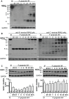
Figure 1
Figure 1. P. gingivalis 381-induced proteolysis of RIPK1 and RIPK2 in HAEC.
HAEC were treated with medium (M) or with P. gingivalis strain 381 (MO1 100) for 0.25, 0.5, 1, 2, 6, 12, 24 or 48 h. Whole cell lysates were analyzed for the detection of A) RIPK1, B) RIPK2 with an anti N′-terminal RIPK2 antibody (left panel) or an anti C′-terminal RIPK2 antibody (right panel), or C) NOD1 (left panel) and NOD2 (right panel). Full-length RIPK1 (74-kDa) and RIPK2 (61-kDa) are indicated with arrows. Prominent P. gingivalis-induced LMW bands are indicated with asterisks. Molecular weight (MW) ladder is indicated on the left in kDa. GAPDH was detected as a loading control. (−) protein levels in medium-treated cells were similar at all time points. Densitometric analysis is presented below respective blots as the mean (+/− SEM) ratio of NOD1 (or NOD2) to GAPDH protein levels (arbitrary densitometric units (A.D.U.) from at least 3 independent membranes. Means are displayed within the bar charts.
Figure 2
Figure 2. P. gingivalis-induced proteolysis of RIPK proteins is dose-dependent and heat labile.
HAEC were treated with medium (M), live P. gingivalis strain 381 (MOI 10) (10), live P. gingivalis (MOI 100) (100), heat-killed (HK) (60°C, 60 min) P. gingivalis 381 (MOI 100 equivalency) (60°), or with HK (80°C, 20 min) P. gingivalis 381 (MOI 100 equivalency) (80°) for 2 h. Whole cell lysates were analyzed for the detection of RIPK1 (left panel) or RIPK2 (right panel). Full-length RIPK1 and RIPK2 are indicated with arrows. Prominent P. gingivalis-induced LMW bands are indicated with asterisk(s). MW ladder is indicated on the left in kDa. GAPDH was detected as a loading control.
Figure 3
Figure 3. RIPK2 levels are stable in HAEC stimulated with TLR or NLR agonists.
HAEC were treated with medium, P. gingivalis 381 (MOI 100), 10 µg/ml Pam3CSK4, 10 µg/ml FSL-1, 10 µg/ml P. gingivalis 381 LPS, 1.0 µg/ml E. coli 0111:B4 LPS, 100 ng/ml recombinant human TNF, 100 µg/ml iE-DAP, 100 µg/ml iE-DAP control, 1000 ng/ml C12-iE-DAP, 0.01% DMSO (C12-iE-DAP vehicle control), 100 µg/ml MDP, 100 µg/ml MDP control, or 1000 ng/ml L18-MDP for 2 h. Whole cell lysates were analyzed for the detection of RIPK2 and GAPDH. Full-length RIPK2 and RIPK2β are indicated with arrows. A prominent P. gingivalis-induced LMW band is indicated with an asterisk. MW ladder is indicated on the left in kDa.
Figure 4
Figure 4. Classical apoptotic stimuli do not induce the proteolysis of RIPK1 or RIPK2 in HUVEC.
HUVEC were treated with medium, P. gingivalis 381 (MOI 100), 2 µM staurosporine (STS) 25 µg/ml cycloheximide (CHX), 10 ng/ml TNFα, or co-treated with 25 µg/ml CHX and 10 ng/ml TNFα for 6 h. Whole cell lysates were analyzed for the detection of RIPK1 (left panel) or RIPK2 (right panel). Full-length RIPK1 and RIPK2 are indicated with arrows. Prominent P. gingivalis-induced LMW bands are indicated with asterisks. MW ladder is indicated on the left in kDa. GAPDH was detected as a loading control.
Figure 5
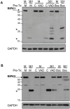
Figure 5. General caspase inhibitors z-VAD-FMK and Boc-D-FMK alter P. gingivalis-induced modification of RIPK1 and RIPK2 in HUVEC.
HUVEC were pretreated (Pre-Tx) with medium (M), 0.25% DMSO vehicle control (C), 25 µM z-VAD-FMK (VAD), or 100 µM Boc-D-FMK (Boc) with for 1.5 h. HUVEC were then treated with medium (M) or P. gingivalis strain 381 (MOI 100, 381) for 2 h. Whole cell lysates were analyzed for (A) RIPK1 or (B) RIPK2 and GAPDH. Full-length RIPK1 and RIPK2 are indicated with arrows. Prominent P. gingivalis-induced LMW bands are indicated with asterisks. MW ladder is indicated on the left in kDa.
Figure 6
Figure 6. P. gingivalis modifies RIPK2 in wild type and caspase-deficient murine bone marrow-derived macrophages.
A) C57BL/6 (wt) or casp1-deficient (casp1−/−) BMDM were untreated (M) or treated with 100 ng/ml E. coli LPS (LPS), live P. gingivalis 381 (MOI 100, Live) or heat-killed (60°C, 60 min) P. gingivalis 381 (MOI 100 equivalency, HK) for 2 h. B) C57BL/6 (wt), casp2-deficient (casp2−/−), casp3-deficient (casp3−/−), or casp7-deficient (casp7−/−) BMDM were untreated () or treated with P. gingivalis 381 (MOI 100) (+) for 2 h. Whole cell lysates were analyzed for RIPK2. Full-length RIPK2 is indicated with an arrow. A prominent P. gingivalis-induced LMW band is indicated with an asterisk. MW ladder is indicated on the left in kDa.
Figure 7
Figure 7. KYT inhibitors specifically inhibit P. gingivalis gingipain activity and do not alter host 3 caspase activity.
Effect of gingipain inhibitors on P. gingivalis A) Rgp or B) Kgp protease activity. P. gingivalis was untreated (none) or pretreated with 10 µM Rgp-specific inhibitor KYT-1, 10 µM Kgp-specific inhibitor KYT-36, 10 µM KYT-1 and 10 µM KYT-36, 1 mM TLCK, or vehicle controls (DMSO or acid water) for 10 min and monitored for arginine-X-specific or lysine-X-specific protease activity. Effect of inhibitors on P. gingivalis is presented as percent Rgp-X activity or Kgp-X activity relative to untreated P. gingivalis. C) HUVEC were untreated or treated with 2 µM staurosporine (STS) for 5 h. Whole cell lysates were analyzed for caspase-3 activity in the presence of KYT-36 gingipain inhibitor (3 µM). Activity is represented as fold change relative to untreated. A reversible caspase inhibitor was included to demonstrate observed fluorescence is specific to caspase-3 like proteases. Statistical analysis was performed using unpaired T-test (α = 0.05), **p<0.001, NS = no significance.
Figure 8
Figure 8. Inhibition of Kgp activity alters P. gingivalis-mediated RIPK1 and RIPK2 cleavage in HAEC.
P. gingivalis strain 381 was pretreated with 10 µM KYT-1, 10 µM KYT-36, 10 µM KYT-1 and 10 µM KYT-36, 1 mM TLCK, or vehicle controls (DMSO or acid water) for 45 min. HAEC were then immediately co-cultured with medium or with pretreated preparations of P. gingivalis 381 (MOI 100) for 2 h. Whole cell lysates were analyzed for A) RIPK1 or B) RIPK2. Full-length RIPK1 and RIPK2 are indicated with arrows. Prominent P. gingivalis-induced LMW bands are indicated with asterisk(s). MW ladder is indicated on the left in kDa. GAPDH was detected as a loading control.
Figure 9
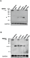
Figure 9. P. gingivalis Kgp mutant is deficient in the induction of RIPK1 and RIPK2 proteolysis in HAEC.
HAEC were untreated or treated with P. gingivalis strain 381, strain ATCC 33277, or isogenic mutants of 33277: YPP1 (rgpA), RgpA/B (rgpA, rgpB), or with YPP2 (kgp) (MOI 100) for 2 h. Whole cell lysates were analyzed for A) RIPK1 or B) RIPK2. Full-length RIPK1 and RIPK2 are indicated with arrows. Prominent P. gingivalis-induced LMW bands are indicated with asterisks. MW ladder is indicated on the left in kDa. GAPDH was detected as a loading control.
Figure 10
Figure 10. Cleavage of recombinant RIPK2 kinase by P. gingivalis in the absence of host cell proteins.
P. gingivalis strain 381 was pretreated with 10 µM KYT-1, 10 µM KYT-36, 10 µM KYT-1 and 10 µM KYT-36, 1 mM TLCK, 100 µM zVAD-fmk, 100 µM BocD-fmk with or vehicle controls (HEPES (none), DMSO or acid water) for 45 min, then immediately co-cultured with 0.1 µg recombinant RIPK2 kinase for 1 h at 37°C. Reactions were stopped by the addition of SDS-PAGE loading dye and analyzed by Western blot analysis with an antibody to the N′-terminal kinase domain of RIPK2. Top panel: reaction with recombinant protein and P. gingivalis; bottom panel: 10% of reaction prior to incubation with P. gingivalis (untreated recombinant protein, i.e., gel loading control).
Figure 11
Figure 11. Model of P. gingivalis innate immune activation/invasion in endothelial cells.
Outcome of innate immune responses is representative as a balance of functional, intact pathways (TLR-left panel) and dysregulated pathways (NLR-right panel). P. gingivalis represents a human pathogen that utilizes fimbriae for attachment and invasion of endothelial cells. Fimbriae are not only expressed on whole bacteria (A), but within outer membrane vesicles (OMV) that are released from the cell (B), as occurs with all Gram-negative bacteria identified to date. Fimbriae bind to TLR2 and MD2 to activate TLR2 and TLR4, resulting in the induction of NF-κB, leading to inflammation, and de novo protein synthesis (C) of cell adhesion molecules (CAM) and TLR (TLR) expressed at the cell surface (D). Many studies have demonstrated invasion of endothelial cells by P. gingivalis (E). However, recent studies have shown that OMV (containing fimbriae and active gingipain activity) gain entry into host cells rapidly in a gingipain-dependent manner (E), independent of whole organism. Upon entry, intracellular gingipain activity degrades RIPK1 and RIPK2 (abrogated by protease inhibitors) (F), resulting in a variety of possible consequences as a function of selective targeting of intracellular NOD1 or NOD2 (NOD1/2) signaling pathways and/or disruption of RIPK1 or RIPK2-mediated cell signaling. Alteration of immune signaling responses may result in decreased host cell death, decreased inflammatory mediator expression, and subsequent enhancement of intracellular bacterial cell survival, all of which contributes to an intracellular niche for P. gingivalis (G).

References

    1. Inohara N, del Peso L, Koseki T, Chen S, Nunez G. RICK, a novel protein kinase containing a caspase recruitment domain, interacts with CLARP and regulates CD95-mediated apoptosis. J Biol Chem. 1998;273:12296–12300. - PubMed
    1. Chin AI, Dempsey PW, Bruhn K, Miller JF, Xu Y. Involvement of receptor-interacting protein 2 in innate and adaptive immune responses. Nature. 2002;416:190–194. - PubMed
    1. Kobayashi K, Inohara N, Hernandez LD, Galan JE, Nunez G. RICK/Rip2/CARDIAK mediates signalling for receptors of the innate and adaptive immune systems. Nature. 2002;416:194–199. - PubMed
    1. Zhang N, Chen Y, Jiang R, Li E, Chen X. PARP and RIP 1 are required for autophagy induced by 11′-deoxyverticillin A, which precedes caspase-dependent apoptosis. Autophagy. 2011;7:598–612. - PubMed
    1. Travassos LH, Carneiro LA, Girardin SE, Boneca IG, Lemos R. Nod1 participates in the innate immune response to Pseudomonas aeruginosa. J Biol Chem. 2005;280:36714–36718. - PubMed

Publication types

MeSH terms

Substances