. 2012;8(6):e1002737.
doi: 10.1371/journal.ppat.1002737. Epub 2012 Jun 7.

Anopheles Imd pathway factors and effectors in infection intensity-dependent anti-Plasmodium action

Affiliations

Anopheles Imd pathway factors and effectors in infection intensity-dependent anti-Plasmodium action

Lindsey S Garver et al. PLoS Pathog. 2012.

Abstract

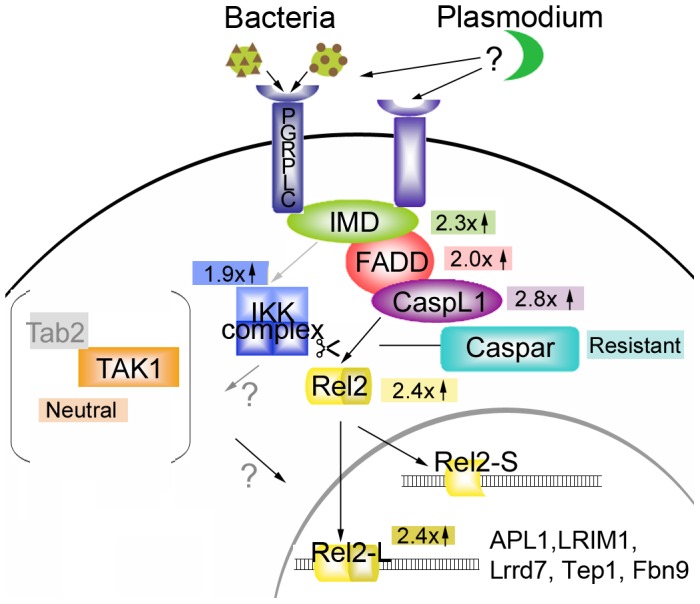
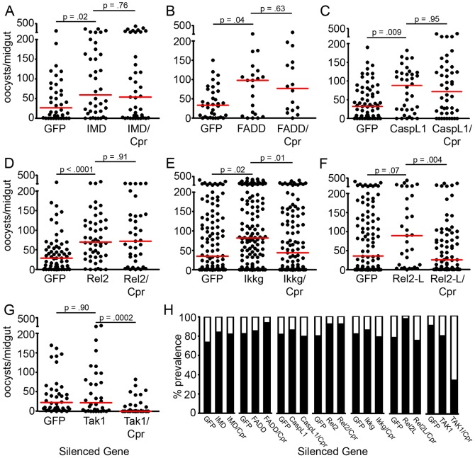
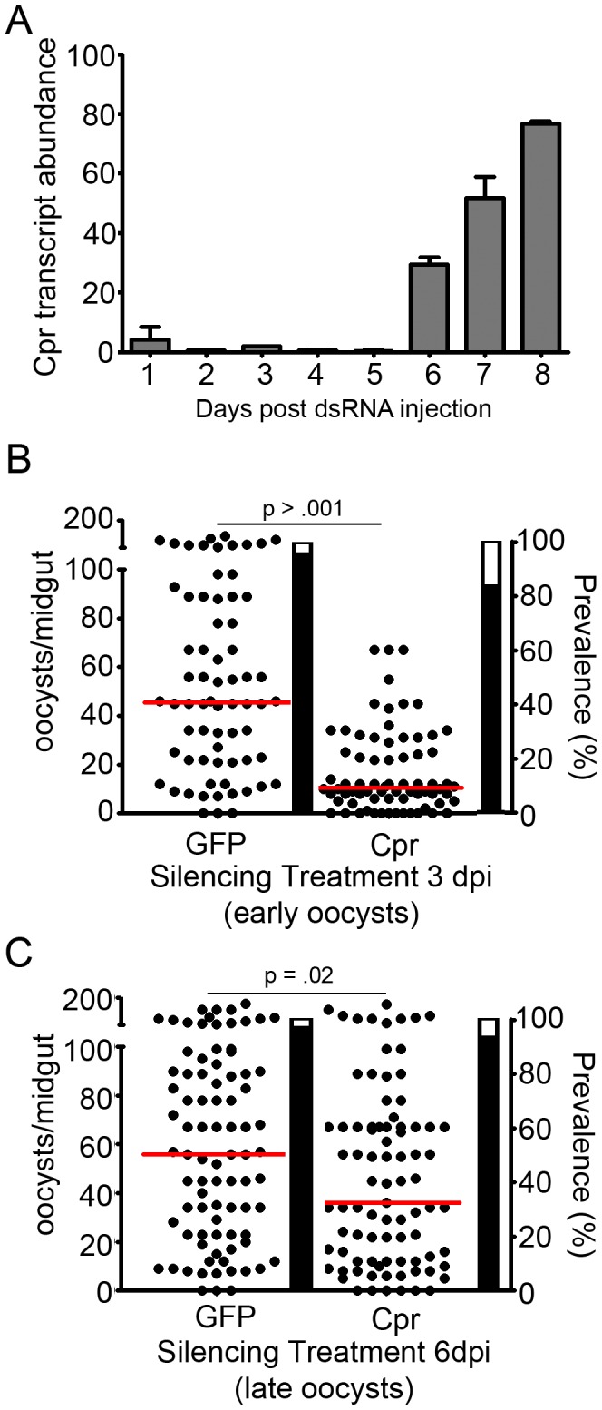
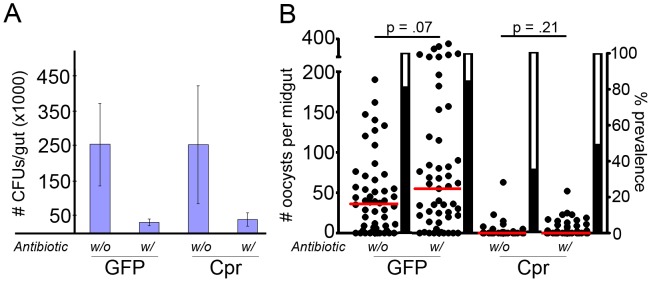
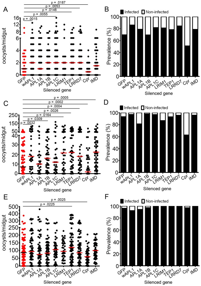
The Anopheles gambiae immune response against Plasmodium falciparum, an etiological agent of human malaria, has been identified as a source of potential anti-Plasmodium genes and mechanisms to be exploited in efforts to control the malaria transmission cycle. One such mechanism is the Imd pathway, a conserved immune signaling pathway that has potent anti-P. falciparum activity. Silencing the expression of caspar, a negative regulator of the Imd pathway, or over-expressing rel2, an Imd pathway-controlled NFkappaB transcription factor, confers a resistant phenotype on A. gambiae mosquitoes that involves an array of immune effector genes. However, unexplored features of this powerful mechanism that may be essential for the implementation of a malaria control strategy still remain. Using RNA interference to singly or dually silence caspar and other components of the Imd pathway, we have identified genes participating in the anti-Plasmodium signaling module regulated by Caspar, each of which represents a potential target to achieve over-activation of the pathway. We also determined that the Imd pathway is most potent against the parasite's ookinete stage, yet also has reasonable activity against early oocysts and lesser activity against late oocysts. We further demonstrated that caspar silencing alone is sufficient to induce a robust anti-P. falciparum response even in the relative absence of resident gut microbiota. Finally, we established the relevance of the Imd pathway components and regulated effectors TEP1, APL1, and LRIM1 in parasite infection intensity-dependent defense, thereby shedding light on the relevance of laboratory versus natural infection intensity models. Our results highlight the physiological considerations that are integral to a thoughtful implementation of Imd pathway manipulation in A. gambiae as part of an effort to limit the malaria transmission cycle, and they reveal a variety of previously unrecognized nuances in the Imd-directed immune response against P. falciparum.


Conflict of interest statement

The authors have declared that no competing interests exist.

Figures

Figure 1
Figure 1. Anopheles Imd pathway model.
Components of the Imd pathway explored in this study or others are represented by different colored shapes. Black arrows or lines indicate known interactions or translocations. Gray arrows indicate potential interactions based on D. melanogaster studies. The gray bracketed area indicates the molecules possibly involved in other responses, but not the responses against P. falciparum. Numbers and arrows within colored blocks indicate the -fold change in P. falciparum infection that results when the corresponding pathway member is silenced. The list of genes inside the nucleus portion of the diagram shows those known to be active against Plasmodium and whose expression has been shown by our studies to be REL2-regulated.
Figure 2
Figure 2. Some members of the Imd pathway have an effect on P. falciparum infection.
(A–G) Dots represent individual oocyst counts following the indicated RNAi treatment; horizontal red bars represent the median number of oocysts per gut. P-values were derived from Mann-Whitney statistical tests and appear above each treatment and refer to that treatment as compared to the GFP dsRNA-treated control. Additional statistical analyses appear in Table S1) Filled portion of bars represent the % of all mosquitoes harboring at least one oocyst; open portion represents those in the group that were uninfected. All assays represent two to three independent biological replicate. Cpr, Caspar. (H) Prevalence of P. falciparum infection following the indicated RNAi treatment.
Figure 3
Figure 3. Caspar silencing also influences early and late oocysts.
(A) Time course of caspar-silencing efficiency, quantified using real-time quantitative PCR. Gray bars represent the % caspar expression at each given time point as an average of two replicates, and error bars reflect the standard error between those replicates. Cpr, Caspar. (B) Infection intensity of mosquitoes silenced for caspar at 3 days post-infection (dpi). Since ookinetes invade the midgut at 24 hours post-infection this time point targets early oocysts. (C) Infection intensity of mosquitoes silenced for caspar at 6 days post-infection. Since sporozoites begin to emerge from the oocyst at 7–8 days post infection, this time point targets late oocysts. For both, dots represent individual oocyst counts following the indicated RNAi treatment; horizontal red bars represent the median number of oocysts per gut. Assays represent two to three independent biological replicates and were subject to Mann-Whitney statistical tests. P-values appear above each treatment and refer to that treatment as compared to the GFP dsRNA-treated control. Additional statistical analyses appear in Table S2. Filled portion of bars represent the % of all mosquitoes harboring at least one oocyst; open portion represents those in the group that were uninfected. Cpr, Caspar.
Figure 4
Figure 4. Caspar-mediated killing of P. falciparum is not dependent on midgut bacteria.
(A) Blue bars represent bacteria colony-forming unit (CFUs) from midguts of mosquitoes undergoing the indicated treatments. Pluses and minuses indicate the presence or absence of antibiotic in GFP or Cpr dsRNA treated group. Each bar represents the average of at least 15 mosquitoes tested, with each mosquito's CFU count determined by averaging counts from three serial dilutions. Bars represent the standard deviation for all mosquitoes in a given treatment group. Cpr, Caspar. (B) Dots represent individual oocyst counts following the indicated RNAi treatment; horizontal red bars represent the median number of oocysts per gut. Pluses and minuses indicate the presence or absence of antibiotic in the GFP or Cpr dsRNA-treated group. Assays represent three independent biological replicates and were subject to Mann-Whitney statistical tests. P-values appear below each treatment and refer to that treatment as compared to the GFP dsRNA-treated control. Additional statistical analyses appear in Table S3. Filled portion of bars represent the % of all mosquitoes harboring at least one oocyst; open portion represents those in the group that were uninfected. Cpr, Caspar.
Figure 5
Figure 5. Imd pathway components and effectors differ in their ability to affect P. falciparum infections of high, medium, and low infection intensities.
Intensity of P. falciparum oocysts in A. gambiae silenced for given genes and subjected to low (A), medium (C) or high (E) infection exposures. Bars represent median numbers of oocysts per midgut, and dots represent individual midgut oocyst counts. Assays represent at least three independent biological replicates and were subject to Mann-Whitney statistical tests. P-values appear above each treatment and refer to that treatment as compared to the GFP dsRNA-treated control. Non-significant p-values were not included in the figure. Additional statistical analyses appear in Table S4. D-F: Prevalence of infection in A. gambiae subjected to low (B), medium (D) and high (F) loads of P. falciparum. Filled portion of bars represent the % of all mosquitoes harboring at least one oocyst; open portion represents those in the group that were uninfected. wAPL1 (whole APL1) dsRNA – dsRNA for a conserved region of APL1 genes, which results in the silencing of all three APL1 proteins (APL1A, APL1B and APL1C).

References

    1. World Health Organization. Global Malaria Control and Elimination: report of a technical review. Geneva, Switzerland. 2008.
    1. World Health Organization. World Malaria Report. Geneva, Switzerland. 2010.
    1. Frolet C, Thoma M, Blandin S, Hoffmann JA, Levashina EA. Boosting NF-κB-dependent basal immunity of Anopheles gambiae aborts development of Plasmodium berghei. Immunity. 2006;25:685. - PubMed
    1. Garver LS, Dong Y, Dimopoulos G. Caspar controls resistance to Plasmodium falciparum in diverse anopheline species. PLoS Pathog. 2009;5:e1000335. - PMC - PubMed
    1. Gupta L, Molina-Cruz A, Kumar S, Rodrigues J, Dixit R. The STAT pathway mediates late-phase immunity against Plasmodium in the mosquito Anopheles gambiae. Cell Host Microbe. 2009;5:507. - PMC - PubMed

Publication types

Substances