

3D reconstruction of VZV infected cell nuclei and PML nuclear cages by serial section array scanning electron microscopy and electron tomography
- PMID: 22685402
- PMCID: PMC3369938
- DOI: 10.1371/journal.ppat.1002740
3D reconstruction of VZV infected cell nuclei and PML nuclear cages by serial section array scanning electron microscopy and electron tomography
Abstract
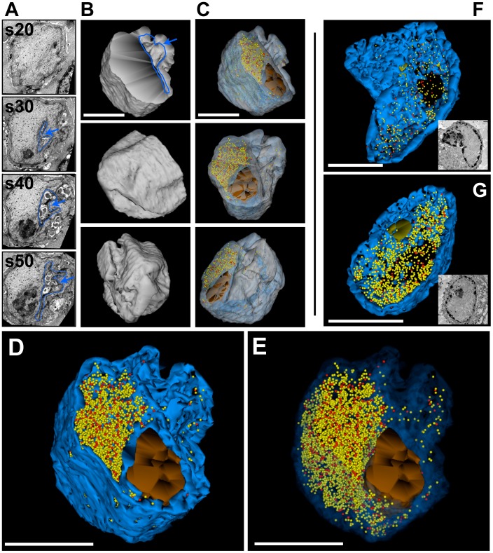
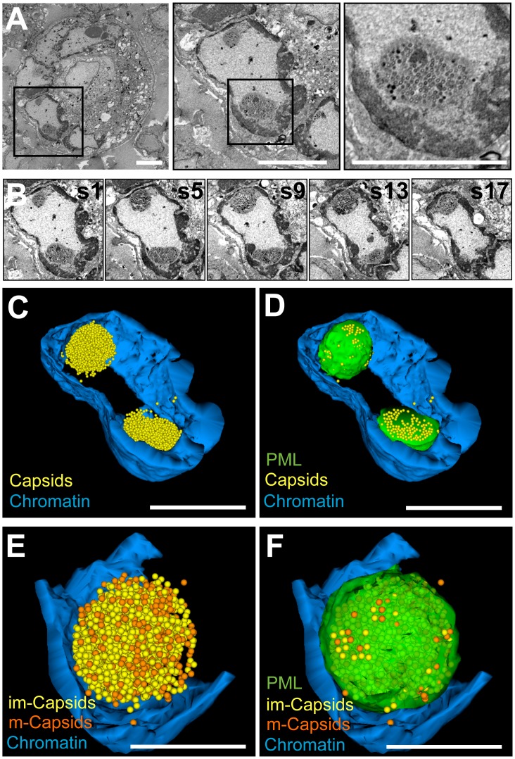
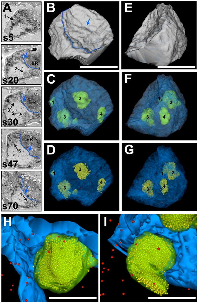
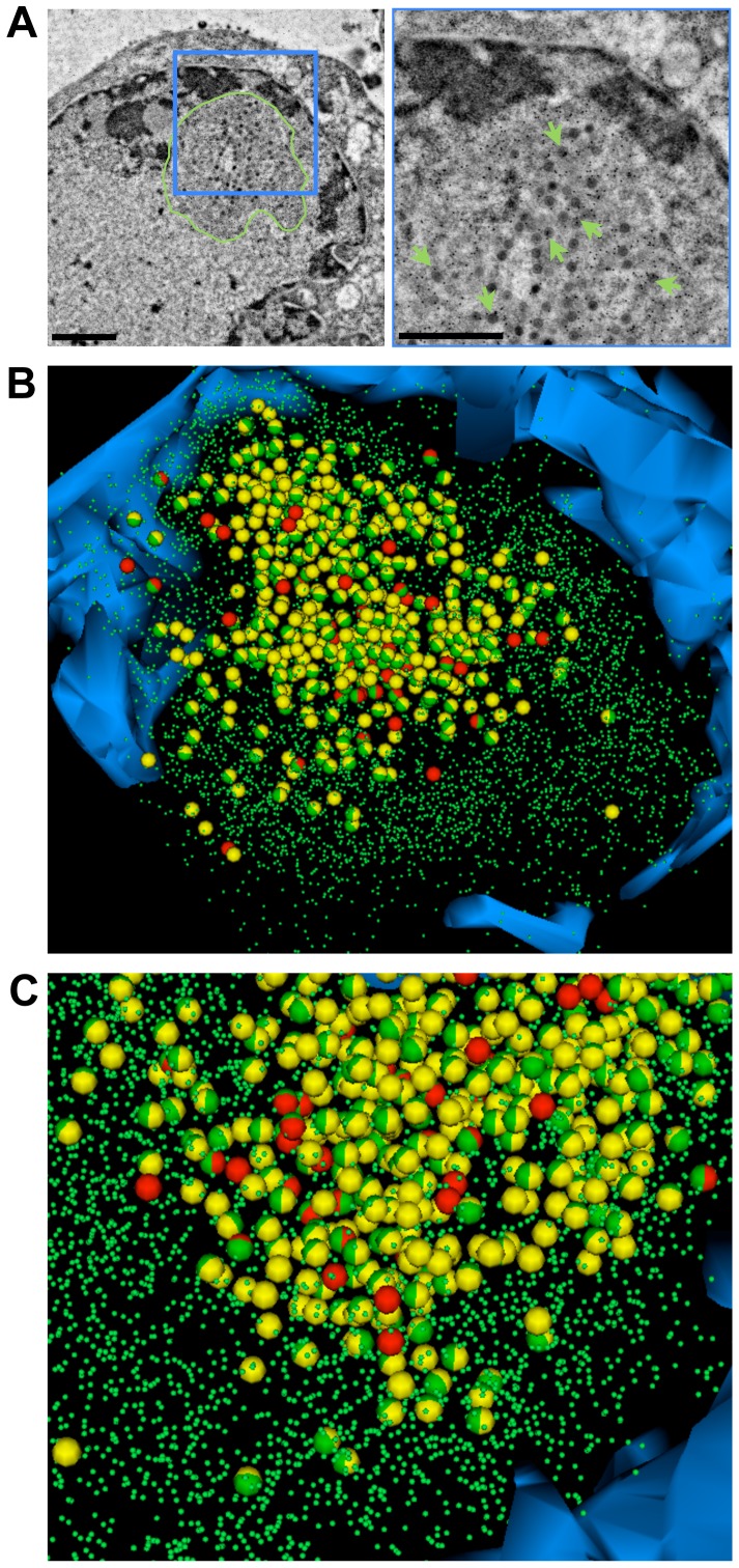
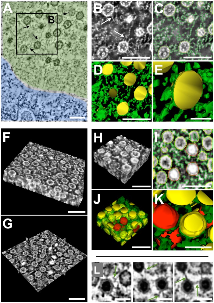
Varicella-zoster virus (VZV) is a human alphaherpesvirus that causes varicella (chickenpox) and herpes zoster (shingles). Like all herpesviruses, the VZV DNA genome is replicated in the nucleus and packaged into nucleocapsids that must egress across the nuclear membrane for incorporation into virus particles in the cytoplasm. Our recent work showed that VZV nucleocapsids are sequestered in nuclear cages formed from promyelocytic leukemia protein (PML) in vitro and in human dorsal root ganglia and skin xenografts in vivo. We sought a method to determine the three-dimensional (3D) distribution of nucleocapsids in the nuclei of herpesvirus-infected cells as well as the 3D shape, volume and ultrastructure of these unique PML subnuclear domains. Here we report the development of a novel 3D imaging and reconstruction strategy that we term Serial Section Array-Scanning Electron Microscopy (SSA-SEM) and its application to the analysis of VZV-infected cells and these nuclear PML cages. We show that SSA-SEM permits large volume imaging and 3D reconstruction at a resolution sufficient to localize, count and distinguish different types of VZV nucleocapsids and to visualize complete PML cages. This method allowed a quantitative determination of how many nucleocapsids can be sequestered within individual PML cages (sequestration capacity), what proportion of nucleocapsids are entrapped in single nuclei (sequestration efficiency) and revealed the ultrastructural detail of the PML cages. More than 98% of all nucleocapsids in reconstructed nuclear volumes were contained in PML cages and single PML cages sequestered up to 2,780 nucleocapsids, which were shown by electron tomography to be embedded and cross-linked by an filamentous electron-dense meshwork within these unique subnuclear domains. This SSA-SEM analysis extends our recent characterization of PML cages and provides a proof of concept for this new strategy to investigate events during virion assembly at the single cell level.
Conflict of interest statement
The authors have declared that no competing interests exist.
Figures


Similar articles
-
Entrapment of viral capsids in nuclear PML cages is an intrinsic antiviral host defense against varicella-zoster virus.PLoS Pathog. 2011 Feb 3;7(2):e1001266. doi: 10.1371/journal.ppat.1001266. PLoS Pathog. 2011. PMID: 21304940 Free PMC article.
-
Three-dimensional organization of promyelocytic leukemia nuclear bodies.J Cell Sci. 2010 Feb 1;123(Pt 3):392-400. doi: 10.1242/jcs.053496. J Cell Sci. 2010. PMID: 20130140
-
Egress of varicella-zoster virus from the melanoma cell: a tropism for the melanocyte.J Virol. 1995 Aug;69(8):4994-5010. doi: 10.1128/JVI.69.8.4994-5010.1995. J Virol. 1995. PMID: 7609070 Free PMC article.
-
Tomography of the cell nucleus using confocal microscopy and medium voltage electron microscopy.Crit Rev Oncol Hematol. 2009 Feb;69(2):127-43. doi: 10.1016/j.critrevonc.2008.07.022. Epub 2008 Sep 10. Crit Rev Oncol Hematol. 2009. PMID: 18783961 Review.
-
Varicella-zoster virus.Clin Microbiol Rev. 1996 Jul;9(3):361-81. doi: 10.1128/CMR.9.3.361. Clin Microbiol Rev. 1996. PMID: 8809466 Free PMC article. Review.
Cited by
-
Automated transmission-mode scanning electron microscopy (tSEM) for large volume analysis at nanoscale resolution.PLoS One. 2013;8(3):e59573. doi: 10.1371/journal.pone.0059573. Epub 2013 Mar 26. PLoS One. 2013. PMID: 23555711 Free PMC article.
-
Replication Compartments of DNA Viruses in the Nucleus: Location, Location, Location.Viruses. 2020 Jan 29;12(2):151. doi: 10.3390/v12020151. Viruses. 2020. PMID: 32013091 Free PMC article. Review.
-
Morphological, Biochemical, and Functional Study of Viral Replication Compartments Isolated from Adenovirus-Infected Cells.J Virol. 2016 Jan 13;90(7):3411-27. doi: 10.1128/JVI.00033-16. J Virol. 2016. PMID: 26764008 Free PMC article.
-
Multilamellar structures and filament bundles are found on the cell surface during bunyavirus egress.PLoS One. 2013 Jun 14;8(6):e65526. doi: 10.1371/journal.pone.0065526. Print 2013. PLoS One. 2013. PMID: 23799021 Free PMC article.
-
The Stanford Nanocharacterization Laboratory (SNL) and Recent Applications of an Aberration-Corrected Environmental Transmission Electron Microscope.Adv Eng Mater. 2014 May;16(5):476-481. doi: 10.1002/adem.201400015. Adv Eng Mater. 2014. PMID: 25364299 Free PMC article.
References
-
- Cohen JI, Straus SE, Arvin AM. Varicella-Zoster Virus. In: Knipe DM, Howley PM, editors. Fields Virology. 5 ed. Philadelphia: Lippincott Williams and Wilkins; 2007. pp. 2773–2818.
-
- Zerboni L, Reichelt M, Arvin A. Varicella-zoster virus neurotropism in SCID mouse-human dorsal root ganglia xenografts. Curr Top Microbiol Immunol. 2010;342:255–276. - PubMed
Publication types
MeSH terms
Substances
Grants and funding
LinkOut - more resources
Full Text Sources
Other Literature Sources
Research Materials
