. 2012;8(6):e1002746.
doi: 10.1371/journal.ppat.1002746. Epub 2012 Jun 7.

Propagation of RML prions in mice expressing PrP devoid of GPI anchor leads to formation of a novel, stable prion strain

Affiliations

Propagation of RML prions in mice expressing PrP devoid of GPI anchor leads to formation of a novel, stable prion strain

Sukhvir Paul Mahal et al. PLoS Pathog. 2012.

Abstract

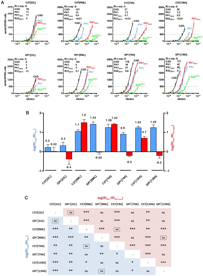
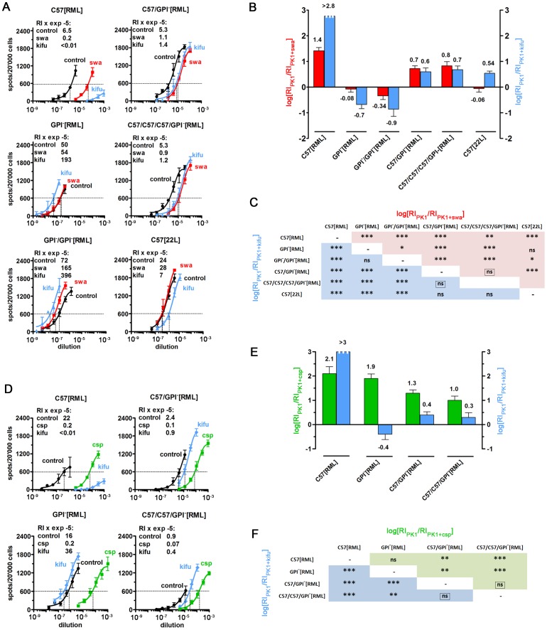
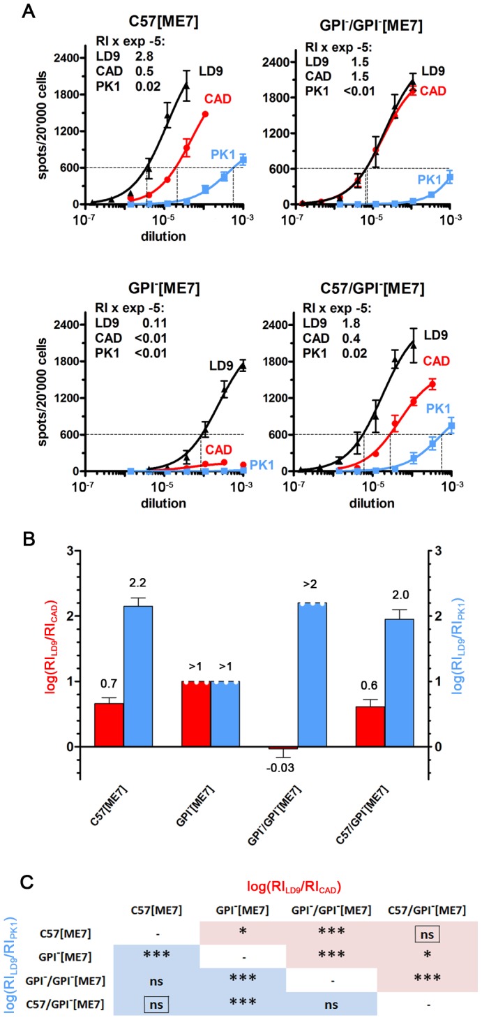
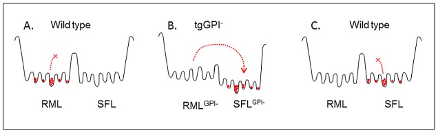
PrP(C), a host protein which in prion-infected animals is converted to PrP(Sc), is linked to the cell membrane by a GPI anchor. Mice expressing PrP(C) without GPI anchor (tgGPI⁻ mice), are susceptible to prion infection but accumulate anchorless PrP(Sc) extra-, rather than intracellularly. We investigated whether tgGPI⁻ mice could faithfully propagate prion strains despite the deviant structure and location of anchorless PrP(Sc). We found that RML and ME7, but not 22L prions propagated in tgGPI⁻ brain developed novel cell tropisms, as determined by the Cell Panel Assay (CPA). Surprisingly, the levels of proteinase K-resistant PrP(Sc) (PrP(res)) in RML- or ME7-infected tgGPI⁻ brain were 25-50 times higher than in wild-type brain. When returned to wild-type brain, ME7 prions recovered their original properties, however RML prions had given rise to a novel prion strain, designated SFL, which remained unchanged even after three passages in wild-type mice. Because both RML PrP(Sc) and SFL PrP(Sc) are stably propagated in wild-type mice we propose that the two conformations are separated by a high activation energy barrier which is abrogated in tgGPI⁻ mice.


Conflict of interest statement

The authors have declared that no competing interests exist.

Figures

Figure 1
Figure 1. PrPres in RML- and ME7-infected wild-type and tgGPI- mouse brains, quantified by Western blot and sandwich ELISA.
(A) Western blot of RML and ME7-infected brain homogenates before and after PK treatment. “µg”, total protein loaded. ‘r’, 2.5 ng recombinant murine PrP (recPrP). GPI[RML] (lanes 5, 7) and GPI/GPI[ME7] (lane 17) brain homogenates gave rise to ladders reflecting multimers with molecular weights extending to >250 kDa, which were reduced to monomers by PK digestion, while GPI[ME7] homogenate gave no detectable signals. (B) Western blot of PK-digested brain homogenates treated or not with PNGase. Lanes 3, 5, 7, 9 show, from top to bottom, diglycosylated, monoglycosylated and unglycosylated, truncated PrP; PNGase-treated samples (lanes 4, 6, 8, 10) show a single band corresponding to unglycosylated, truncated PrP. Samples from tgGPI mice (lanes 1, 11) show two bands, corresponding to truncated, monoglycosylated (top) and unglycosylated PrP, and, after PNGase treatment, a single band corresponding to truncated, deglycosylated PrP (lanes 2, 12). Because anchorless PrP is retained inefficiently by PVDF membranes , 24 times more total protein was loaded for GPI than for C57 samples, to give about the same signal strength. (C) Sandwich ELISA of PK-treated samples. Absorbance of quadruplicate samples is plotted against log[input protein]. Samples a to g are identified in panel D; h, uninfected C57 brain homogenate. The abundance of a sample relative to that of RML can be read off by comparing the amounts of protein required to give the same absorbance. For example, an absorbance of 0.1 is given by 0.01 µg GPI[RML] and 0.5 µg C57[RML] brain homogenate (total protein prior to PK treatment), therefore the abundance of GPI[RML] is about 50 times higher than that of C57[RML] PrPres. In Figure S2 absorbance of the same samples is plotted against input protein on a linear scale, to show that the response is almost linear up to a protein input of 1.5 µg/well. (D) The PrPres signals (“pix”) from the western blots of Figure 1A were quantified relative to C57[RML] and the log of the ratio was plotted (red bars). For the sandwich ELISA, the plot shows the log of the absorbance (A) relative to that of C57[RML] (blue bars).
Figure 2
Figure 2. The cell tropism of various prion strains changes after propagation in tgGPI mice.
Homogenates of GPI or C57 brains infected with the strains indicated were subjected to the CPA. (A) The patterns elicited by 22L from both sources were very similar, however RML, 79A and 139A prions from wild-type brain were swa sensitive on PK1 cells and R332H11 incompetent, while those from tgGPI brain were swa resistant and R332H1 competent. The RI600 (Response Index for 600 spots) on CAD, PK1, PK1+swa and R332H11 cells is given within the graphs (left upper corner) and the logarithm ± SD of the ratios RICAD/RIPK1 (blue) and RIPK1/RIPK1+swa (red) is plotted in the bar graph (B). The matrix (C) gives the p values for the pairwise comparison of two strains on the basis of their log[RICAD/RIPK1] (blue) and log[RIPK1/RIPK1+swa] (red) values. The framed “ns” indicates p values>0.1 for both log[ratios]. For example, C57[RML] and GPI[RML] prions are significantly different (p = 0.0097 for log[RICAD/RIPK1] and p = 0.0001 for log[RIPK1/RIPK1+swa]), as are C57[79A] and GPI[79A] prions, whereas C57[22L] and GPI[22L] prions do not show a significant difference (framed “ns”; p>0.1) for both logRI ratios. By the same token C57[79A] and C57[RML], and GPI[139A] and GPI[RML] prions are not distinguishable, while C57[139A] and C57[RML] prions differ.
Figure 3
Figure 3. RML prions propagated in tgGPI mice and returned to wild-type mice emerge as a novel, stable strain.
Analysis of authentic RML prions propagated in C57BL/6 brain (C57[RML]), and of RML prions first propagated in GPI brain (GPI[RML]) and then once (C57/GPI[RML]), twice (C57/C57/GPI[RML]) or three times (C57/C57/C57/GPI[RML]) in C57BL/6 brain. (A) shows the SSCA performed on PK1 cells in the presence or absence of kifunensine (kifu; 5 µg/ml) or swainsonine (swa; 2 µg/ml) for the samples indicated; C57[22L] was added as control. RI's were determined at 600 spots (RI600). Kifu strongly inhibited the propagation of C57[RML], but not of C57/GPI[RML] and C57/C57/C57/GPI[RML] prions on PK1 cells. (B) The bar graph shows the log[RIPK1/RIPK1+kifu] and log[RIPK1/RIPK1+swa] values for the samples listed in (A). The pairwise comparison in panel (C) shows that C57/GPI[RML] and C57/C57/C57/GPI[RML] prions do not differ significantly, but are vastly different from C57[RML] and GPI[RML] prions. The framed “ns” indicates high p values (>0.1) for both log[ratios], indicating no significant difference between the samples. (D) Effect of castanospermine (csp; 50 µg/ml) or kifu (5 µg/ml) on propagation of the samples indicated on PK1 cells. As in (A), kifu strongly inhibits the propagation of C57[RML], but not C57/GPI[RML] and C57/C57/GPI[RML] prions. The same is true for csp, however to a lesser extent. (E) The bar graph depicts the log[RIPK1/RIPK1+kifu] (blue) and log[RIPK1/RIPK1+csp] (green) values. The RIPK1/RIPK1+kifu ratio was >500-fold lower for C57/GPI[RML] and C57/C57/GPI[RML] than for C57[RML] prions, again underscoring the difference between RML prions and the novel strain. The matrix (F) also shows that C57/GPI[RML] and C57/C57/GPI[RML] prions do not differ from each other, but that both differ significantly from C57[RML] prions. In summary, the figure sustains the conclusion that a new strain, designated SFL, emerged after RML prions were passaged through GPI brain and returned C57 brain, and that they remained unchanged after two further passages.
Figure 4
Figure 4. ME7 prions propagated in tgGPI mice for one or two passages acquire novel characteristics.
(A) Serially diluted brain homogenates were analyzed on LD9, CAD and PK1 cells by the CPA. After the first passage of ME7 prions in GPI mice (GPI[ME7]), the RI on CAD cells dropped 20-fold, most likely reflecting low titers, but increased about 30-fold after the second passage (GPI/GPI[ME7]), indicating adaptation to the GPI environment. Returning prions from the first passage in GPI brain to wild-type brain (C57/GPI[ME7]) restored the original CPA pattern. (B) Log[RILD9/RICAD] (red) and log[RILD9/RIPK1] (blue) are plotted as a bar graph. (C) The matrix shows that C57[ME7] and C57/GPI[ME7] prions do not differ, while both differ in at least one log[ratio] from GPI[ME7] and GPI/GPI[ME7] prions.
Figure 5
Figure 5. Conjectural free energy profile.
(A) The RML quasi-species in wild-type brain are confined to a set of wells separated from those of the SFL quasi-species by a high activation energy barrier. (B) In tgGPI brain the transition to the “SFLGPI-” set of conformations is enabled by a lower activation energy barrier. (C) SFLGPI- prions introduced into wild-type brain can now occupy, and are trapped in, the set of “SFL wells” which was not accessible to RML prions in (A).

Similar articles

Cited by

References

    1. Endo T, Groth D, Prusiner SB, Kobata A. Diversity of oligosaccharide structures linked to asparagines of the scrapie prion protein. Biochemistry. 1989;28:8380–8388. - PubMed
    1. Rudd PM, Endo T, Colominas C, Groth D, Wheeler SF, et al. Glycosylation differences between the normal and pathogenic prion protein isoforms. Proc Natl Acad Sci U S A. 1999;96:13044–13049. - PMC - PubMed
    1. Rudd PM, Wormald MR, Wing DR, Prusiner SB, Dwek RA. Prion glycoprotein: structure, dynamics, and roles for the sugars. Biochemistry. 2001;40:3759–3766. - PubMed
    1. Beringue V, Mallinson G, Kaisar M, Tayebi M, Sattar Z, et al. Regional heterogeneity of cellular prion protein isoforms in the mouse brain. Brain. 2003;126:2065–2073. - PubMed
    1. Somerville RA, Hamilton S, Fernie K. Transmissible spongiform encephalopathy strain, PrP genotype and brain region all affect the degree of glycosylation of PrPSc. J Gen Virol. 2005;86:241–246. - PubMed

Publication types