. 2012;8(6):e1002752.
doi: 10.1371/journal.ppat.1002752. Epub 2012 Jun 7.

Structure-function analysis of barley NLR immune receptor MLA10 reveals its cell compartment specific activity in cell death and disease resistance

Affiliations

Structure-function analysis of barley NLR immune receptor MLA10 reveals its cell compartment specific activity in cell death and disease resistance

Shiwei Bai et al. PLoS Pathog. 2012.

Abstract

Plant intracellular immune receptors comprise a large number of multi-domain proteins resembling animal NOD-like receptors (NLRs). Plant NLRs typically recognize isolate-specific pathogen-derived effectors, encoded by avirulence (AVR) genes, and trigger defense responses often associated with localized host cell death. The barley MLA gene is polymorphic in nature and encodes NLRs of the coiled-coil (CC)-NB-LRR type that each detects a cognate isolate-specific effector of the barley powdery mildew fungus. We report the systematic analyses of MLA10 activity in disease resistance and cell death signaling in barley and Nicotiana benthamiana. MLA10 CC domain-triggered cell death is regulated by highly conserved motifs in the CC and the NB-ARC domains and by the C-terminal LRR of the receptor. Enforced MLA10 subcellular localization, by tagging with a nuclear localization sequence (NLS) or a nuclear export sequence (NES), shows that MLA10 activity in cell death signaling is suppressed in the nucleus but enhanced in the cytoplasm. By contrast, nuclear localized MLA10 is sufficient to mediate disease resistance against powdery mildew fungus. MLA10 retention in the cytoplasm was achieved through attachment of a glucocorticoid receptor hormone-binding domain (GR), by which we reinforced the role of cytoplasmic MLA10 in cell death signaling. Together with our data showing an essential and sufficient nuclear MLA10 activity in disease resistance, this suggests a bifurcation of MLA10-triggered cell death and disease resistance signaling in a compartment-dependent manner.


Conflict of interest statement

The authors have declared that no competing interests exist.

Figures

Figure 1
Figure 1. MLA10 CC is the cell death signaling domain whose activity is regulated by other domains.
(A) Schematic diagram of the MLA10 domain structure and the derived fragments expressed in N. benthamiana. Individual domains of MLA10 are represented by colored boxes, and the relative positions of relevant amino acids and motifs (in parentheses) are indicated on top (upper panel). Lower panel: MLA10 fragments consisting only of the CC domain or the CC combined with other domains are drawn schematically in solid lines. All fragments were expressed as C-terminal -3×HA fusions in N. benthamiana leaves using Agrobacterium-mediated transient transformation (Agro-infiltration). (B) Analysis of cell death inducing activity of MLA10 fragments. MLA10 fragments or full-length(FL) protein fused with a C-terminal 3×HA tag were expressed by Agro-infiltration in N. benthamiana (left), and cell-death triggered by each protein was visualized by trypan blue staining at 42 hrs post Agro-infiltration (hpi) (right). Red circles indicate cell death; white circles indicate no obvious cell death in the infiltrated area. (C) Quantification of cell-death inducing activity of MLA10 fragments. Upon expression of the same fragment fusions as indicated in (B) by Agro-infiltration in N. benthamiana, electrolyte leakage was measured each hour from 28 to 34 hpi. Error bars were calculated from three replicates per time point and per construct. Experiments were done at least twice with similar results. Letters (a–d) represent groups with significant differences [p<0.05, Tukey's honest significant difference (HSD) test]. (D) Protein expression of MLA10 fragments. Total protein was extracted from N. benthamiana leaves at 40 hpi and MLA10-HA was detected by immunoblotting using an anti-HA antibody. Asterisk indicates non-specific signals throughout this article except specified. Ponceau staining of Rubisco small subunit was used to show equal loading throughout this article except specified. EV: empty vector.
Figure 2
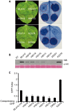
Figure 2. Glutamate substitution analyses in the MLA10 CC domain identify the F99E autoactive mutation.
(A) Analysis of cell death inducing activity of MLA10 CC mutant variants. MLA10 CC wild-type or mutant variants harboring indicated amino acid substitution were expressed in N. benthamiana leaves, and cell death induced by each protein was visualized by trypan blue staining at 42 hpi (upper panel). (B) Protein expression levels of each CC variant shown by Western blotting. Proteins were extracted from N. benthamiana leaves at 40 hpi and detection was done by immunoblotting with anti-HA antibody. (C) Comparison of cell death inducing activity of MLA10 FL and the FL(F99E) variant. FL and FL(F99E) were expressed on the same N. benthamiana leaf, and the amount of cell death induced by each protein was shown by Trypan blue staining at 24 hpi (upper panel); protein expression levels of FL and FL(F99E) were shown by protein immunoblotting analysis using anti-HA antibody (bottom panel), protein extracts were obtained at ∼22 hpi. (D) Quantification of cell-death inducing activity of FL and FL(F99E). Upon expression of FL or FL(F99E) by Agro-infiltration in N. benthamiana, electrolyte leakage was measured each hour from 22 to 30 hpi, and then every two hours from 30 to 34 hpi; empty vector (EV) was included as a negative control. Error bars representing standard error (SE) were calculated from three replicates per time point and per construct. Similar experiments were repeated at least twice with similar results. Letters (a–c) represent significant differences [p<0.05, Tukey's honest significant difference (HSD) test].
Figure 3
Figure 3. Cell death activity analyses of MLA10 MHD or P-loop mutant variants in N. benthamiana and barley.
(A) Analysis of cell death Inducing activity of MLA10 WT and its mutant variants in N. benthamiana. Individual C-terminal 3×HA tagged MLA10 WT and mutant proteins were expressed by Agro-infiltration in N. benthamiana, and cell-death triggered by each protein was scored by trypan blue staining at 24 hpi. (B) Protein expression of MLA10 WT and its mutant variants. Total proteins were extracted from N. benthamiana leaves at 22 hpi and followed by immunoblotting. MLA10 was detected using an anti-HA antibody. (C) Analysis of cell death Inducing activity of MLA10 mutant variants in barley. Plasmids of MLA10 MHD motif mutants (H501R/G/Q/V/A, D502V) and a P-loop mutant (K207R) were co-expressed with a GFP maker plasmid in barley epidermal cells using biolistic delivery. The histogram bars represent the number of GFP expressing cell of each co-expression experiment standardized to the MLA10-YFP as control (see Materials and Methods). GFP expressing cells were scored as vital cells at 36–42 hrs post bombardment. The error bars represented SE of three representing experiments from more than five replicates.
Figure 4
Figure 4. Tightly regulated MLA10 cell death inducing activity.
(A) Analysis of cell death triggering activity of MLA10 variants harboring the K207R P-loop mutation. The indicated N-terminal MLA10 fragments containing the K207R mutation, or FL variants harboring K207R alone or K207R combined with an MHD mutation (H501, D502V), were expressed in N. benthamiana leaves by Agro-infiltration. Cell death triggered by each fusion was assessed by trypan blue staining at 42 hpi (upper panel). Protein expression levels of indicated MLA10 mutant fragments are shown (lower panel). Proteins were extracted at 40 hpi and detection was done by immunoblotting with anti-HA antibody. (B) Analysis of cell death activity of MLA10 variants harboring mutations in the EDVID motif. The EDVID motif (EDVVD in MLA10) was mutated to AAVVD, and the indicated MLA10 fragments or FL variants harboring indicated mutation(s) were expressed in N. benthamiana leaves by Agro-infiltration. Cell death triggered by each variant was assessed by trypan blue staining at 42 hpi (upper panel). Protein expression levels of indicated MLA10 mutant fragments are shown (lower panel). Proteins were extracted at 40 hpi and detection was done by immunoblotting with anti-HA antibody. (C) Analysis of cell death activity of F99E containing MLA10 fragments or FL variants. Indicated MLA10 fragments or FL variants harboring indicated mutation(s) were expressed in N. benthamiana leaves by Agro-infiltration. Cell death triggered by each protein was scored by trypan blue staining at 42 hpi (upper panel); Protein were extracts from N. benthamiana leaves and subjected to immunoblotting with anti-HA antibody (lower panel).
Figure 5
Figure 5. Manipulation of MLA10 subcellular localization and the relevance to MLA10 cell death signaling and disease resistance.
(A) Confocal images of barley leaf epidermal cells expressing MLA10 fusion proteins. Indicated fusion proteins were expressed in barley leaf epidermal cells upon biolistic delivery, the confocal images were taken at 36 hrs post bombardment. Upper panel: A representative barley cell coexpressing MLA10-YFP-NLS and a nucleus marker CFP-WRKY2 (2D z plane). Lower panel: a cell expressing MLA10-YFP-nls alone (2D z plane). NLS: nuclear localization signal; nls: mutated nuclear localization signal. Arrowheads mark the nucleus and scale bar is 50 µm. (B) Analyses of disease resistance activity of MLA10 fusions or mutant variants. Relative single cell resistance/susceptibility is shown by fungal haustorium index upon biolistic delivery of plasmids expressing indicated protein and a GUS reporter into the barley leaf epidermal cells of a susceptible barley line (Golden promise). Bombarded leaves were inoculated with B. graminis fungal spores expressing AVRA10, and the fungal haustorium index was microscopically scored at 36 hrs post spore inoculation. Histogram bar represents average of three independent experiments and error bar represents SD. In the case of MLA10(F99E) expression, the number of cells expressing GUS reporter were extremely low. (C) Analysis of cell death triggering activity of MLA10 fusion proteins. Indicated MLA10 fusion proteins were expressed in N. benthamiana leaves by Agro-infiltration, and cell-death triggered by each fusion protein was scored by trypan blue staining at 40 hpi. NES: nuclear exclusion signal; nes: mutated nuclear exclusion signal. (D) Protein expression of indicated MLA10 fusions. Proteins were extracted at 23 hpi and MLA was detected by immunoblotting using an anti-MLA27 monoclonal antibody. Asterisk indicates non-specific signals. (E) Quantification of cell-death inducing activity of MLA10 fusion proteins. Upon expression of indicated MLA10 fusion proteins by Agro-infiltration in N. benthamiana, ion leakage was measured each hour from 23 to 34 hpi. Error bars (SE) were calculated from three replicates per time point and construct. Experiment was done at least twice with similar result. Letters (a–c) represent groups with significant differences [p<0.05, Tukey's honest significant difference (HSD) test].
Figure 6
Figure 6. MLA10 CC-NB or full-length trigger cell death signaling in the cytoplasm.
(A) Analysis of cell death inducing activity of MLA10 CC-NB fusion proteins. MLA10 CC-NB fusion proteins, CC-NB-YFP, CC-NB-YFP-NES and CC-NB-YFP-NLS, were expressed in N. benthamiana leaves by Agro-infiltration; confocal images were taken at ∼22 hpi (upper panel) and cell-death triggered by each fusion protein was scored by trypan blue staining at ∼48 hpi (lower panel). Scale bar is 50 µm. (B) Analysis of cell death phenotype upon expression of MLA10 CC-NB-YFP-GR or co-expression of MLA10 CC-NB-YFP-NLS and CC-NB-YFP-GR fusions before and after Dex treatment. The indicated fusion(s) were expressed in N. benthamiana leaves by Agro-infiltration. Buffer with/or without Dex was sprayed onto N. benthamiana leafs 20 hpi before the confocal images were taken (upper panel). The cell-death phenotype of each fusion protein was scored by trypan blue staining at ∼48 hrs post treatment with/or without Dex (lower panel). GR: Steroid binding domain of the mammalian glucocorticoid receptor. Scale bar is 50 µm. (C) Analysis of cell death activity of GR fusions of MLA10 WT and autoactive variants. Indicated MLA10-GR fusion, and the C-terminal YFP-GR fusions of MLA10 WT and two autoactive mutant variants were expressed in N. benthamiana leaves by Agro-infiltration. The cell-death phenotype was revealed by trypan blue staining at ∼24 hrs post infiltration without Dex induction.

References

    1. Jones JD, Dangl JL. The plant immune system. Nature. 2006;444:323–329. - PubMed
    1. Dodds PN, Rathjen JP. Plant immunity: towards an integrated view of plant-pathogen interactions. Nat Rev Genet. 2010;11:539–548. - PubMed
    1. Maekawa T, Kufer TA, Schulze-Lefert P. NLR functions in plant and animal immune systems: so far and yet so close. Nat Immunol. 2011;12:817–826. - PubMed
    1. Shirasu K, Schulze-Lefert P. Regulators of cell death in disease resistance. Plant Mol Biol. 2000;44:371–385. - PubMed
    1. Tao Y, Xie Z, Chen W, Glazebrook J, Chang H-S, et al. Quantitative nature of Arabidopsis responses during compatible and incompatible interactions with the bacterial pathogen Pseudomonas syringae. Plant Cell. 2003;15:317–330. - PMC - PubMed

Publication types

MeSH terms

Substances

LinkOut - more resources