. 2012 Jun 6:5:71.
doi: 10.3389/fnmol.2012.00071. eCollection 2012.

Semaphorin signaling in vertebrate neural circuit assembly

Affiliations

Semaphorin signaling in vertebrate neural circuit assembly

Yutaka Yoshida. Front Mol Neurosci. .

Abstract

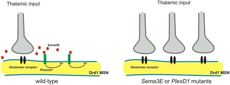
Neural circuit formation requires the coordination of many complex developmental processes. First, neurons project axons over long distances to find their final targets and then establish appropriate connectivity essential for the formation of neuronal circuitry. Growth cones, the leading edges of axons, navigate by interacting with a variety of attractive and repulsive axon guidance cues along their trajectories and at final target regions. In addition to guidance of axons, neuronal polarization, neuronal migration, and dendrite development must be precisely regulated during development to establish proper neural circuitry. Semaphorins consist of a large protein family, which includes secreted and cell surface proteins, and they play important roles in many steps of neural circuit formation. The major semaphorin receptors are plexins and neuropilins, however other receptors and co-receptors also mediate signaling by semaphorins. Upon semaphorin binding to their receptors, downstream signaling molecules transduce this event within cells to mediate further events, including alteration of microtubule and actin cytoskeletal dynamics. Here, I review recent studies on semaphorin signaling in vertebrate neural circuit assembly, with the goal of highlighting how this diverse family of cues and receptors imparts exquisite specificity to neural complex connectivity.

Keywords: axon guidance; neuropilin; plexin; semaphorin; synapse formation.


Figures

Figure 1
Figure 1
Semaphorins and their receptors (plexins and neuropilins). Semaphorins consist of secreted (class 3), glycosylphosphatidylinositol (GPI)-anchored (class 7), or transmembrane (class 4–6) family members. Neuropilins consist of two transmembrane molecules (Npn1–2), and plexins consist of transmembrane A (1–4), B (1–3), C1, and D1 family members. Most class 3 semaphorins require an obligate neuropilin co-receptor. Sema3E binds to PlexD1 without neuropilins. Class 4 and 5 semaphorins interact with plexinBs. Class 6 semaphorins interact with plexinAs. Sema7A interacts with PlexC1. CUB indicates complement binding. One to seven indicate the interactions between semaphorins and their receptors; 1: class 3 semaphorins and neuropilin1/2, 2: Sema3E and PlexD1, 3: Sema4D and PlexB1/B2 or Sema4C/4G and PlexB1, 4: Sema5A/5B and PlexA1/A3, 5: Sema5A and PlexB3, 6: Sema6A/6B and PlexA2/A3 or Sema6C/6D and PlexA1, 7: Sema7A and PlexC1.
Figure 2
Figure 2
Sema6D controls retinal midline crossing through its interaction with PlexA1 and Nr-CAM. Sema6D and Nr-CAM are expressed by glial cells in the chiasm, and PlexA1 is expressed by SSEA1+ neurons in the chiasm. Nr-CAM and PlexA1 are expressed by crossed RGCs. The receptor complex in cis on RGCs and Sema6D alone leads to repulsion of RGCs, whereas the complex on chiasm cells acting in trans with RGC receptors promotes axonal attraction.
Figure 3
Figure 3
Class 5 and class 6 transmembrane semaphorin signaling through plexinAs governs laminar targeting of inner retinal neuron subtypes. (A) Sema6A and PlexA4 show complementary protein expression in the ON and OFF layers of the inner plexiform layer, and this repulsive signaling confines neurite extension of dopaminergic amacrine cells (TH+) within the S1 sublamina in wild-type mice. In Sema6A or PlexA4 mutant mice, dopaminergic amacrine cells extend their aberrant processes to the S4/S5 sublaminae. (B) During early postnatal retinal development, Sema5A/5B and PlexA1/A3 expressions are found in a complementary pattern in the retina. Loss of Sema5A/5B or plexA1/A3 results in neurite mistargeting of multiple inner retinal neuron subtypes into the outer retina.
Figure 4
Figure 4
Roles of semaphorin-plexin signaling in sensory-motor circuitry. (A) Cross-sectional diagrams depicting the dorsal spinal cords of wild-type, PlexA1, and Sema6D, and oligodendrocyte-deleted Sema6D mutant mice used in two studies (Yoshida et al., ; Leslie et al., 2011). Proprioceptive axons (red lines), oligodendrocytes (blue circles), cutaneous axons (green lines), cutaneous synapses (green wavy lines). Blue areas show Sema6D-expressing region. PlexA1 is expressed by proprioceptive sensory neurons but not cutaneous sensory neurons. Cutaneous synapses are disrupted when oligodendrocytes aberrantly enter the dorsal horn, whereas genetic deletion of oligodendrocytes from Sema6D mutants rescues these synaptic defects. (B) Cross-sectional diagrams depicting the spinal cords of wild-type, PlexD1, and Sema3E mutant mice as well as motor neuron-specific Sema3E-expressing mice (Pecho-Vrieseling et al., 2009). Motor neurons innervating triceps (Tri) muscle receive monosynaptic inputs from Tri sensory afferents, whereas cutaneous maximus (CM) motor neurons lack monosynaptic inputs from CM sensory afferents. Sema3E is expressed by CM motor neurons but not Tri motor neurons. PlexD1 is expressed by ∼80% of CM proprioceptive sensory neurons, and ∼50% of Tri proprioceptive sensory neurons. Absence of Sema3E-PlexD1 signaling causes aberrant monosynaptic sensory-motor connections between CM sensory and CM motor neurons. Ectopic expression of Sema3E in Tri motor neurons reduces monosynaptic connections between Tri afferents and Tri motor neurons.
Figure 5
Figure 5
Sema3E-PlexD1 signaling regulates pathway-specific synapse formation in the striatum. Direct and indirect pathway MSNs are functionally and molecularly distinct. Direct pathway MSNs express type 1 dopamine receptors (Drd1) and indirect pathway MSNs express type 2 dopamine receptors (Drd2). PlexD1 is exprsssed by Drd1on-direct pathway MSNs in the striatum, whereas Sema3E is expressed by subsets of neurons in the main thalamic nuclei that project to the striatum. Loss of Sema3E-PlexD1 signaling causes functional and anatomical rearrangement of thalamostriatal synapses specifically in direct pathway MSNs without effects on corticostriatal synapses.

References

    1. Artigiani S., Conrotto P., Fazzari P., Gilestro G. F., Barberis D., Giordano S., Comoglio P. M., Tamagnone L. (2004). Plexin-B3 is a functional receptor for semaphorin 5A. EMBO Rep. 5, 710–71410.1038/sj.embor.7400189 - DOI - PMC - PubMed
    1. Bagnard D., Lohrum M., Uziel D., Puschel A. W., Bolz J. (1998). Semaphorins act as attractive and repulsive guidance signals during the development of cortical projections. Development 125, 5043–5053 - PubMed
    1. Bagri A., Cheng H. J., Yaron A., Pleasure S. J., Tessier-Lavigne M. (2003). Stereotyped pruning of long hippocampal axon branches triggered by retraction inducers of the semaphorin family. Cell 113, 285–29910.1016/S0092-8674(03)00267-8 - DOI - PubMed
    1. Baudet M. L., Zivraj K. H., Abreu-Goodger C., Muldal A., Armisen J., Blenkiron C., Goldstein L. D., Miska E. A., Holt C. E. (2011). miR-124 acts through CoREST to control onset of Sema3A sensitivity in navigating retinal growth cones. Nat. Neurosci. 15, 29–3810.1038/nn.2979 - DOI - PMC - PubMed
    1. Behar O., Golden J. A., Mashimo H., Schoen F. J., Fishman M. C. (1996). Semaphorin III is needed for normal patterning and growth of nerves, bones and heart. Nature 383, 525–52810.1038/383525a0 - DOI - PubMed

LinkOut - more resources