. 2012;7(6):e37920.
doi: 10.1371/journal.pone.0037920. Epub 2012 Jun 7.

Prospective monitoring reveals dynamic levels of T cell immunity to Mycobacterium tuberculosis in HIV infected individuals

Affiliations

Prospective monitoring reveals dynamic levels of T cell immunity to Mycobacterium tuberculosis in HIV infected individuals

Jessica E Mitchell et al. PLoS One. 2012.

Abstract

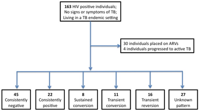
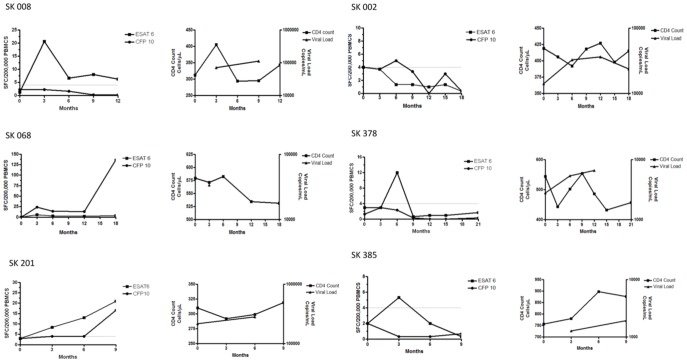
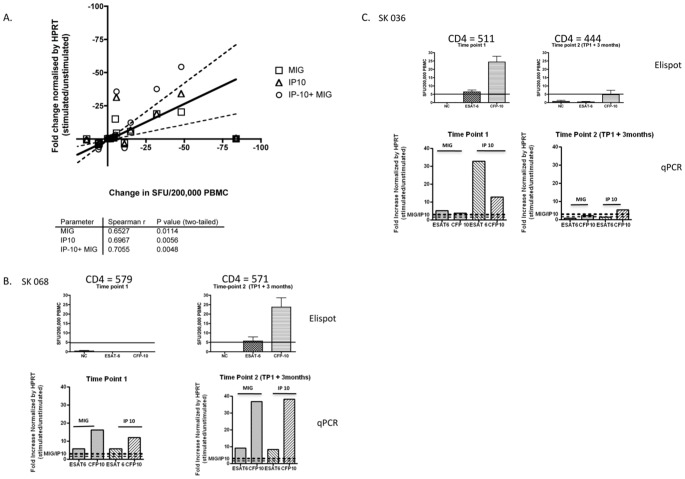
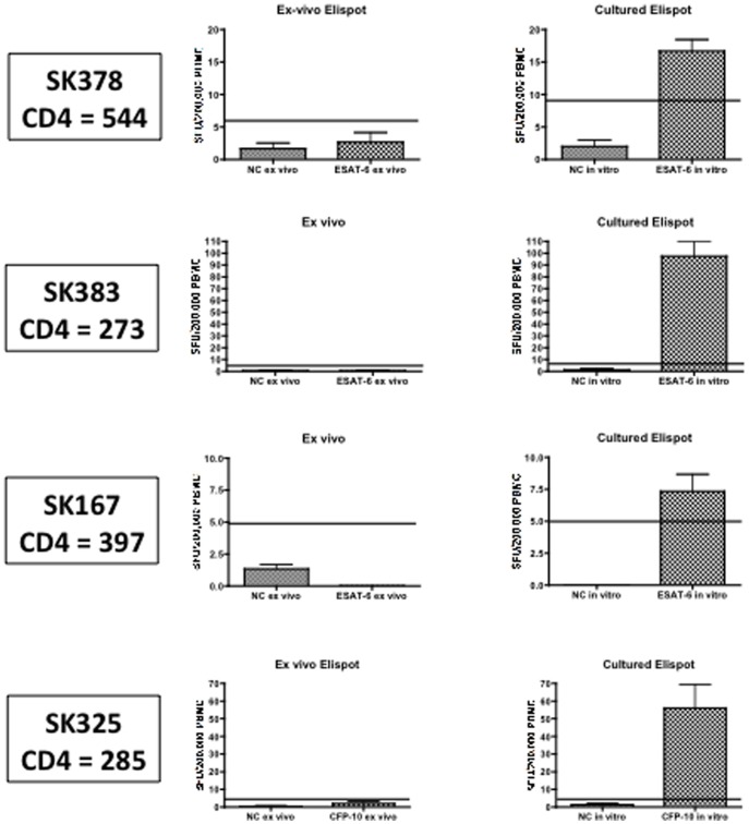
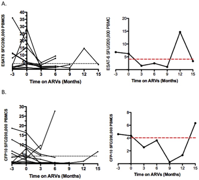
Monitoring of latent Mycobacterium tuberculosis infection may prevent disease. We tested an ESAT-6 and CFP-10-specific IFN-γ Elispot assay (RD1-Elispot) on 163 HIV-infected individuals living in a TB-endemic setting. An RD1-Elispot was performed every 3 months for a period of 3-21 months. 62% of RD1-Elispot negative individuals were positive by cultured Elispot. Fluctuations in T cell response were observed with rates of change ranging from -150 to +153 spot-forming cells (SFC)/200,000 PBMC in a 3-month period. To validate these responses we used an RD1-specific real time quantitative PCR assay for monokine-induced by IFN-γ (MIG) and IFN-γ inducible protein-10 (IP10) (MIG: r=0.6527, p=0.0114; IP-10: r=0.6967, p=0.0056; IP-10+MIG: r=0.7055, p=0.0048). During follow-up 30 individuals were placed on ARVs and 4 progressed to active TB. Fluctuations in SFC did not correlate with CD4 count, viral load, treatment initiation, or progression to active TB. The RD1-Elispot appears to have limited value in this setting.


Conflict of interest statement

Competing Interests: The authors have declared that no competing interests exist.

Figures

Figure 1
Figure 1. Schematic representation of study results.
163 chronically HIV infected (ARV naïve) individuals (CD4 = 5–1142 cells/ul) were recruited in Durban, South Africa. An RD1-Elispot was performed on PBMC every 3 months for a period of 3–21 months. During the period of follow-up 30 individuals were placed on ARVs and 4 individuals progressed to active TB. Of the 129 individuals who were not placed on ARVs or TB treatment during follow-up, 5 categories of Elispot kinetics were observed: 45 (35%) individuals were consistently negative at each time-point tested, 22 (17%) individuals were consistently positive at each time-point tested, 8 (6%) individuals displayed sustained conversions (three or more positive Elispots after at least one negative Elispot), 11 (9%) individuals displayed transient conversions (one or two positive Elispots between negative plates), and 16 (12%) individuals displayed transient reversions (one or two negative Elispots between positive plates). For 27 (21%) individuals the category is unknown due to either a lack of interpretable results (e.g. SK 179 had indeterminate Elispots at all 3 time points tested), or the absence of either pre-baseline samples or further follow-up to determine if observed reversions and conversions are transient or sustained.
Figure 2
Figure 2. Longitudinal MTB-specific T cell profiles and correlation with CD4 count and HIV viral load.
Longitudinal Elispot data from individuals who were not on ARVs for the entire period of follow-up. Figure 2A: Examples of responses from 3 individuals who were ex vivo RD1-Elispot positive at all time-points during follow-up. Figure 2B: Examples of responses from 3 individuals who displayed an RD1-Elispot conversion that was sustained during follow-up and examples of responses from 3 individuals who displayed a transient RD1-Elispot conversion during follow-up. Figure 2C: Examples of responses from 3 individuals who displayed a transient RD1-Elispot reversion during follow-up. Left-hand panel =  Elispot data, right-hand panel  =  CD4 counts and viral loads.
Figure 3
Figure 3. Longitudinal comparison of the ex vivo RD1- Elispot and the RD1-qPCR assay.
A highly sensitive assay of RD1-specific IFN-γ production using real time quantitative PCR (qPCR) for two reporters - monokine-induced by IFN-γ (MIG) and the IFN-γ inducible protein-10 (IP10) was compared longitudinally with the ex vivo RD1-Elispot. Figure 3A: A head-to–head comparison was performed on 14 individuals on samples from 2 time-points 3 months apart. A significant correlation was observed between fold change on the RD1 qPCR and change in SFC using the RD1-Elispot. Figure 3B: Data from SK068. Figure 3C: Data from SK036. Top panel displays RD1-Elispot data and bottom-panel displays RD1-qPCR data.
Figure 4
Figure 4. In- vitro culture reveals RD1-specific T cell responses.
Left-hand panel displays ex vivo IFN-gamma Elispot data from 4 individuals (SK378, SK383, SK167, and SK325) who were ex vivo Elispot negative at 2 time-points 3 months apart. Right-hand panel displays data from the same individuals following a cultured Elispot (14 day in vitro culture of PBMC with the ESAT-6 or CFP-10 peptides followed by IFN-gamma Elispot). All individuals had CD4 counts above 250.
Figure 5
Figure 5. Impact of ARVS on the RD1-specific T cell kinetic profile.
Figure 5 A–B: Summary of longitudinal data from 11 individuals who were placed on ARVs and had a consistent cut-off at all time-points tested. Figure 5A: Summary of ESAT-6-specific responses. Figure 5B: Summary of CFP-10-specific responses. Left-hand panel =  data from all 11 individuals. Right-hand panel =  Mean response plotted. The mean ESAT-6 spot-forming cells (SFC) at 3 months prior to ARV initiation was 7, and that for CFP-10 was 5 SFC. These responses declined to 2 SFC for ESAT-6 specific responses and 3 SFC for CFP-10 specific responses. Overall the mean response declined after the initiation of ARV therapy, however this did not reach statistical significance (ESAT-6: p = −0.1133, CFP-10: p = 0.1109 (paired t-test)).
Figure 6
Figure 6. Prospective monitoring of RD1- specific IFN-gamma Elispot responses does not predict progression to active TB.
Longitudinal Elispots are shown for SK 169, SK 236, SK325 and SK 351 all of whom progressed to active TB during the follow-up period and 2 of which (SK 325 and SK 351) were also subsequently placed on ARVS. TB Treatment was initiated shortly after diagnosis of active TB. Longitudinal CD4 and viral load data is also shown for all 4 individuals.

References

    1. Ringshausen FC, Nienhaus A, Torres Costa J, Knoop H, Schlosser S. Within-Subject Variability of Mycobacterium tuberculosis-Specific Gamma Interferon Responses in German Health Care Workers. Clin Vaccine Immunol. 2011;18:1182. - PMC - PubMed
    1. WHO (2010 guidelines under development) Policy statement on preventive therapy against TB in people living with HIV.
    1. Shafer RW, Edlin BR. Tuberculosis in patients infected with human immunodeficiency virus: perspective on the past decade. Clin Infect Dis. 1996;22:704. - PubMed
    1. Hanifa Y, Grant AD, Lewis J, Corbett EL, Fielding K. Prevalence of latent tuberculosis infection among gold miners in South Africa. Int J Tuberc Lung Dis. 2009;13:46. - PubMed
    1. Talati NJ, Seybold U, Humphrey B, Aina A, Tapia J. Poor concordance between interferon-gamma release assays and tuberculin skin tests in diagnosis of latent tuberculosis infection among HIV-infected individuals. BMC Infect Dis. 2009;9:15. - PMC - PubMed

Publication types

MeSH terms