

Routine use of color Doppler in fetal heart scanning in a low-risk population
- PMID: 22685669
- PMCID: PMC3363954
- DOI: 10.5402/2012/496935
Routine use of color Doppler in fetal heart scanning in a low-risk population
Abstract
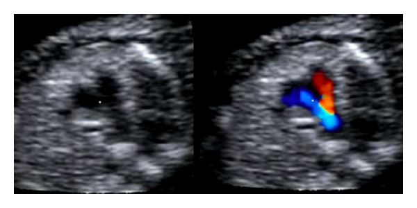
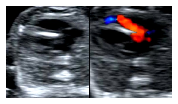
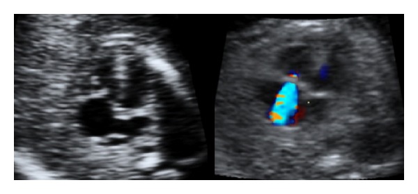
Objectives. To investigate the detection rate of major fetal heart defects in a low-risk population implementing routine use of color Doppler. Material and Methods. In a prospective observational study, all women undergoing fetal heart scanning (including 6781 routine examinations in the second trimester) during a three-year period were included. First a gray-scale scanning was performed including assessment of the four-chamber view and the great vessels. Thereafter three cross-sectional planes through the fetal thorax were assessed with color Doppler. Results. Thirty-nine fetuses had major heart defects, and 26 (67%) were prenatally detected. In 9/26 (35%) of cases the main ultrasound finding was related to the use of color Doppler. The survival rate of live born children was 91%. Conclusions. Routine use of color Doppler in fetal heart scanning in a low-risk population may be helpful in the detection of major heart defects; however, still severe malformations were missed prenatally.
Figures
Similar articles
-
Early fetal ultrasound screening for major congenital heart defects without Doppler.Eur J Obstet Gynecol Reprod Biol. 2019 Feb;233:93-97. doi: 10.1016/j.ejogrb.2018.11.030. Epub 2018 Dec 14. Eur J Obstet Gynecol Reprod Biol. 2019. PMID: 30580230
-
First-trimester fetal cardiac examination using spatiotemporal image correlation, tomographic ultrasound and color Doppler imaging for the diagnosis of complex congenital heart disease in high-risk patients.Ultrasound Obstet Gynecol. 2014 Nov;44(5):562-7. doi: 10.1002/uog.13341. Epub 2014 Oct 15. Ultrasound Obstet Gynecol. 2014. PMID: 24585667
-
Three-dimensional (3D) and 4D color Doppler fetal echocardiography using spatio-temporal image correlation (STIC).Ultrasound Obstet Gynecol. 2004 Jun;23(6):535-45. doi: 10.1002/uog.1075. Ultrasound Obstet Gynecol. 2004. PMID: 15170792
-
Three cross-sectional planes for fetal color Doppler echocardiography.Ultrasound Obstet Gynecol. 2003 Jan;21(1):81-93. doi: 10.1002/uog.5. Ultrasound Obstet Gynecol. 2003. PMID: 12528169 Review.
-
The role of fetal echocardiography in genetic sonography.Semin Perinatol. 2003 Apr;27(2):160-72. doi: 10.1053/sper.2003.50015. Semin Perinatol. 2003. PMID: 12769202 Review.
Cited by
-
A pictorial guide for the second trimester ultrasound.Australas J Ultrasound Med. 2013 Aug;16(3):98-113. doi: 10.1002/j.2205-0140.2013.tb00106.x. Epub 2015 Dec 31. Australas J Ultrasound Med. 2013. PMID: 28191183 Free PMC article.
-
Impact of a standardized training program on midwives' ability to assess fetal heart anatomy by ultrasound.BMC Med Imaging. 2014 Jun 2;14:20. doi: 10.1186/1471-2342-14-20. BMC Med Imaging. 2014. PMID: 24889837 Free PMC article.
References
-
- Dolk H, Loane M, Garne E. Congenital heart defects in Europe: prevalence and perinatal mortality, 2000 to 2005. Circulation. 2011;123(8):841–849. - PubMed
-
- Allan L, Benacerraf B, Copel JA, et al. Isolated major congenital heart disease. Ultrasound in Obstetrics & Gynecology. 2001;17(5):370–379. - PubMed
-
- Blyth M, Howe D, Gnanapragasam J, Wellesley D. The hidden mortality of transposition of the great arteries and survival advantage provided by prenatal diagnosis. International Journal of Obstetrics and Gynaecology. 2008;115(9):1096–1100. - PubMed
-
- Copel JA, Tan ASA, Kleinman CS. Does a prenatal diagnosis of congenital heart disease alter short-term outcome? Ultrasound in Obstetrics and Gynecology. 1997;10(4):237–241. - PubMed
-
- Sharland G. Fetal cardiac screening: why bother? Archives of Disease in Childhood. 2010;95(1):F64–F68. - PubMed
LinkOut - more resources
Full Text Sources
Medical