. 2012 Jun 11:11:75.
doi: 10.1186/1475-2859-11-75.

Decolorization of industrial synthetic dyes using engineered Pseudomonas putida cells with surface-immobilized bacterial laccase

Affiliations

Decolorization of industrial synthetic dyes using engineered Pseudomonas putida cells with surface-immobilized bacterial laccase

Wei Wang et al. Microb Cell Fact. .

Abstract

Background: Microbial laccases are highly useful in textile effluent dye biodegradation. However, the bioavailability of cellularly expressed or purified laccases in continuous operations is usually limited by mass transfer impediment or enzyme regeneration difficulty. Therefore, this study develops a regenerable bacterial surface-displaying system for industrial synthetic dye decolorization, and evaluates its effects on independent and continuous operations.

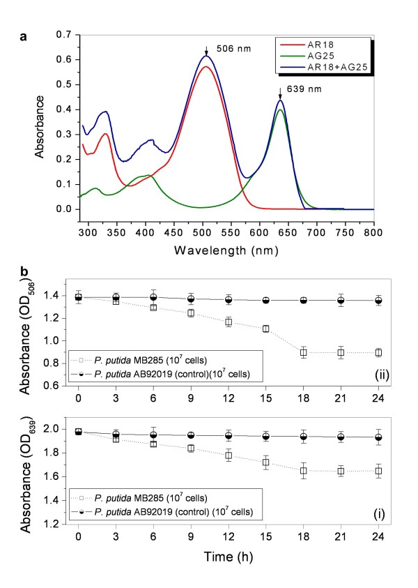
Results: A bacterial laccase (WlacD) was engineered onto the cell surface of the solvent-tolerant bacterium Pseudomonas putida to construct a whole-cell biocatalyst. Ice nucleation protein (InaQ) anchor was employed, and the ability of 1 to 3 tandemly aligned N-terminal repeats to direct WlacD display were compared. Immobilized WlacD was determined to be surface-displayed in functional form using Western blot analysis, immunofluorescence microscopy, flow cytometry, and whole-cell enzymatic activity assay. Engineered P. putida cells were then applied to decolorize the anthraquinone dye Acid Green (AG) 25 and diazo-dye Acid Red (AR) 18. The results showed that decolorization of both dyes is Cu(2+)- and mediator-independent, with an optimum temperature of 35°C and pH of 3.0, and can be stably performed across a temperature range of 15°C to 45°C. A high activity toward AG25 (1 g/l) with relative decolorization values of 91.2% (3 h) and 97.1% (18 h), as well as high activity to AR18 (1 g/l) by 80.5% (3 h) and 89.0% (18 h), was recorded. The engineered system exhibited a comparably high activity compared with those of separate dyes in a continuous three-round shake-flask decolorization of AG25/AR18 mixed dye (each 1 g/l). No significant decline in decolorization efficacy was noted during first two-rounds but reaction equilibriums were elongated, and the residual laccase activity eventually decreased to low levels. However, the decolorizing capacity of the system was easily retrieved via a subsequent 4-h cell culturing.

Conclusions: This study demonstrates, for the first time, the methodology by which the engineered P. putida with surface-immobilized laccase was successfully used as regenerable biocatalyst for biodegrading synthetic dyes, thereby opening new perspectives in the use of biocatalysis in industrial dye biotreatment.


Figures

Figure 1
Figure 1
Map of recombinant plasmids. Plasmid pYMBP was used as the parent vector to construct pMB281, pMB282, and pMB283. PoprL, a constitutive promoter in P. putida; Cbr, carbenicillin-resistant gene; ori, replication origin of Pseudomonas sp.; inaQ-N, N-terminal domain of inaQ; and wlacD, a mutated bacterial laccase gene.
Figure 2
Figure 2
SDS-PAGE analysis of recombinantP. putidastrains (a) and Western blot analysis of recombinantP. putidastrain cell fractions (b). Panels (a) and (b): lane 1, P. putida AB92019 (negative control); lanes 2–4, whole cell fraction (WC), cytoplasmic fraction (CP), and outer membrane fraction (OM) of P. putida MB284 expressing InaQ-N/WlacD, respectively; lanes 5–7, WC, CP, and OM of P. putida MB285 expressing (InaQ-N)2/WlacD, respectively; lanes 8–10, WC, CP, and OM of P. putida MB286 expressing (InaQ-N)3/WlacD, respectively.
Figure 3
Figure 3
Micrographs (a) and flow cytometric analysis (b) of recombinantP. putidastrains. The cells were treated with anti-WlacD polyclonal antiserum followed by secondary Cy3-conjugated goat anti-mouse IgG for immunofluorescence microscopic examination or with goat anti-mouse Cy5-conjugate antibody for flow cytometric analysis. Panel (b): (i) P. putida AB92019 (negative control); (ii) P. putida MB284; (iii) P. putida MB285; and (iv) P. putida MB286. A total of 100,000 cells were analyzed for each flow cytometry experiment.
Figure 4
Figure 4
Measurement of whole-cell laccase activity of recombinantP. putidastrains. The recipient strain, P. putida AB92019, was used as the negative control. Each value and error bar represents the mean and standard deviation of three independent experiments.
Figure 5
Figure 5
Dye decolorization ofP. putidaMB285 cells towards independent AG25 and AR18. (a) Full wavelength (290 nm to 800 nm) scanning curves of AG25 and AR18 indicate the maximal adsorbent peaks, which were used as OD values for dye absorbance measurement. (b) A final 107 cells in each reaction mixture was measured. (i) Effect on AG25; (ii) Effect on AR18.
Figure 6
Figure 6
Effect of Cu2+(a), temperature (b), and pH value (c) on AG25 and AR18 decolorization. A final concentration of 1 × 10⁹ cells and 1 g/l of dye substrate in each reaction mixture were measured. (i) Effect on AR18; (ii) Effect on AG25.
Figure 7
Figure 7
Decolorization values of AG25 and AR18 withP. putidaMB285 cells. Decolorization was catalyzed by 1 × 10⁹ cells (each reaction) for 3 h or 18 h at 35°C and pH 3.0. P. putida AB92019 strain was used as control.
Figure 8
Figure 8
Continuous decolorization of AG18/AG25 mixed dyes (each 1 g/l at final concentration). Panels (a) and (b), continuous first- and second-round decolorization reactions; Panel (c), third-round decolorization after 4-h cultivation of cultures by directly adding LB medium into the flask; Panel (d), residual who-cell laccase activity of P. putida MB285 cells after each-round decolorization reaction.
Figure 9
Figure 9
Chemical structures of laccase substrate compounds.

Similar articles

Cited by

References

    1. Kosman DJ. Multicopper oxidases: a workshop on copper coordination chemistry, electron transfer, and metallophysiology. J Biol Inorg Chem. 2010;15:15–28. doi: 10.1007/s00775-009-0590-9. - DOI - PubMed
    1. Francis CA, Tebo BM. cumA multicopper oxidase genes from diverse Mn(II)-oxidizing and non-Mn(II)-oxidizing Pseudomonas strains. Appl Environ Microbiol. 2001;67:4272–4278. doi: 10.1128/AEM.67.9.4272-4278.2001. - DOI - PMC - PubMed
    1. Martins LO, Soares CM, Pereira MM, Teixeira M, Costa T, Jones GH, Henriques AO. Molecular and biochemical characterization of a highly stable bacterial laccase that occurs as a structural component of the Bacillus subtilis endospore coat. J Biol Chem. 2002;277:18849–18859. doi: 10.1074/jbc.M200827200. - DOI - PubMed
    1. Sanchez-Amat A, Lucas-Elio P, Fernandez E, Garcia-Borron JC, Solano F. Molecular cloning and functional characterization of a unique multipotent polyphenol oxidase from Marinomonas mediterranea. Biochim Biophys Acta. 2001;1547:104–116. doi: 10.1016/S0167-4838(01)00174-1. - DOI - PubMed
    1. Dube E, Shareck F, Hurtubise Y, Beauregard M, Daneault C. Decolourization of recalcitrant dyes with a laccase from Streptomyces coelicolor under alkaline conditions. J Ind Microbiol Biotechnol. 2008;35:1123–1129. doi: 10.1007/s10295-008-0391-0. - DOI - PubMed

Publication types