

Diversity of intervertebral disc cells: phenotype and function
- PMID: 22686699
- PMCID: PMC3512276
- DOI: 10.1111/j.1469-7580.2012.01521.x
Diversity of intervertebral disc cells: phenotype and function
Abstract
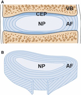
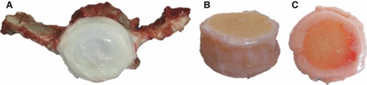
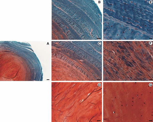
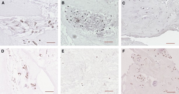
The intervertebral disc (IVD) is a moderately moving joint that is located between the bony vertebrae and provides flexibility and load transmission throughout the spinal column. The disc is composed of different but interrelated tissues, including the central highly hydrated nucleus pulposus (NP), the surrounding elastic and fibrous annulus fibrosus (AF), and the cartilaginous endplate (CEP), which provides the connection to the vertebral bodies. Each of these tissues has a different function and consists of a specific matrix structure that is maintained by a cell population with distinct phenotype. Although the healthy IVD is able to balance the slow matrix turnover of synthesis and degradation, this balance is often disturbed, leading to degenerative disorders. Successful therapeutic management of IVD degeneration requires a profound understanding of the cellular and molecular characteristics of the functional IVD. Hence, the phenotype of IVD cells has been of significant interest from multiple perspectives, including development, growth, remodelling, degeneration and repair. One major challenge that complicates our understanding of the disc cells is that both the cellular phenotype and the extracellular matrix strongly depend on disc maturity and health and as a consequence are continuously evolving. This review delineates the diversity of the cell types found in the intervertebral disc, with emphasis on human, but with reference to other species. The cells of the NP appear rounded and express a proteoglycan-rich matrix, whereas the more elongated AF cells are embedded in a collagen fibre matrix and the CEPs represent a layer of cartilage. Even though all disc cells have often been referred to as 'intervertebral disc chondrocytes', distinct phenotypical differences in comparison with articular chondrocytes exist and have been reported recently. The availability of more specific markers has also improved our understanding of progenitor cell differentiation towards an IVD cell phenotype. Ultimately, new cell- and tissue-engineering approaches to regenerative therapies will only be successful if the specific characteristics of the individual tissues and their context in the function of the whole organ, are taken into consideration.
© 2012 The Authors. Journal of Anatomy © 2012 Anatomical Society.
Figures
References
-
- Adams MA, Hutton WC. The effect of posture on the lumbar spine. J Bone Joint Surg Br. 1985;67:625–629. - PubMed
-
- Adams DS, Keller R, Koehl MA. The mechanics of notochord elongation, straightening and stiffening in the embryo of Xenopus laevis. Development. 1990;110:115–130. - PubMed
-
- Adams MA, Dolan P, McNally DS. The internal mechanical functioning of intervertebral discs and articular cartilage, and its relevance to matrix biology. Matrix Biol. 2009;28:384–389. - PubMed
-
- Aguiar DJ, Johnson SL, Oegema TR. Notochordal cells interact with nucleus pulposus cells: regulation of proteoglycan synthesis. Exp Cell Res. 1999;246:129–137. - PubMed
Publication types
MeSH terms
LinkOut - more resources
Full Text Sources
Other Literature Sources
Medical
Miscellaneous
