. 2013 Jan;222(1):147-60.
doi: 10.1111/j.1469-7580.2012.01526.x. Epub 2012 Jun 11.

Evolution of the mammalian middle ear and jaw: adaptations and novel structures

Affiliations

Evolution of the mammalian middle ear and jaw: adaptations and novel structures

Neal Anthwal et al. J Anat. 2013 Jan.

Abstract

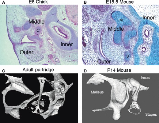
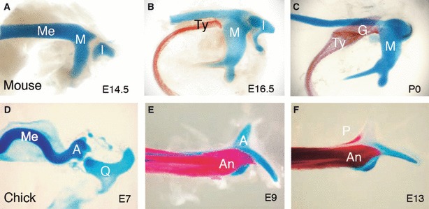
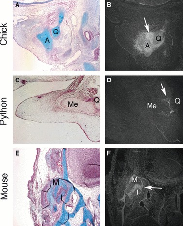
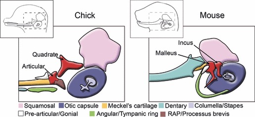
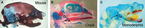
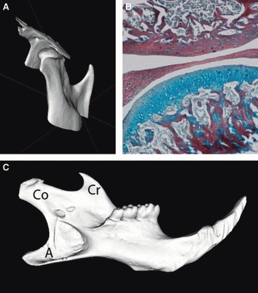
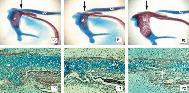
Having three ossicles in the middle ear is one of the defining features of mammals. All reptiles and birds have only one middle ear ossicle, the stapes or columella. How these two additional ossicles came to reside and function in the middle ear of mammals has been studied for the last 200 years and represents one of the classic example of how structures can change during evolution to function in new and novel ways. From fossil data, comparative anatomy and developmental biology it is now clear that the two new bones in the mammalian middle ear, the malleus and incus, are homologous to the quadrate and articular, which form the articulation for the upper and lower jaws in non-mammalian jawed vertebrates. The incorporation of the primary jaw joint into the mammalian middle ear was only possible due to the evolution of a new way to articulate the upper and lower jaws, with the formation of the dentary-squamosal joint, or TMJ in humans. The evolution of the three-ossicle ear in mammals is thus intricately connected with the evolution of a novel jaw joint, the two structures evolving together to create the distinctive mammalian skull.


Figures

Fig. 1
Fig. 1
Middle ear ossicles in mammals and birds. (A) Frontal section through the developing middle ear of a chick showing the columella (c) spanning the gap between the external and internal ear at E (embryonic day) 6. (B) Sagittal section through the developing murine middle ear showing three ossicles, the malleus (M), incus (I) and stapes (S), between the external and inner ear at E15.5. (C,D) MicroCT images. (C) Footplate of the columella (c) inserting into the oval window of the inner ear in an adult partridge. The shaft and footplate of the columella are ossified while the extracolumella arms, which interact with the tympanic membrane, remain cartilaginous and are not picked up by microCT. (D) Three ossicles form a chain in a P (postnatal day) 14 mouse.
Fig. 2
Fig. 2
Comparison of membranous bone ossification in the middle ear and jaw joint. Alcian Blue and Alizarin Red-stained skeletal preparations. (A–C) Development of the cartilages and bones of the murine middle ear, side view. (D–F).Development of the cartilages and bones of the chick jaw joint, dorsal view. (A) Formation of the malleus and incus at E14.5. The malleus develops at the proximal end of Meckel’s cartilage. There is no bone ossification at this stage. (B) Ossification of the tympanic ring at the base of the malleus at E16.5. (C) Ossification of the gonium in between the malleus and tympanic ring at birth (P0). (D) Formation of the jaw joint between the articular and the quadrate at E7. The articular lies at the proximal end of Meckel’s cartilage. (E) Ossification of the angular under the articular and Meckel’s at E9. (F) Ossification of the prearticular next to the angular at E13. I, incus; M, malleus; Me, Meckel’s cartilage; Ty, tympanic ring; G, gonium; A, articular; Q, quadrate; An, angular; P, prearticular. (Chick images taken from Tucker et al. 2004).
Fig. 3
Fig. 3
Conservation of Bapx1 expression between the quadrate and articular and malleus and incus in a bird, reptile and mammal. Sagittal sections through the developing jaw joint in (A,B) Chick and (C,D) Python, and through the middle ear in the mouse (E,F). (A,C,E) Histology sections. (B,D,F) Serial sections in situ hybridization for Bapx1 (silver grains). Arrows point to joint. A, articular; Q, quadrate; Me, Meckel’s cartilage; M, malleus; I, Incus.
Fig. 4
Fig. 4
Schematic of a chick and mouse head during late development. Homologous structures are shown in the same colour.
Fig. 5
Fig. 5
Jaw joint comparison in the mouse, chick and the marsupial Monodelphis. Skeletal preps. Red – bone stained by Alizarin Red. Blue – cartilage stained by Alcian Blue. (A) Mouse postnatal day (P) 0. (B) Chick embryonic day (E) 13. (C) Monodelphis P2. Arrows joints to the articulation point for the upper and lower jaws. Q, quadrate; A, articular; M, malleus; I, incus; S, squamosal; D, dentary.
Fig. 6
Fig. 6
The mammalian jaw articulation (A,C) MicroCT images of an adult mouse. (A) Condylar head of the dentary bone fitting into the glenoid fossa of the upper jaw. (B) Frontal section through an adult mouse jaw joint. The disc is sandwiched between the glenoid fossa (above) and condylar (below). (C) Dentary bone. Cr, coronoid process; A, angular process; Co, Condylar process.
Fig. 7
Fig. 7
Breakdown of Meckel’s cartilage (A–C) Skeletal preps of the mouse malleus and Meckel’s cartilage. Red – bone stained by Alizarin Red. Blue – cartilage stained by Alcian Blue. (A) P0. (B) P1. (C) P3. (D–F) Histology sagittal sections through the transforming Meckel’s cartilage. (D) P0. (E) P1. (F) P2. Arrows points to region where breakdown initiates. Me, Meckel’s cartilage; M, malleus.

References

    1. Allin EF, Hopson JA. Evolution of the auditory system in Synapsida (‘mammal-like reptiles’ and primitive mammals) as seen in the fossil record. In: Webster DB, Fay RR, Popper AN, editors. The Evolutionary Biology of Hearing. New York: Springer-Verlag; 1992. pp. 587–614.
    1. Amin S, Tucker AS. Joint formation in the middle ear: lessons from the mouse and guinea pig. Dev Dyn. 2006;235:1326–1333. - PubMed
    1. Anthwal N, Tucker AS. Molecular biology of the mammalian dentary: insights into how complex skeletal elements can be shaped during development and evolution. In: Asher RJ, Muller J, editors. Clone to Bone: The Synergy of Morphological and Molecular Tools in Palaeobiology. Cambridge: Cambridge University Press; 2012. pp. 396–426. Chapter 8.
    1. Anthwal N, Chai Y, Tucker AS. The role of transforming growth factor-beta signalling in the patterning of the proximal processes of the murine dentary. Dev Dyn. 2008;237:1604–1613. - PubMed
    1. Archer CW, Buxton P, Hall BK, et al. Mechanical regulation of secondary chondrogenesis. Biorheology. 2006;43:355–370. - PubMed

Publication types

LinkOut - more resources