

Magnetic resonance spectroscopy in the evaluation of treatment efficacy in unipolar major depressive disorder: a review of the literature
- PMID: 22687162
- PMCID: PMC3812759
Magnetic resonance spectroscopy in the evaluation of treatment efficacy in unipolar major depressive disorder: a review of the literature
Abstract
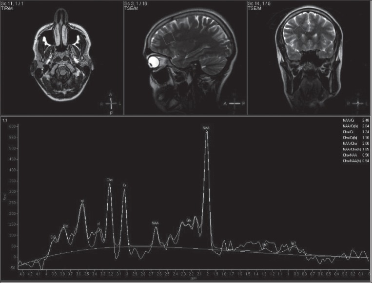
More and more neuroimaging studies are using in vivo proton magnetic resonance spectroscopy (1H-MRS) to explore correlates of response to therapy in major depressive disorder (MDD). Their aim is to further understanding of the effects of neurotransmitter changes in areas involved in MDD and the mechanisms underlying a good treatment response. We set out to summarise the literature from the past fifteen years on biochemical correlates of treatment response in MDD patients, reflected in pre- and post-therapy changes in 1H-MRS measurements. Our literature search identified fifteen articles reporting 1H-MRS studies in MDD treatment; no study used 1P-MRS. Despite the wide diversity of 1H-MRS methods applied, brain regions studied, and metabolite changes found, there emerged strong evidence of a correlation between changes in neurometabolite concentrations, in particular glutamate, N-acetylaspartate and choline, and a good treatment response to pharmacotherapy or antidepressant stimulation techniques.
Figures
References
-
- Griffith HR, Stewart CC, den Hollander JA. Proton magnetic resonance spectroscopy in dementias and mild cognitive impairment. Int Rev Neurobiol. 2009;84:105–31. - PubMed
-
- Charles HC, Lazeyras F, Krishnan KR, Boyko OB, Payne M, Moore D. Brain choline in depression: in vivo detection of potential pharmacodynamic effects of antidepressant therapy using hydrogen localized spectroscopy. Prog Neuropsychopharmacol Biol Psychiatry. 1994;18:1121–1127. - PubMed
-
- Sonawalla SB, Renshaw PF, Moore CM, et al. Compounds containing cytosolic choline in the basal ganglia: a potential biological marker of true drug response to fluoxetine. Am J Psychiatry. 1999;156:1638–1640. - PubMed
Publication types
MeSH terms
Substances
LinkOut - more resources
Full Text Sources
Medical
Miscellaneous