

Rotavirus RNA polymerases resolve into two phylogenetically distinct classes that differ in their mechanism of template recognition
- PMID: 22687427
- PMCID: PMC3381288
- DOI: 10.1016/j.virol.2012.05.011
Rotavirus RNA polymerases resolve into two phylogenetically distinct classes that differ in their mechanism of template recognition
Abstract
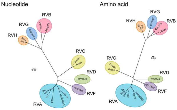
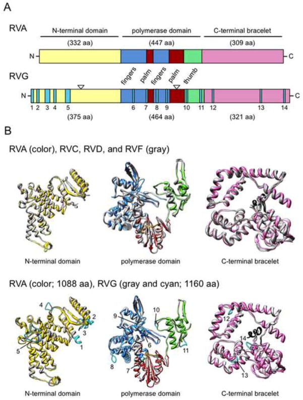
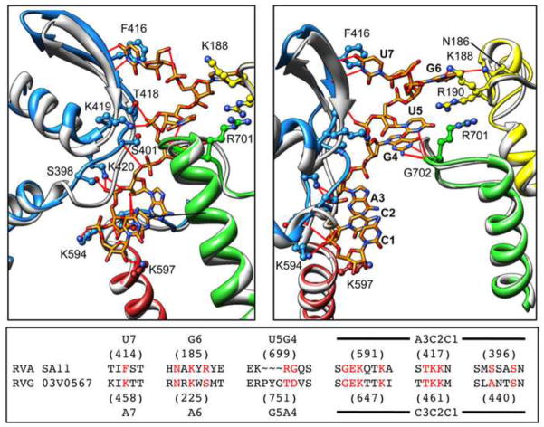
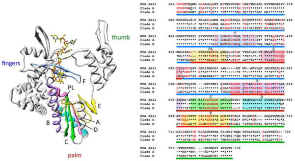
Rotaviruses (RVs) are segmented double-stranded RNA viruses that cause gastroenteritis in mammals and birds. Within the RV genus, eight species (RVA-RVH) have been proposed. Here, we report the first RVF and RVG sequences for the viral RNA polymerase (VP1)-encoding segments and compare them to those of other RV species. Phylogenetic analyses indicate that the VP1 RNA segments and proteins resolve into two major clades, with RVA, RVC, RVD and RVF in clade A, and RVB, RVG and RVH in clade B. Plus-strand RNA of clade A viruses, and not clade B viruses, contain a 3'-proximal UGUG cassette that serves as the VP1 recognition signal. VP1 structures for a representative of each RV species were predicted using homology modeling. Structural elements involved in interactions with the UGUG cassette were conserved among VP1 of clade A, suggesting a conserved mechanism of viral RNA recognition for these viruses.
Published by Elsevier Inc.
Figures
Similar articles
-
Shared and group-specific features of the rotavirus RNA polymerase reveal potential determinants of gene reassortment restriction.J Virol. 2009 Jun;83(12):6135-48. doi: 10.1128/JVI.00409-09. Epub 2009 Apr 8. J Virol. 2009. PMID: 19357162 Free PMC article.
-
Genetic relatedness of VP1 genes of Australian and Taiwanese rotavirus isolates.FEMS Microbiol Lett. 2001 May 1;198(2):147-50. doi: 10.1111/j.1574-6968.2001.tb10634.x. FEMS Microbiol Lett. 2001. PMID: 11430406
-
Mechanism for coordinated RNA packaging and genome replication by rotavirus polymerase VP1.Structure. 2008 Nov 12;16(11):1678-88. doi: 10.1016/j.str.2008.09.006. Structure. 2008. PMID: 19000820 Free PMC article.
-
Assortment and packaging of the segmented rotavirus genome.Trends Microbiol. 2011 Mar;19(3):136-44. doi: 10.1016/j.tim.2010.12.002. Epub 2010 Dec 31. Trends Microbiol. 2011. PMID: 21195621 Free PMC article. Review.
-
Orthoreovirus and Aquareovirus core proteins: conserved enzymatic surfaces, but not protein-protein interfaces.Virus Res. 2004 Apr;101(1):15-28. doi: 10.1016/j.virusres.2003.12.003. Virus Res. 2004. PMID: 15010214 Review.
Cited by
-
Rotavirus Species B Encodes a Functional Fusion-Associated Small Transmembrane Protein.J Virol. 2019 Sep 30;93(20):e00813-19. doi: 10.1128/JVI.00813-19. Print 2019 Oct 15. J Virol. 2019. PMID: 31375572 Free PMC article.
-
Predicted structure and domain organization of rotavirus capping enzyme and innate immune antagonist VP3.J Virol. 2014 Aug;88(16):9072-85. doi: 10.1128/JVI.00923-14. Epub 2014 Jun 4. J Virol. 2014. PMID: 24899176 Free PMC article.
-
Mutational analysis of residues involved in nucleotide and divalent cation stabilization in the rotavirus RNA-dependent RNA polymerase catalytic pocket.Virology. 2012 Sep 15-30;431(1-2):12-20. doi: 10.1016/j.virol.2012.05.009. Epub 2012 Jun 2. Virology. 2012. PMID: 22664357 Free PMC article.
-
Recent advances in rotavirus reverse genetics and its utilization in basic research and vaccine development.Arch Virol. 2021 Sep;166(9):2369-2386. doi: 10.1007/s00705-021-05142-7. Epub 2021 Jul 3. Arch Virol. 2021. PMID: 34216267 Free PMC article. Review.
-
Reassortment in segmented RNA viruses: mechanisms and outcomes.Nat Rev Microbiol. 2016 Jul;14(7):448-60. doi: 10.1038/nrmicro.2016.46. Epub 2016 May 23. Nat Rev Microbiol. 2016. PMID: 27211789 Free PMC article. Review.
References
-
- Ahmed MU, Kobayashi N, Wakuda M, Sanekata T, Taniguchi K, Kader A, Naik TN, Ishino M, Alam MM, Kojima K, Mise K, Sumi A. Genetic analysis of group B human rotaviruses detected in Bangladesh in 2000 and 2001. J Med Virol. 2004;72:149–155. - PubMed
-
- Attoui H, Becnel J, Belaganahalli S, Bergoin M, Brussaard CP, Chappell JD, Ciarlet M, del Vas M, Dermody TS, Dormitzer PR, Duncan R, Fcang Q, Graham R, Guglielmi KM, Harding RM, Hillman B, Makkay A, Marzachià C, Matthijnssens J, Mertens PPC, Milne RG, Mohd Jaafar F, Mori H, Noordeloos AA, Omura T, Patton JT, Rao S, Maan M, Stoltz D, Suzuki N, Upadhyaya NM, Wei C, Zhou H. Part II: The Viruses – The Double Stranded RNA Viruses - Family Reoviridae. In: King AMQ, Adams MJ, Carstens EB, Lefkowitz EJ, editors. Virus taxonomy: classification and nomenclature: Ninth Report of the International Committee on Taxonomy of Viruses. Elsevier Academic Press; San Diego: 2012. pp. 541–637.
-
- Chen Z, Lambden PR, Lau J, Caul EO, Clarke IN. Human group C rotavirus: completion of the genome sequence and gene coding assignments of a non-cultivatable rotavirus. Virus Res. 2002;83:179–187. - PubMed
-
- Drummond AJ, Ashton B, Buxton S, Cheung M, Cooper A, Duran C, Field M, Heled J, Kearse M, Markowitz S, Moir R, Stones-Havas S, Sturrock S, Thierer T, Wilson A. Geneious v5.4.4. 2010 Available from http://www.geneious.com. - PubMed
-
- Estes MK, Kapikian AZ. Rotaviruses. In: Howley PM, editor. Fields Virology. 5. Lippincott, Williams and Wilkins; Philadelphia: 2007. pp. 1918–1974.
Publication types
MeSH terms
Substances
Associated data
- Actions
- Actions
Grants and funding
LinkOut - more resources
Full Text Sources
