

Bifunctional CD4-DC-SIGN fusion proteins demonstrate enhanced avidity to gp120 and inhibit HIV-1 infection and dissemination
- PMID: 22687513
- PMCID: PMC3421874
- DOI: 10.1128/AAC.00623-12
Bifunctional CD4-DC-SIGN fusion proteins demonstrate enhanced avidity to gp120 and inhibit HIV-1 infection and dissemination
Abstract
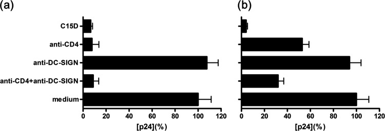
Early stages of mucosal infection are potential targets for HIV-1 prevention. CD4 is the primary receptor in HIV-1 infection whereas DC-SIGN likely plays an important role in HIV-1 dissemination, particularly during sexual transmission. To test the hypothesis that an inhibitor simultaneously targeting both CD4 and DC-SIGN binding sites on gp120 may provide a potent anti-HIV strategy, we designed constructs by fusing the extracellular CD4 and DC-SIGN domains together with varied arrangements of the lengths of CD4, DC-SIGN and the linker. We expressed, purified and characterized a series of soluble CD4-linker-DC-SIGN (CLD) fusion proteins. Several CLDs, composed of a longer linker and an extra neck domain of DC-SIGN, had enhanced affinity for gp120 as evidenced by molecular-interaction analysis. Furthermore, such CLDs exhibited significantly enhanced neutralization activity against both laboratory-adapted and primary HIV-1 isolates. Moreover, CLDs efficiently inhibited HIV-1 infection in trans via a DC-SIGN-expressing cell line and primary human dendritic cells. This was further strengthened by the results from the human cervical explant model, showing that CLDs potently prevented both localized and disseminated infections. This is the first time that soluble DC-SIGN-based bifunctional proteins have demonstrated anti-HIV potency. Our study provides proof of the concept that targeting both CD4 and DC-SIGN binding sites on gp120 represents a novel antiviral strategy. Given that DC-SIGN binding to gp120 increases exposure of the CD4 binding site and that the soluble forms of CD4 and DC-SIGN occur in vivo, further improvement of CLDs may render them potentially useful in prophylaxis or therapeutics.
Figures


Similar articles
-
Role of the carbohydrate-binding sites of griffithsin in the prevention of DC-SIGN-mediated capture and transmission of HIV-1.PLoS One. 2013 May 31;8(5):e64132. doi: 10.1371/journal.pone.0064132. Print 2013. PLoS One. 2013. PMID: 23741304 Free PMC article.
-
Human seminal plasma abrogates the capture and transmission of human immunodeficiency virus type 1 to CD4+ T cells mediated by DC-SIGN.J Virol. 2007 Dec;81(24):13723-34. doi: 10.1128/JVI.01079-07. Epub 2007 Oct 3. J Virol. 2007. PMID: 17913809 Free PMC article.
-
Short Communication: Inhibition of DC-SIGN-Mediated HIV-1 Infection by Complementary Actions of Dendritic Cell Receptor Antagonists and Env-Targeting Virus Inactivators.AIDS Res Hum Retroviruses. 2016 Jan;32(1):93-100. doi: 10.1089/aid.2015.0184. Epub 2015 Sep 18. AIDS Res Hum Retroviruses. 2016. PMID: 26383762 Free PMC article.
-
DC-SIGN: a novel HIV receptor on DCs that mediates HIV-1 transmission.Curr Top Microbiol Immunol. 2003;276:31-54. doi: 10.1007/978-3-662-06508-2_2. Curr Top Microbiol Immunol. 2003. PMID: 12797442 Review.
-
Bitter-sweet symphony: defining the role of dendritic cell gp120 receptors in HIV infection.J Clin Virol. 2001 Oct;22(3):229-39. doi: 10.1016/s1386-6532(01)00194-9. J Clin Virol. 2001. PMID: 11564587 Review.
Cited by
-
Herpes Simplex Virus Type 2 Immediate Early Protein ICP27 Inhibits IFN-β Production in Mucosal Epithelial Cells by Antagonizing IRF3 Activation.Front Immunol. 2019 Feb 26;10:290. doi: 10.3389/fimmu.2019.00290. eCollection 2019. Front Immunol. 2019. PMID: 30863402 Free PMC article.
-
HIV-1 Trans Infection of CD4(+) T Cells by Professional Antigen Presenting Cells.Scientifica (Cairo). 2013;2013:164203. doi: 10.1155/2013/164203. Epub 2013 May 7. Scientifica (Cairo). 2013. PMID: 24278768 Free PMC article. Review.
-
CCL19 and CCL28 Assist Herpes Simplex Virus 2 Glycoprotein D To Induce Protective Systemic Immunity against Genital Viral Challenge.mSphere. 2021 Apr 28;6(2):e00058-21. doi: 10.1128/mSphere.00058-21. mSphere. 2021. PMID: 33910988 Free PMC article.
-
Dendritic cell-specific intercellular adhesion molecule-3 grabbing nonintegrin mediates HIV-1 infection of and transmission by M2a-polarized macrophages in vitro.AIDS. 2013 Mar 13;27(5):707-16. doi: 10.1097/QAD.0b013e32835cfc82. AIDS. 2013. PMID: 23211775 Free PMC article.
-
Neutralization of Virus Infectivity by Antibodies: Old Problems in New Perspectives.Adv Biol. 2014;2014:157895. doi: 10.1155/2014/157895. Epub 2014 Sep 9. Adv Biol. 2014. PMID: 27099867 Free PMC article.
References
-
- Abdiche Y, Malashock D, Pinkerton A, Pons J. 2008. Determining kinetics and affinities of protein interactions using a parallel real-time label-free biosensor, the Octet. Anal. Biochem. 377:209–217 - PubMed
-
- Allaway GP, et al. 1995. Expression and characterization of CD4-IgG2, a novel heterotetramer that neutralizes primary HIV type 1 isolates. AIDS Res. Hum. Retroviruses 11:533–539 - PubMed
-
- Baleux F, et al. 2009. A synthetic CD4-heparan sulfate glycoconjugate inhibits CCR5 and CXCR4 HIV-1 attachment and entry. Nat. Chem. Biol. 5:743–748 - PubMed
Publication types
MeSH terms
Substances
LinkOut - more resources
Full Text Sources
Other Literature Sources
Medical
Research Materials
