

Deciphering actin cytoskeletal function in the contractile vascular smooth muscle cell
- PMID: 22687615
- PMCID: PMC3473273
- DOI: 10.1113/jphysiol.2012.232306
Deciphering actin cytoskeletal function in the contractile vascular smooth muscle cell
Abstract
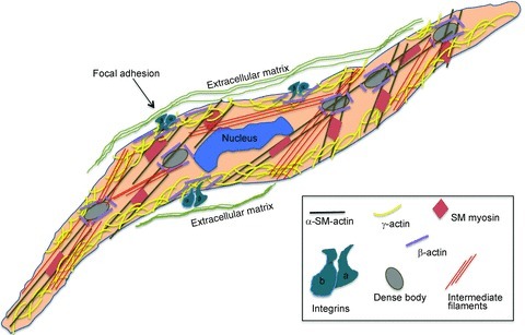
This review focuses on the vascular smooth muscle cells present in the medial layer of the blood vessels wall in the fully differentiated state (dVSMCs). The dVSMC contractile phenotype enables these cells to respond in a highly regulated manner to changes in extracellular stimuli. Through modulation of vascular contractile force and vascular compliance dVSMCs regulate blood pressure and blood flow. The cellular and molecular mechanisms by which vascular smooth muscle contractile functions are regulated are not completely elucidated. Recent studies have documented a critical role for actin polymerization and cytoskeletal dynamics in the regulation of contractile function. Here we will review the current understanding of actin cytoskeletal dynamics and focal adhesion function in dVSMCs in order to better understand actin cytoskeleton connections to the extracellular matrix and the effects of cytoskeletal remodelling on vascular contractility and vascular stiffness in health and disease.
Figures


Similar articles
-
Cortical actin regulation modulates vascular contractility and compliance in veins.J Physiol. 2015 Sep 1;593(17):3929-41. doi: 10.1113/JP270845. Epub 2015 Jul 26. J Physiol. 2015. PMID: 26096914 Free PMC article.
-
Non-receptor tyrosine kinases and the actin cytoskeleton in contractile vascular smooth muscle.J Physiol. 2015 Sep 1;593(17):3807-14. doi: 10.1113/jphysiol.2014.284174. Epub 2014 Dec 23. J Physiol. 2015. PMID: 25433074 Free PMC article. Review.
-
Invited review: focal adhesion and small heat shock proteins in the regulation of actin remodeling and contractility in smooth muscle.J Appl Physiol (1985). 2001 Aug;91(2):963-72. doi: 10.1152/jappl.2001.91.2.963. J Appl Physiol (1985). 2001. PMID: 11457815 Review.
-
The Dynamic Actin Cytoskeleton in Smooth Muscle.Adv Pharmacol. 2018;81:1-38. doi: 10.1016/bs.apha.2017.06.001. Epub 2017 Aug 24. Adv Pharmacol. 2018. PMID: 29310796 Review.
-
Physiologic properties and regulation of the actin cytoskeleton in vascular smooth muscle.J Cardiovasc Pharmacol Ther. 2008 Jun;13(2):130-40. doi: 10.1177/1074248407313737. Epub 2008 Jan 22. J Cardiovasc Pharmacol Ther. 2008. PMID: 18212360 Free PMC article. Review.
Cited by
-
Icariside II Restores Vascular Smooth Muscle Cell Contractile Phenotype by Enhancing the Focal Adhesion Signaling Pathway in the Rat Vascular Remodeling Model.Front Pharmacol. 2022 Jun 13;13:897615. doi: 10.3389/fphar.2022.897615. eCollection 2022. Front Pharmacol. 2022. PMID: 35770073 Free PMC article.
-
Elastic fibers and biomechanics of the aorta: Insights from mouse studies.Matrix Biol. 2020 Jan;85-86:160-172. doi: 10.1016/j.matbio.2019.03.001. Epub 2019 Mar 15. Matrix Biol. 2020. PMID: 30880160 Free PMC article. Review.
-
β1-Subunit of the calcium-sensitive potassium channel modulates the pulmonary vascular smooth muscle cell response to hypoxia.Am J Physiol Lung Cell Mol Physiol. 2018 Aug 1;315(2):L265-L275. doi: 10.1152/ajplung.00060.2018. Epub 2018 Apr 12. Am J Physiol Lung Cell Mol Physiol. 2018. PMID: 29644895 Free PMC article.
-
The intermediate filament protein nestin serves as a molecular hub for smooth muscle cytoskeletal signaling.Respir Res. 2023 Jun 14;24(1):157. doi: 10.1186/s12931-023-02473-8. Respir Res. 2023. PMID: 37316833 Free PMC article.
-
Severe Molecular Defects Exhibited by the R179H Mutation in Human Vascular Smooth Muscle α-Actin.J Biol Chem. 2016 Oct 7;291(41):21729-21739. doi: 10.1074/jbc.M116.744011. Epub 2016 Aug 22. J Biol Chem. 2016. PMID: 27551047 Free PMC article.
References
-
- Albinsson S, Nordstrom I, Hellstrand P. Stretch of the vascular wall induces smooth muscle differentiation by promoting actin polymerization. J Biol Chem. 2004;279:34849–34855. - PubMed
Publication types
MeSH terms
Substances
Grants and funding
LinkOut - more resources
Full Text Sources