Review
. 2013 Feb;59(2):188-98.
doi: 10.1016/j.ymeth.2012.05.013. Epub 2012 Jun 8.

In vitro capping and transcription of rhabdoviruses

Affiliations
Review

In vitro capping and transcription of rhabdoviruses

Tomoaki Ogino. Methods. 2013 Feb.

Abstract

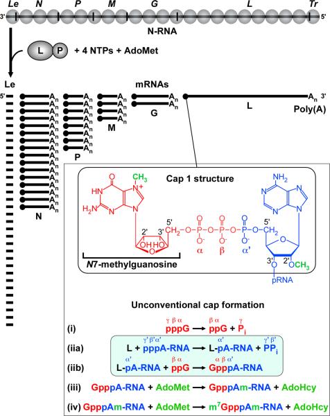
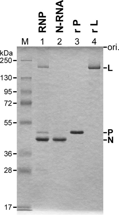
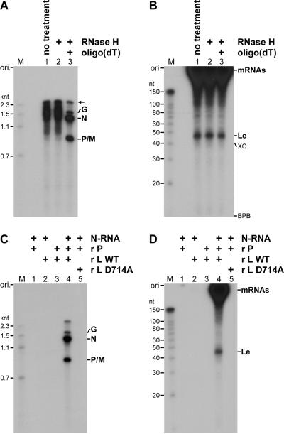
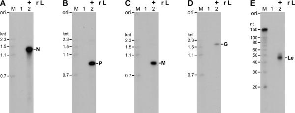
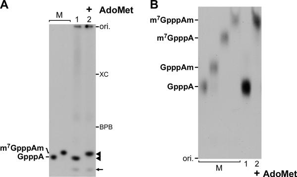
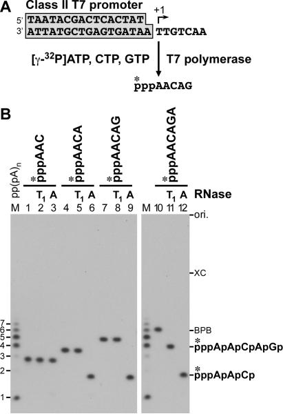
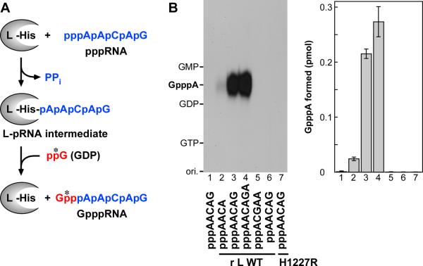
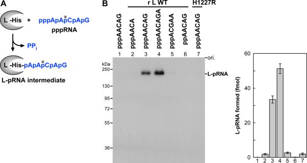
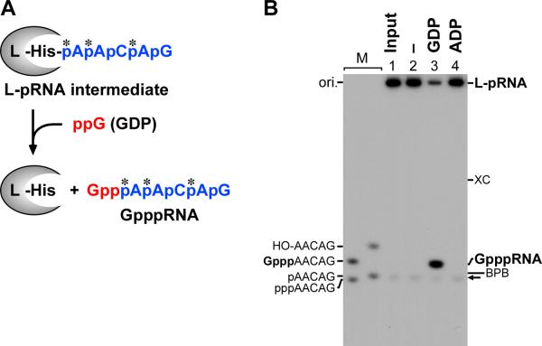
The RNA-dependent RNA polymerase L protein of vesicular stomatitis virus (VSV), a prototypic nonsegmented negative strand (NNS) RNA virus classified into the Rhabdoviridae family, has been used to investigate the fundamental molecular mechanisms of NNS RNA viral mRNA synthesis and processing. In vitro studies on mRNA cap formation with the VSV L protein eventually led to the discovery of the unconventional mRNA capping pathway catalyzed by the guanosine 5'-triphosphatase and RNA:GDP polyribonucleotidyltransferase (PRNTase) activities. The PRNTase activity is a novel enzymatic activity, which transfers 5'-monophosphorylated (p-) RNA from 5'-triphosphorylated (ppp-) RNA to GDP to form 5'-capped RNA (GpppRNA) in a viral mRNA-start sequence-dependent manner. This unconventional capping (pRNA transfer) reaction with PRNTase can be experimentally distinguished from the conventional capping (GMP transfer) reaction with eukaryotic GTP:RNA guanylyltransferase (GTase) on the basis of the following differences in their substrate specificity for the cap formation: PRNTase uses GDP and pppRNA, but not ppRNA, whereas GTase employs GTP, but not GDP, and ppRNA. The pRNA transfer reaction with PRNTase proceeds through a covalent enzyme-pRNA intermediate with a phosphoamide bond. Hence, to prove the PRNTase activity, it is necessary to demonstrate the following consecutive steps separately: (1) the enzyme forms a covalent enzyme-pRNA intermediate, and (2) the intermediate transfers pRNA to GDP. This article describes the methods for in vitro transcription and capping with the recombinant VSV L protein, which permit detailed characterization of its enzymatic reactions and mapping of active sites of its enzymatic domains. It is expected that these systems are adaptable to rhabdoviruses and, by extension, other NNS RNA viruses belonging to different families.


Figures

Fig. 1
Fig. 1. Synthesis of VSV mRNAs with the cap 1 structure
The RdRp complex composed of the L and P proteins sequentially transcribes the genomic RNA wrapped with the N proteins (N-RNA template) into the leader (Le) RNA (~47 nt) and five 5'-capped and 3'-polyadenylated mRNAs. The cap 1 (m7GpppAm-) structure is co-transcriptionally formed on the 5'-ends of mRNAs by the (i) guanosine 5'-triphosphatase, (iia and iib) RNA:GDP polyribonucleotidyltransferase (highlighted), (iii) mRNA cap (nucleoside-2'-O-)-methyltransferase, and (iv) mRNA cap (guanine-N7)-methyltransferase activities. Moieties derived from GTP, RNA, and S-adenosyl-l-methionine (AdoMet) are shown in red, blue, and green, respectively. Pi and PPi indicates inorganic phosphate and pyrophosphate, respectively. AdoHcy denotes S-adenosyl-l-homocysteine.
Fig. 2
Fig. 2. Analysis of purified VSV transcription factors
The native VSV RNP (1.5 μg protein), N-RNA complex (1 μg), recombinant P protein (r P, 1 μg), and recombinant L protein (r L, 1 μg) were analyzed by 10% SDS-PAGE followed by Coomassie Brilliant Blue staining.
Fig. 3
Fig. 3. In vitro VSV transcription system reconstituted with purified factors
(A and B) In vitro transcription was performed with the N-RNA complex, recombinant P protein, and recombinant L protein. Transcripts containing incorporated [α-32P]GMP (lane 1, no treatment) were incubated with RNase H in the absence (lane 2) or presence (lane 3) of oligo(dT), and analyzed by 5% (A) or 15% (B) urea-PAGE followed by autoradiography. Lanes M show 32P-labeled marker RNAs with indicated lengths. The positions of the origin (ori.) and the VSV transcripts (N, P/M, G, and Le) are indicated. An arrow indicates an unidentified transcript. XC and BPB show the positions of xylene cyanol and bromophenol blue, respectively. (C and D) In vitro transcription was carried out with the N-RNA complex, recombinant P protein (r P), recombinant wild-type L protein (r L WT), and/or recombinant D714A mutant L protein (r L D714A), as indicated. After poly(A) removal, transcripts containing incorporated [α-32P]GMP were analyzed by 5% (C) or 15% (D) urea-PAGE followed by autoradiography.
Fig. 4
Fig. 4. Northern blot analysis of in vitro synthesized VSV transcripts
In vitro transcription was performed with the N-RNA complex and recombinant P protein in the absence (lane 1) or presence (lane 2) of the recombinant L protein (r L). After poly(A) removal, transcripts were separated by 5% urea-PAGE, and analyzed by Northern blotting with 32P-labeled oligo-DNA probes complementary to the N (A), P (B), M (C), and G (D) mRNAs. Transcripts were separated by 15% urea-PAGE to analyze the leader (Le) RNA by Northern blotting (E). Lanes M show marker RNAs with indicated lengths.
Fig. 5
Fig. 5. Identification of cap structures formed on in vitro synthesized VSV transcripts
(A) In vitro transcription was performed with the N-RNA complex, recombinant P protein, and recombinant wild-type L protein in the absence (lane 1) or presence (lane 2) of S-adenosyl-l-methionine (AdoMet). Transcripts containing incorporated [α-32P]GMP were digested with nuclease P1 and CIAP, and the resulting digests were analyzed by 20% urea-PAGE followed by autoradiography. Arrowheads indicate major nuclease P1 and CIAP-resistant products. The minor 32P-labeled product (marked by an arrow) was identified as undigested GMP (not shown). Lanes M indicate the positions of 32P-labeled GpppA and m7GpppAm. (B) The major nuclease P1 and CIAP-resistant products (see panel A, lanes 1 and 2) co-migrating with GpppA and m7GpppAm, respectively, were purified, and reanalyzed by PEI-cellulose TLC followed by autoradiography. The 32P-labeled cap structures (GpppA, GpppAm, m7GpppA, and m7GpppAm) were enzymatically synthesized, and used as standards (lanes M).
Fig. 6
Fig. 6. Synthesis of oligo-RNA substrates for the VSV mRNA capping enzyme
(A) [γ-32P]ATP-labeled oligo-RNAs (e.g., pppAACAG) were synthesized from unique synthetic DNA templates by T7 RNA polymerase. (B) Indicated oligo-RNAs ([32P]phosphate groups marked by asterisks) were purified and incubated in the absence (lanes 1, 4, 7, and 10) or presence of RNase T1 or A, as indicated. The resulting digests were analyzed by 20% urea-PAGE followed by autoradiography. Lane M indicates [γ-32P]ATP-initiated poly(A) RNAs with indicated lengths. The positions of cleaved RNA products are shown on the right.
Fig. 7
Fig. 7. In vitro VSV RNA capping assay with the recombinant L protein
(A) The PRNTase activity of the L protein can be measured using pppAACAG (blue) and [α-32P]GDP (red, a [32P]phosphate group marked by an asterisk) as the substrates. The L protein transfers pRNA from pppRNA to GDP through a covalent L-pRNA intermediate to yield a capped RNA (GpppRNA). His indicates the histidine residue at position 1,227 (H1227) in the VSV L protein, which serves as a nucleophile forming the intermediate. (B) The recombinant wild-type L protein (r L WT) or the H1227R mutant L protein (r L H1227R) was incubated with the indicated oligo-RNAs and [α-32P]GDP. Nuclease P1 and CIAP-resistant products were analyzed by PEI-cellulose TLC followed by autoradiography. The positions of standard compounds are shown the left. The right panel shows the amounts of GpppA cap structure formed, which are determined by counting their 32P radioactivities. The mean values of three independent experiments are shown with standard deviation indicated by error bars.
Fig. 8
Fig. 8. L-pRNA intermediate formation assay with the recombinant L protein
(A) When the L protein is incubated with pppAACAG (blue, a [32P]phosphate group marked by an asterisk), a covalent L-pRNA intermediate is formed. (B) The recombinant wild-type L protein (r L WT) or the H1227R mutant L protein (r L H1227R) was incubated with the indicated oligo-RNAs containing incorporated [α-32P]CMP. The resulting L-pRNA intermediates were analyzed by 7.5% SDS-PAGE followed by autoradiography. The right panel shows amounts of the L-pRNA intermediate formed, which are determined by counting their 32P radioactivities. The mean values of three independent experiments are shown with standard deviation indicated by error bars.
Fig. 9
Fig. 9. pRNA transfer assay with the purified L-pRNA intermediate
(A) When the purified L-pRNA (pAACAG, blue, [32P]phosphate groups marked by asterisks) intermediate is incubated with GDP (red), pRNA is transferred from the intermediate to GDP, resulting in the release of a capped RNA (GpppRNA). (B) The purified L-pRNA (containing incorporated [α-32P]AMP) intermediate (lane 1, input) was incubated in the absence (lane 2) or presence of GDP (lane 3) or ADP (lane 4). After the reaction, released RNAs were analyzed by 20% urea-PAGE followed by autoradiography. The positions of the L-pRNA intermediate and GpppRNA are shown on the right. Lanes M indicate the positions of 5'-hydroxyl (HO-), capped (Gppp-), monophosphorylated (p-), and triphosphorylated (ppp-) AACAG RNAs. The position of free pAACAG co-purified with the L-pRNA intermediate is marked by an arrow.

Similar articles

Cited by

References

    1. Lyles DS, Rupprecht CE. In: Fields Virology Lippincott. Knipe DM, Howley PM, editors. Williams & Wilkins; Philadelphia: 2007. pp. 1363–1408.
    1. Briese T, Schneemann A, Lewis AJ, Park YS, Kim S, Ludwig H, Lipkin WI. Proc. Natl. Acad. Sci. USA. 1994;91:4362–4366. - PMC - PubMed
    1. Poch O, Blumberg BM, Bougueleret L, Tordo N. J. Gen. Virol. 1990;71:1153–1162. - PubMed
    1. Volchkov VE, Volchkova VA, Chepurnov AA, Blinov VM, Dolnik O, Netesov SV, Feldmann H. J. Gen. Virol. 1999;80:355–362. - PubMed
    1. Baltimore D, Huang AS, Stampfer M. Proc. Natl. Acad. Sci. USA. 1970;66:572–576. - PMC - PubMed

Publication types

MeSH terms

LinkOut - more resources