. 2012 Aug:101:60-71.
doi: 10.1016/j.exer.2012.05.013. Epub 2012 Jun 8.

Pathological consequences of long-term mitochondrial oxidative stress in the mouse retinal pigment epithelium

Affiliations

Pathological consequences of long-term mitochondrial oxidative stress in the mouse retinal pigment epithelium

Soo-jung Seo et al. Exp Eye Res. 2012 Aug.

Abstract

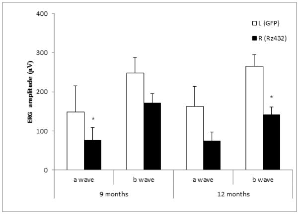
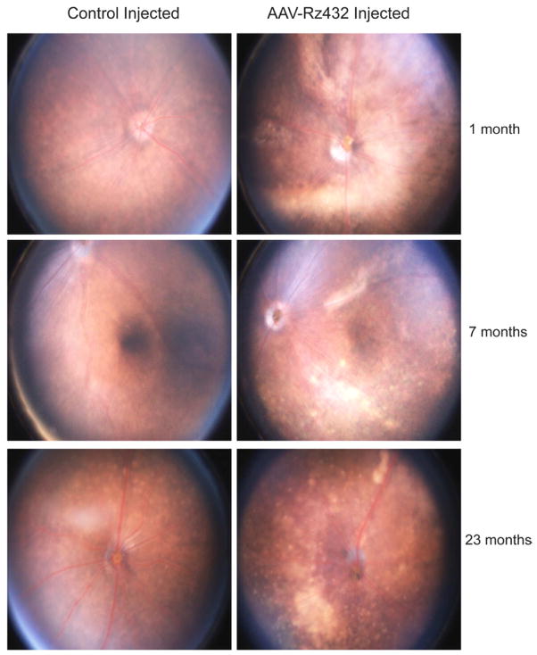
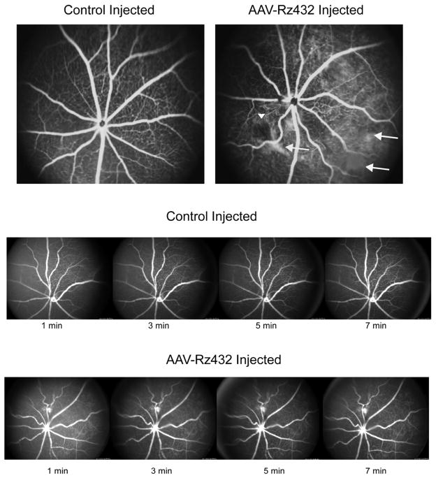
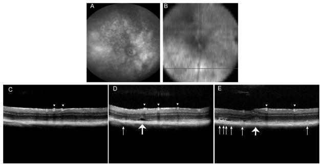
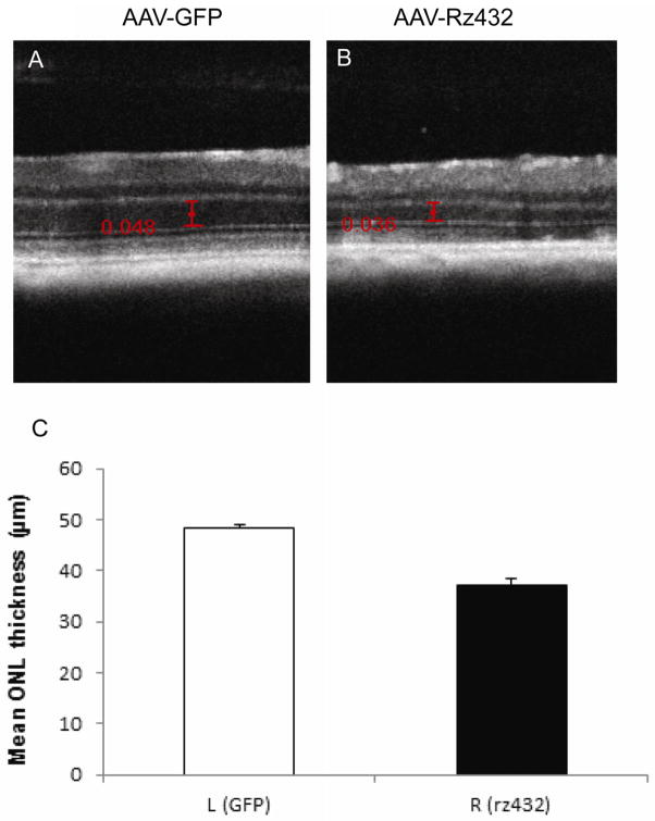
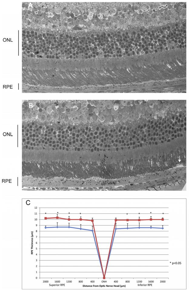
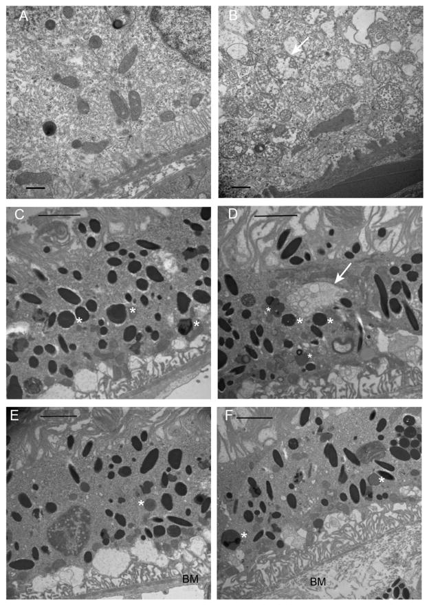
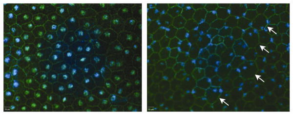
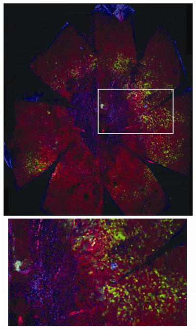
Oxidative stress in the retinal pigment epithelium (RPE) is hypothesized to be a major contributor to the development of age-related macular degeneration (AMD). Mitochondrial manganese superoxide dismutase (MnSOD) is a critical antioxidant protein that scavenges the highly reactive superoxide radical. We speculated that specific reduction of MnSOD in the RPE will increase the level of reactive oxygen species in the retina/RPE/choroid complex leading to pathogenesis similar to geographic atrophy. To test this hypothesis, an Sod2-specific hammerhead ribozyme (Rz), delivered by AAV2/1 and driven by the human VMD2 promoter was injected subretinally into C57BL/6J mice. Dark-adapted full field electroretinogram (ERG) detected a decrease in the response to light. We investigated the age-dependent phenotypic and morphological changes of the outer retina using digital fundus imaging and SD-OCT measurement of ONL thickness. Fundus microscopy revealed pigmentary abnormalities in the retina and these corresponded to sub-retinal and sub-RPE deposits seen in SD-OCT B-scans. Light and electron microscopy documented the localization of apical deposits and thickening of the RPE. In RPE flat-mounts we observed abnormally displaced nuclei and regions of apparent fibrosis in the central retina of the oldest mice. This region was surrounded by enlarged and irregular RPE cells that have been observed in eyes donated by AMD patients and in other mouse models of AMD.


Figures

Fig. 1
Fig. 1
Scotopic electroretinogram (ERG) amplitudes decrease following subretinal injection of AAV-SOD2 Rz432. Scotopic full-field ERGs of C57BL/6 mice injected with AAV1-VMD2-Rz432 or AAV-GFP control vector. ERGs were measured at 9 and 12 months post injection. Mice treated with Rz432 showed loss of ERG a-wave response that was significant at both time points and a decrease in the b-wave response that was significant at 12 months. Note: Different cohorts of mice were used at the two intervals. n= 5 at 9 months post injection and n= 10 at 12 months * = p<0.05,). Error bars represent standard error of the mean.
Fig. 2
Fig. 2
Funduscopic changes of Rz432-injected eyes. Hypopigmented regions of the enteral retina were observed as early as one month post injection of AAV-VMD2-Rz432, but not in control injected eyes. At later time points both regions of pigment loss, often in regions underlying the vasculature, and discrete punctate deposits were observed.
Fig. 3
Fig. 3
Vascular abnormalities revealed by fluorescein angiography. Top. Fluorescein angiography was performed 10 min post injection of fluorescein. Increased tortuosity of retinal blood vessels was observed by seven months and areas of diffuse fluorescence suggested hemorrhaging. However, time-lapse imaging (bottom) indicated no change in the fluorescence pattern with time, indicating that the diffuse fluorescence was not caused by an active hemorrhage, but perhaps by scarring.
Fig. 4
Fig. 4
SD-OCT imagining reveals subretinal deposits of undefined origin in eyes treated with AAV-Rz432. (A) Digital fundus image of retina taken at 12 months post injection. (B) An en face SD-OCT image of the deeper layer of the same retina made at the same time. (C–D) B-scans taken at the position in the retina indicated by the black line in (B). The arrowheads point to retinal blood vessels and the small arrows point to subretinal hyper-reflective material. The large arrow in (D) points to what may be a fluid filled space beneath the retina and the large arrow in (E) indicates a deformation affecting both outer and the inner retina.
Fig. 5
Fig. 5
Thinning of the ONL revealed by SD-OCT. (A) SD-OCT b-scan of the central retina in an eye 12 months after injection with AAV-GFP. Calipers are in millimeters. (B) A similar scan in an eye 12 months after treatment with AAV-VMD2-Rz432. This is the contralateral eye of the same mouse. (C) Mean ONL thickness measured in six mice, left eyes treated with AAV-GFP, and right eyes treated with AAV-VMD2-Rz432 (**p<0.001).
Fig. 6
Fig. 6
Subretinal deposit and thickening of the RPE in eyes treated with AAV-VMD2-Rz432. At 7 months post injection, AAV-GFP treated eyes and AAV-VMD2-Rz432, were fixed with glutaraldehyde, dehydrated and embedded in epoxy resin. The thickness of the the RPE layer was measured along the vertical meridian by an investigator who did not know the treatment group. (A) Retina treated with AAV-GFP; (B) Retina treated with AAV-VMD2-Rz432; the arrow indicates a subretinal deposit. (C) Measurement of RPE thickness at 10 locations equally spaced relative to the optic nerve head (ONH) n=4, *=p<0.05. Squares= AAV-VMD2-Rz432 eyes; diamonds=AAV-GFP treated eyes.
Fig. 7
Fig. 7
Mitochondrial damage and Bruch’s membrane injury following knockdown of MnSOD. (A) Image from the AAV-GFP injected eye, 7 months post injection. (B) The AAV-VMD2-Rz432 injected eye of the same mouse. In regions of VMD2-Rz432 transduction, we noted increase vacuolization of the RPE (this can also be seen in the light micrographs), thickening and disorganization of Bruch’s membrane (BM), and distension and loss of cristae from mitochondria (arrow). The scale bar is 500 nm. In both AAV-GFP treated eyes (C) and AAV-Rz432 treated eyes (D), we observed lipofuscin granules (marked with asterisks), but in the ribozyme-treated eyes we frequently observed autophagosomes containing vesicles (arrow in D). In ribozyme treated eyes (F), Bruch’s membrane (BM) was typically distended and lacking in its normal laminar organization compared to control injected retinas (E).
Fig. 8
Fig. 8
Nucelar dislocation in the RPE following delivery of AAV-VMD2-Rz432. RPE flat mounts were stained using antibody to ZO1 and DAPI. RPE derived from Rz432 treated eye (right) showed enlarged cells (arrows), an increased number of binucleated cells and displacement of nuclei to the periphery of cells (blue: DAPI, green: ZO1). These are from the left and right eyes of the same mouse.
Fig. 9
Fig. 9
MnSOD knockdown leads to central fibrosis and irregular RPE morphology. RPE flat mounts were prepared 23 months after injection of AAV-VMD2-Rz432. Top: 48 RGB confocal stacks were made at an original magnification of 10X merged and stitched together using Fiji software. Large irregular cells and RPE atrophy (loss of phalloidin-stained boundaries) are apparent with concomitant appearance of closely packed nuclei, presumably fibrotic cells. Bottom: enlargement of the boxed region, possibly indicating fibrosis surrounding the optic nerve head, with an adjacent transitional zone containing enlarged, irregular and multinucleate RPE cells. Interestingly, surrounding RPE cells appear normal, even though some cells express GFP derived from the AAV vector (Supplemental Fig. 1), and this was confirmed by scanning confocal microscopy (data not shown).

References

    1. Al-Hussaini H, Schneiders M, Lundh P, Jeffery G. Drusen are associated with local and distant disruptions to human retinal pigment epithelium cells. Exp Eye Res. 2009;88:610–2. - PubMed
    1. Beatty S, Koh H, Phil M, Henson D, Boulton M. The role of oxidative stress in the pathogenesis of age-related macular degeneration. Surv Ophthalmol. 2000;45:115–34. - PubMed
    1. Cai J, Nelson KC, Wu M, Sternberg P, Jr, Jones DP. Oxidative damage and protection of the RPE. Prog Retin Eye Res. 2000;19:205–21. - PubMed
    1. Cano M, Thimmalappula R, Fujihara M, Nagai N, Sporn M, Wang AL, Neufeld AH, Biswal S, Handa JT. Cigarette smoking, oxidative stress, the anti-oxidant response through Nrf2 signaling, and Age-related Macular Degeneration. Vision Res. 2010;50:652–64. - PMC - PubMed
    1. Cheung CM, Tai ES, Kawasaki R, Tay WT, Lee JL, Hamzah H, Wong TY. Prevalence of and Risk Factors for Age-Related Macular Degeneration in a Multiethnic Asian Cohort. Arch Ophthalmol 2011 - PubMed

Publication types

MeSH terms