

A novel system to accelerate the progression of nerve degeneration in transgenic mouse models of neuropathies
- PMID: 22688009
- PMCID: PMC3418409
- DOI: 10.1016/j.expneurol.2012.05.021
A novel system to accelerate the progression of nerve degeneration in transgenic mouse models of neuropathies
Abstract
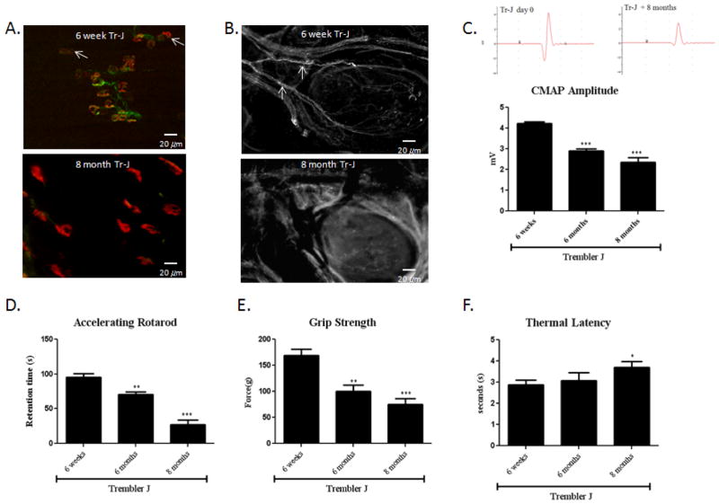
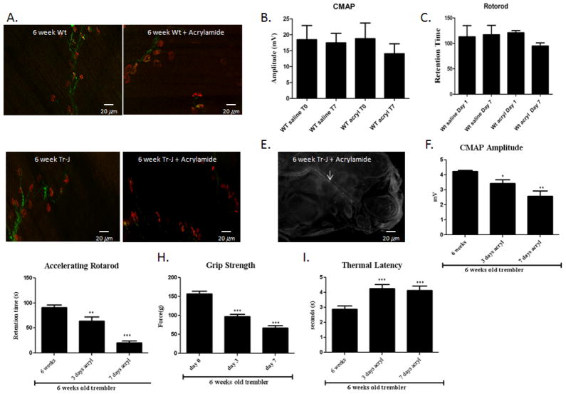
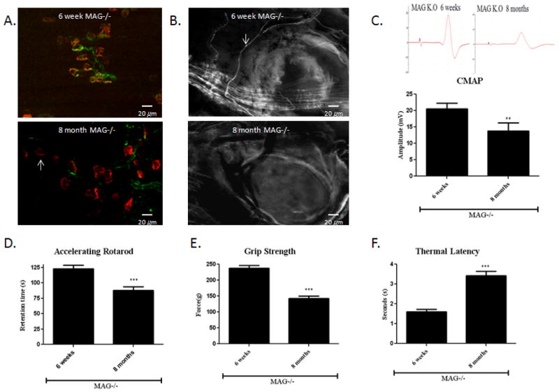
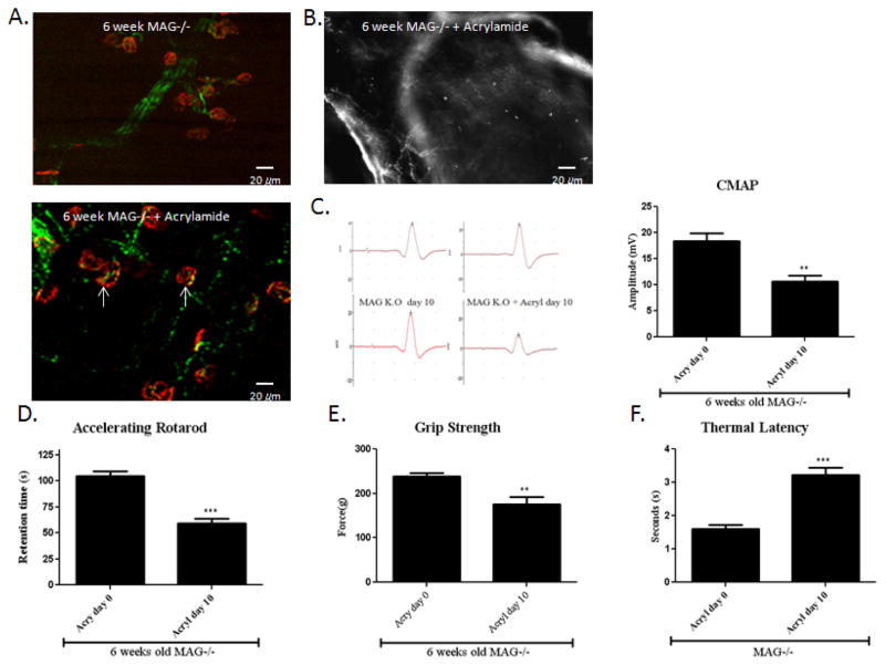
Axon degeneration is a common hallmark of many neurodegenerative diseases. There is now an abundance of spontaneous and genetically engineered mice available to study the mechanisms of axonal degeneration and to screen for axonal protective agents. However, many of these mouse models exhibit slow progressive axonal loss which can span over many months. Consequently, there is a pressing need to accelerate the pace of axonal loss over a short interval for high-throughput screening of pharmacological and genetic therapies. Here, we present a novel technique using acrylamide, an axonal neurotoxin, to provoke rapid axonal degeneration in murine models of neuropathies. The progressive axonal loss which typically occurs over 8 months was reproduced within 7 to 10 days of the acrylamide intoxication. This approach was successfully applied to Myelin Associated Glycoprotein knockout (MAG-/-) mouse and Trembler-J mouse, a popular murine model of Charcot-Marie-Tooth disease type 1 (CMT-1). Acrylamide intoxication in transgenic mouse models offers a novel experimental approach to accelerate the rate of axonal loss over short intervals for timely in vivo studies of nerve degeneration. This report also provides for the first time an animal model for medication or toxin-induced exacerbation of pre-existing neuropathies, a phenomenon widely reported in patients with neuropathies.
Copyright © 2012 Elsevier Inc. All rights reserved.
Conflict of interest statement
The authors declare that they have no competing financial interests.
Figures
Similar articles
-
Axonal degeneration in the Trembler-j mouse demonstrated by stimulated single-fiber electromyography.Muscle Nerve. 2007 Jul;36(1):81-6. doi: 10.1002/mus.20786. Muscle Nerve. 2007. PMID: 17443662
-
Axonal protective effects of the myelin-associated glycoprotein.J Neurosci. 2009 Jan 21;29(3):630-7. doi: 10.1523/JNEUROSCI.5204-08.2009. J Neurosci. 2009. PMID: 19158290 Free PMC article.
-
Limiting multiple sclerosis related axonopathy by blocking Nogo receptor and CRMP-2 phosphorylation.Brain. 2012 Jun;135(Pt 6):1794-818. doi: 10.1093/brain/aws100. Epub 2012 Apr 28. Brain. 2012. PMID: 22544872 Free PMC article.
-
Role of immune cells in animal models for inherited peripheral neuropathies.Neuromolecular Med. 2006;8(1-2):175-90. doi: 10.1385/nmm:8:1-2:175. Neuromolecular Med. 2006. PMID: 16775375 Review.
-
Genetic mechanisms of peripheral nerve disease.Neurosci Lett. 2021 Jan 18;742:135357. doi: 10.1016/j.neulet.2020.135357. Epub 2020 Nov 26. Neurosci Lett. 2021. PMID: 33249104 Review.
Cited by
-
Loss of the E3 ubiquitin ligase LRSAM1 sensitizes peripheral axons to degeneration in a mouse model of Charcot-Marie-Tooth disease.Dis Model Mech. 2013 May;6(3):780-92. doi: 10.1242/dmm.010942. Epub 2013 Mar 8. Dis Model Mech. 2013. PMID: 23519028 Free PMC article.
-
DHTKD1 Deficiency Causes Charcot-Marie-Tooth Disease in Mice.Mol Cell Biol. 2018 Jun 14;38(13):e00085-18. doi: 10.1128/MCB.00085-18. Print 2018 Jul 1. Mol Cell Biol. 2018. PMID: 29661920 Free PMC article.
-
Distinct Changes in Calpain and Calpastatin during PNS Myelination and Demyelination in Rodent Models.Int J Mol Sci. 2022 Dec 6;23(23):15443. doi: 10.3390/ijms232315443. Int J Mol Sci. 2022. PMID: 36499770 Free PMC article.
-
The Gdap1 knockout mouse mechanistically links redox control to Charcot-Marie-Tooth disease.Brain. 2014 Mar;137(Pt 3):668-82. doi: 10.1093/brain/awt371. Epub 2014 Jan 29. Brain. 2014. PMID: 24480485 Free PMC article.
-
Alterations in Galanin-Like Immunoreactivity in the Enteric Nervous System of the Porcine Stomach Following Acrylamide Supplementation.Int J Mol Sci. 2019 Jul 8;20(13):3345. doi: 10.3390/ijms20133345. Int J Mol Sci. 2019. PMID: 31288386 Free PMC article.
References
-
- Chaudhry V, Chaudhry M, Crawford TO, Simmons-O’Brien E, Griffin JW. Toxic neuropathy in patients with pre-existing neuropathy. Neurology. 2003;60:337–40. - PubMed
-
- Dickerhoff R, Lindner W, Scheiber W. Severe vincristine neurotoxicity in a patient with Charcot-Marie-Tooth disease. Pediatr Hematol Oncol. 1988;5:61–4. - PubMed
-
- Graf WD, Chance PF, Lensch MW, Eng LJ, Lipe HP, Bird TD. Severe vincristine neuropathy in Charcot-Marie-Tooth disease type 1A. Cancer. 1996;77:1356–62. - PubMed
Publication types
MeSH terms
Substances
Grants and funding
LinkOut - more resources
Full Text Sources
Medical
Molecular Biology Databases
Research Materials
