. 2012 Aug;32(16):3253-64.
doi: 10.1128/MCB.06791-11. Epub 2012 Jun 11.

EphrinA1 is released in three forms from cancer cells by matrix metalloproteases

Affiliations

EphrinA1 is released in three forms from cancer cells by matrix metalloproteases

Amanda Beauchamp et al. Mol Cell Biol. 2012 Aug.

Abstract

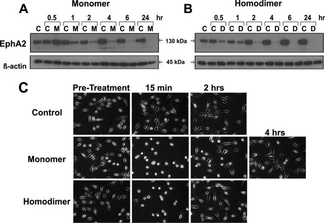
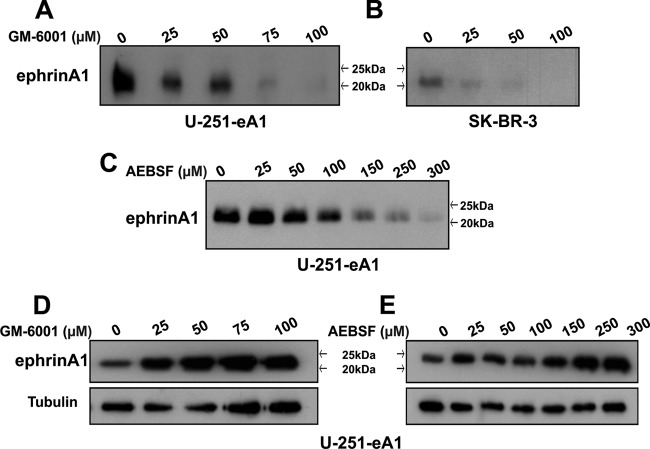
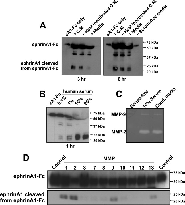
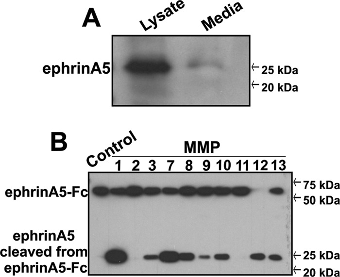
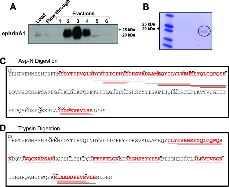
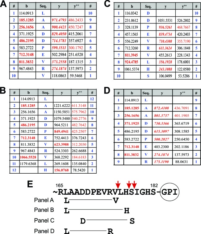
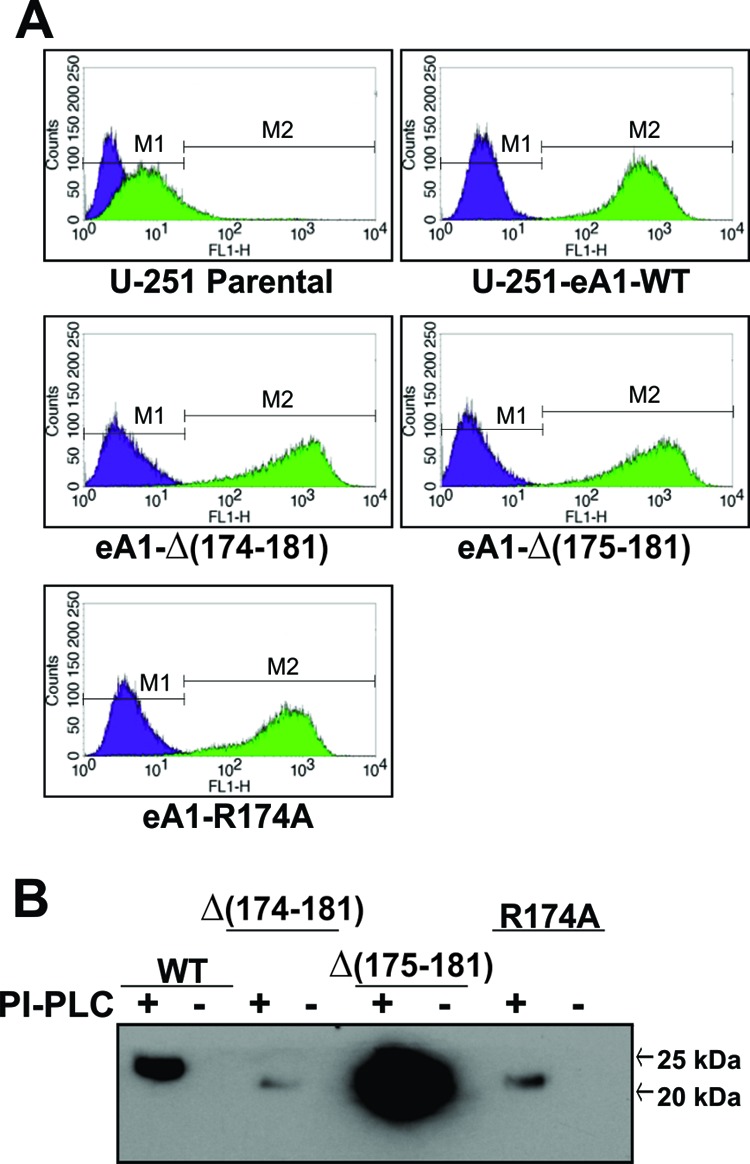
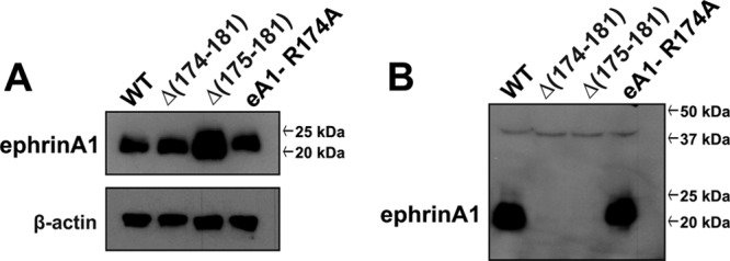
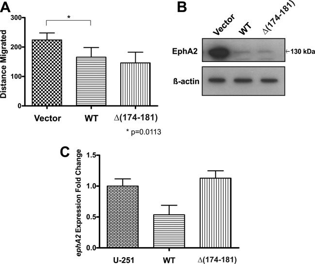
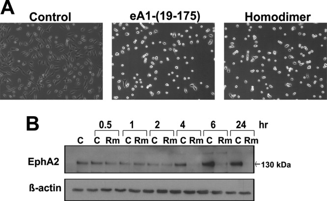
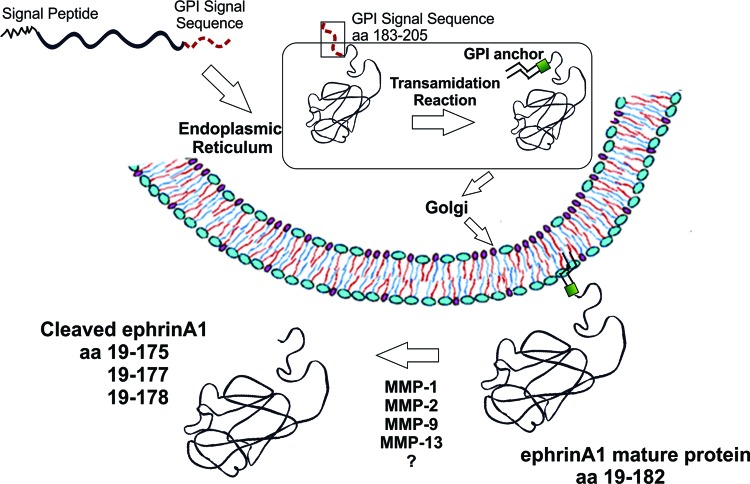
EphrinA1 is a glycosylphosphatidylinositol (GPI)-linked ligand for the EphA2 receptor, which is overexpressed in glioblastoma (GBM), among other cancers. Activation of the receptor by ephrinA1 leads to a suppression of oncogenic properties of GBM cells. We documented that a monomeric functional form of ephrinA1 is released from cancer cells and thus explored the mechanism of ephrinA1 release and the primary protein sequence. We demonstrate here that multiple metalloproteases (MMPs) are able to cleave ephrinA1, most notably MMP-1, -2, -9, and -13. The proteolytic cleavage that releases ephrinA1 occurs at three positions near the C terminus, producing three forms ending in valine-175, histidine-177, or serine-178. Moreover, deletion of amino acids 174 to 181 or 175 to 181 yields ephrinA1 that is still GPI linked but not released by proteolysis, underlining the necessity of amino acids 175 to 181 for release from the membrane. Furthermore, recombinant ephrinA1 ending at residue 175 retains activity toward the EphA2 receptor. These findings suggest a mechanism of release and provide evidence for the existence of several forms of monomeric ephrinA1. Moreover, ephrinA1 should be truncated at a minimum at amino acid 175 in fusions or conjugates with other molecules in order to prevent likely proteolysis within physiological and pathobiological environments.


Figures

Fig 1
Fig 1
Monomeric ephrinA1 downregulates the EphA2 receptor and causes morphological changes in GBM cells comparable to that seen upon treatment with homodimeric ephrinA1-Fc. U-251 MG cells were serum starved for 24 h and treated with 4 μg/ml monomeric ephrinA1 (A) or 1 μg/ml homodimeric ephrinA1-Fc or PBS as a control (B), and whole-cell lysates were collected at the time points indicated. M, monomer treated; D, dimer treated; C, control. (C) For cell rounding assay, cells were treated as for panels A and B and 20× phase-contrast microscopy was performed at the time points shown. The 2-h morphology for the dimer returned to the pretreatment morphology; therefore, no images were taken at the 4-h time point.
Fig 2
Fig 2
The broad-spectrum MMP inhibitor GM-6001 and the serine protease inhibitor AEBSF cause a dose-dependent decrease in the amount of ephrinA1 released into the medium of cancer cells while increasing the total amount of ephrinA1 within the cell. EphrinA1-producing GBM cells (A and C) or SK-BR-3 cells, which endogenously express ephrinA1 (B), were treated with increasing concentrations of GM-6001 (A and B) or AEBSF (C). Medium was collected after 24 h and Western blot analysis performed for ephrinA1. Total-cell lysates were collected from GM-6001-treated cells (D) and AEBSF-treated cells (E), and Western blot analysis was performed for ephrinA1.
Fig 3
Fig 3
Recombinant homodimeric ephrinA1-Fc is cleaved by proteases present in conditioned medium and in human serum and by multiple MMPs. (A) EphrinA1-Fc was incubated in 1 ml PBS, U-251 MG cell-conditioned medium (C.M.), heat-inactivated conditioned medium, regular medium, or serum-free medium at 37°C for either 3 h or 6 h and Western blot analysis performed for ephrinA1. (B) EphrinA1-Fc was incubated at 37°C for 1 h in human serum diluted in PBS to the concentrations listed and Western blot analysis performed for ephrinA1. (C) Gelatin zymography of serum-free medium, serum, and GBM cell-conditioned medium. Degradation of gelatin indicates the presence of MMP-2 and MMP-9 in serum and in conditioned medium from GBM cells. (D) EphrinA1-Fc was incubated with various recombinant MMPs for 1 h at 37°C and Western blot analysis performed for ephrinA1. Multiple MMPs cleaved recombinant ephrinA1-Fc, but MMP-1, -2, -9, and -13 caused the most robust cleavage.
Fig 4
Fig 4
EphrinA5 is released from cells into the medium and recombinant ephrinA5-Fc is susceptible to cleavage by multiple MMPs. (A) Western blot analysis for ephrinA5 in the lysate and the medium of U87 GBM cells. (B) EphrinA5-Fc was incubated with various recombinant MMPs for 1 h at 37°C, and Western blot analysis performed for ephrinA5 demonstrated cleavage of ephrinA5 from ephrinA5-Fc by multiple MMPs.
Fig 5
Fig 5
Mass spectrometry analysis of ephrinA1 released into the medium. (A) Proteins recognized by a specific ephrinA1 antibody immobilized on agarose were isolated from the medium of U-251-eA1cells by affinity chromatography, fractions collected using FPLC, and ephrinA1-containing fractions determined by Western blot analysis. (B) Eluted proteins were pooled, concentrated, and subjected to SDS-PAGE. The Coomassie blue-stained protein bands were subjected to in-gel digestion and analyzed by mass spectrometry. The visible in-gel band of protein is circled. Peptide mass spectra obtained from digests of the soluble ephrinA1 proteins with Asp-N (C) and trypsin (D) were searched against the UniProtKB protein sequence database using Mascot (see Materials and Methods). The lines represent predicted cleavages by Asp-N or trypsin. Peptides in bold red and underlined designate fragments matched experimentally to MS-MS spectra.
Fig 6
Fig 6
MS-MS fragmentation of C-terminal ephrinA1 peptides. (A to D) Summary of the results of Mascot analysis of peptide mass spectra matching four different versions of the C-terminal sequence of ephrinA1. The tables list the singly charged masses of “b,” “y,” and doubly charged “y++” ion peptide fragments theoretically possible during collision-induced dissociation of the peptide sequences listed in the center column of each table. The m/z values shown in red font were observed experimentally and matched the theoretical values within 0.01 Da. (A) Peptide ending in valine-175 (LAADDPEVRV) with a monoisotopic mass of neutral peptide [Mr(calc)] of 1,165.57. (B) Peptide ending in histidine-177 (LAADDPEVRVLH) with an Mr(calc) of 1,333.70. (C) Peptide ending in serine-178 (DDPEVRVLHS) with an Mr(calc) of 1,165.57. (D) Peptide ending in arginine-174 with an Mr(calc) of 984.49. (E) Schematic of the C terminus of ephrinA1 with peptides from panels A to D listed below and with the GPI anchor site at amino acid 182. Cleavage sites localized by mass spectrometry corresponding to each peptide are indicated by arrows.
Fig 7
Fig 7
EphrinA1 mutants are membrane localized via a GPI anchor. (A) Flow cytometry was performed on parental cells, wild-type ephrinA1-transfected cells, and mutants and exhibited membrane localization of all mutants. Purple peaks represent IgG isotype control, and green peaks represent cells incubated with ephrinA1 antibody. (B) Mutant-transfected cells were treated with PI-PLC, which releases GPI-linked proteins, to demonstrate that the mutants retain the ability to be GPI linked to the membrane, just as the wild-type ephrinA1 is attached via a GPI linkage.
Fig 8
Fig 8
EphrinA1 mutants with amino acids 174 to 181 deleted are not cleaved into the medium. (A) Wild-type ephrinA1-transfected U-251 MG cells (WT), 173Δ, 174Δ, and R174A mutants have similar levels of ephrinA1 present in total-cell lysates. (B) However, only WT and R174A mutants show ephrinA1 cleaved from the cell after Western blot analysis was performed on the medium.
Fig 9
Fig 9
Proteolytic-site-deficient mutants behave in a similar fashion to wild-type ephrinA1 transfectants with regard to migratory potential. (A) Quantification of the distance migrated by each transfectant 10 h after wounds were created in confluent cell monolayers. (B) Lysates from vector-transfected, wild-type-transfected, or proteolytic-site-deficient mutant [eA1-Δ(174–181)]-transfected cells were analyzed by Western blotting for the presence of the EphA2 receptor. (C) Analysis of ephA2 levels by real-time PCR, graphed as fold changes over control values, indicates an increase in gene expression of ephA2 in proteolytic-site-deficient mutants compared to WT-transfected cells.
Fig 10
Fig 10
Recombinant ephrinA1 truncated at amino acid 175 [eA1-(19–175)] retains full functionality, indicated by morphological changes and EphA2 downregulation in GBM cells. (A) U-251 MG cells were treated with 1 μg/ml eA1-(19–175) or 1 μg/ml homodimeric ephrinA1-Fc, and 20× phase-contrast microscopy was performed at 30 min. (B) U-251 MG cells were treated with 1 μg/ml ephrinA1 19–175 or 1 μg/ml homodimeric ephrinA1-Fc and whole-cell lysates collected at the time points indicated, followed by Western blot analysis for EphA2, with β-actin as a loading control. C, control; Rm, eA1-(19–175) treated.
Fig 11
Fig 11
Schematic of ephrinA1 arrival on the cell membrane and subsequent cleavage. The signal peptide (amino acids [aa] 1 to 18) and GPI linkage sequences (aa 183 to 205) are removed in the endoplasmic reticulum and ephrinA1 is transported through the Golgi apparatus to the plasma membrane, where it is susceptible to cleavage by multiple MMPs.

Similar articles

Cited by

References

    1. Adibhatla RM, Hatcher JF. 2008. Tissue plasminogen activator (tPA) and matrix metalloproteinases in the pathogenesis of stroke: therapeutic strategies. CNS Neurol. Disord. Drug Targets 7:243–253 - PMC - PubMed
    1. Alford S, et al. 2010. Soluble ephrin a1 is necessary for the growth of HeLa and SK-BR3 cells. Cancer Cell Int. 10:41. - PMC - PubMed
    1. Alford SC, Bazowski J, Lorimer H, Elowe S, Howard PL. 2007. Tissue transglutaminase clusters soluble A-type ephrins into functionally active high molecular weight oligomers. Exp. Cell Res. 313:4170–4179 - PubMed
    1. Anand M, Van Meter T, Fillmore H. 2011. Epidermal growth factor induces matrix metalloproteinase-1 (MMP-1) expression and invasion in glioma cell lines via the MAPK pathway. J. Neurooncol. 104:679–687 - PubMed
    1. Bartley TD, et al. 1994. B61 is a ligand for the ECK receptor protein-tyrosine kinase. Nature 368:558–560 - PubMed

Publication types

MeSH terms

LinkOut - more resources