

doi: 10.1007/978-1-61779-848-1_18.
Microarray-based gene profiling analysis of Müller glia-derived retinal stem cells in light-damaged retinas from adult zebrafish
Affiliations
- PMID: 22688712
- PMCID: PMC6038822
- DOI: 10.1007/978-1-61779-848-1_18
Item in Clipboard
Microarray-based gene profiling analysis of Müller glia-derived retinal stem cells in light-damaged retinas from adult zebrafish
Methods Mol Biol.
2012.
Abstract
Microarray-based gene profiling has become an important technique to measure changes in gene expression on a genome-wide scale. Recently, cell-specific microarrays have been reported to study changes in gene expression of a particular cell type in several model organisms. Here, we describe a protocol to prepare RNA samples for microarray analysis of isolated Müller glia-derived retinal stem cells from light-damaged adult zebrafish expressing a fluorescent marker in Müller cells using enzymatic retinal dissociation followed by fluorescence-activated cell sorting (FACS).
Figures
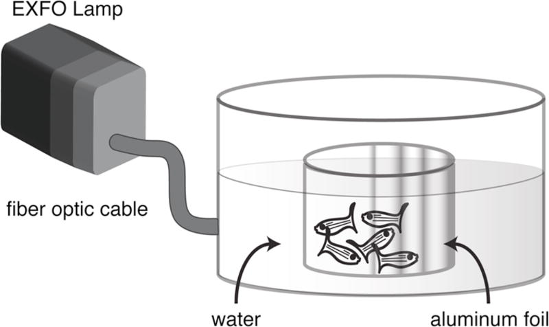
Apparatus for delivering high intensity light to freely-swimming adult zebrafish. See text for details.
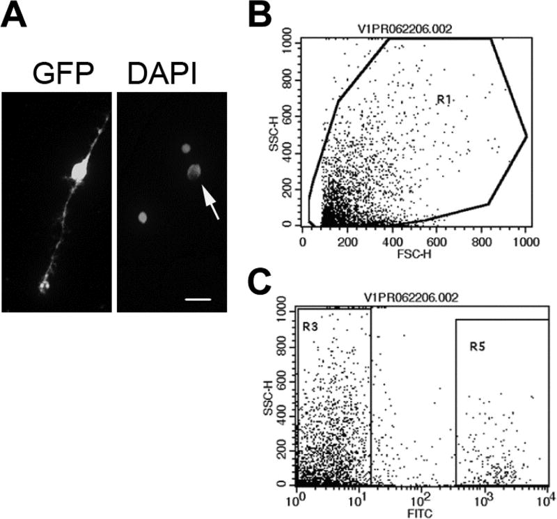
Isolation of GFP+ Müller glia. (A) Left panel: Dissociated GFP+ Müller glial cell. Right panel: Same field, counterstained with DAPI. Arrow indicates the Müller glial cell. Scale bar: 10 µ m. (B, C) Flow cytometry scatter plots; forward scatter-height (FSC-H); side scatter-height (SSC-H). Dissociated cells from adult Tg(gfap:GFP)mi2002 zebrafish retinas were gated by forward and side scatter (B). GFP+ Müller glia were isolated based on fluorescence in the FITC channel (R5 in C). [Modified from (2)]
References
Publication types
MeSH terms
Grants and funding
LinkOut - more resources
Full Text Sources
Medical
