Review
. 2012 Jun;76(2):229-61.
doi: 10.1128/MMBR.05014-11.

Cell biology of cnidarian-dinoflagellate symbiosis

Affiliations
Review

Cell biology of cnidarian-dinoflagellate symbiosis

Simon K Davy et al. Microbiol Mol Biol Rev. 2012 Jun.

Abstract

The symbiosis between cnidarians (e.g., corals or sea anemones) and intracellular dinoflagellate algae of the genus Symbiodinium is of immense ecological importance. In particular, this symbiosis promotes the growth and survival of reef corals in nutrient-poor tropical waters; indeed, coral reefs could not exist without this symbiosis. However, our fundamental understanding of the cnidarian-dinoflagellate symbiosis and of its links to coral calcification remains poor. Here we review what we currently know about the cell biology of cnidarian-dinoflagellate symbiosis. In doing so, we aim to refocus attention on fundamental cellular aspects that have been somewhat neglected since the early to mid-1980s, when a more ecological approach began to dominate. We review the four major processes that we believe underlie the various phases of establishment and persistence in the cnidarian/coral-dinoflagellate symbiosis: (i) recognition and phagocytosis, (ii) regulation of host-symbiont biomass, (iii) metabolic exchange and nutrient trafficking, and (iv) calcification. Where appropriate, we draw upon examples from a range of cnidarian-alga symbioses, including the symbiosis between green Hydra and its intracellular chlorophyte symbiont, which has considerable potential to inform our understanding of the cnidarian-dinoflagellate symbiosis. Ultimately, we provide a comprehensive overview of the history of the field, its current status, and where it should be going in the future.


Figures
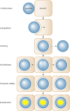
Fig 1
Fig 1
The six phases of symbiosis establishment and persistence in cnidarian-algal symbiosis. 1, initial surface contact between the algal symbiont and cnidarian host cell; 2, symbiont engulfment by the host cell; 3, dynamic sorting of the symbionts (now enclosed by a membrane of host origin), leading either to rejection of the symbiont (dashed arrow) or acceptance; 4, proliferation of the symbiont via cell division within the host tissues; 5, dynamic stability, where the symbiont population is maintained at a steady density; and 6, symbiosis dysfunction and breakdown (for example, in response to environmental stress). For simplicity, not all the possible cellular events are represented here; for more detailed descriptions of these events, see the relevant sections in the text.
Fig 2
Fig 2
Host-microbe signaling during microbial invasion and host phagocytosis. Animal innate immunity acts to detect and manage microbial invaders, both negative and positive. Whatever the quality of the interaction, the host needs to recognize the presence of the microbe and then launch downstream effector pathways to either destroy negative invaders or foster the growth of mutualistic ones. There are many excellent reviews of host-microbe signaling that cover these events in great detail (173, 233, 377). For direct detection of microbes, hosts express a dizzying array of proteins, termed pattern recognition receptors (PRRs) (Table 1), either secreted or on cell surfaces that recognize signature microbial compounds termed microbe-associated molecular patterns (MAMPs). (PRRs depicted: C3R, complement 3 receptor; Nods, nucleotide-binding oligomerization domain proteins; SRs, scavenger receptors; TLRs, toll-like receptors). MAMPs are a variety of sugar, protein, lipid, and nucleic acid compounds that are essential to microbial survival and often unique to certain microbe groups. They include lipopolysaccharide (LPS), peptidoglycan (PG), glycans, and glycosylphosphatidylinositol (GPI) anchors. Host cells can also detect the presence of microbes indirectly through the process of opsonization that can amplify a host response. Invading microbes become coated with secreted host compounds or opsonins, such as complement protein (C3) or immunoglobulins (in the case of vertebrates). Like MAMPs, opsonins then bind PRRs on host cell surfaces. The binding of PRRs to MAMPs or opsonins starts a signaling cascade, often involving the activation of the master immunity regulator nuclear factor κB (NF-κB), which then launches a large array of host responses. In the case of invading pathogens, these responses can include phagocytosis of the microbe, an inflammatory response, antimicrobial killing mechanisms, and initiation of host cell apoptosis or autophagy. The three processes shown within the box are described in more detail in Appendix 1.
Fig 3
Fig 3
Diagrammatic representation of individual cnidarian host gastrodermal cells with resident symbionts. Each symbiont is surrounded by a symbiosome membrane complex, depicted as a single black line. Note the dramatically different host cell sizes, with the hydroids green Hydra and Myrionema sp. having very large cells compared to the typical anthozoan cell. The larger hydroid cells harbor more symbionts per cell than the anthozoan. Chlorella cells are elliptoid in shape. Symbiodinium cells are spherical in shape and approximately 10 μm in diameter. Reports of Chlorella cell size vary, and so they are depicted here as approximately 5 to 8 μm in diameter, which is within the range described in many studies.
Fig 4
Fig 4
Potential mechanisms for the regulation of host-symbiont biomass in cnidarian-alga symbiosis. 1, expulsion of symbiont cells in either detached whole host cells or pinched-off portions of host cells (i.e., aposomes). 2, expulsion of symbiont cells either via active exocytosis or as a result of host cell apoptosis. 3, intracellular degradation of the symbiont, as a result of programmed cell death of the symbiont, reengagement of the phagosomal maturation process in the host, or autophagic digestion of the symbiont by the host cell. 4, control of progression through the symbiont cell cycle by the host. G0, G1, S, G2, and M are the phases of the eukaryotic cell cycle (see Appendix 2), with G1 often being the longest phase and M always being the shortest (as generalized in the schematic). The host may render the intracellular environment unfavorable or signal to the symbiont in such a way that the cell cycle does not, for example, pass through the G1/S checkpoint; in this case, the cell could enter the G0 resting state. 5, control of host cell proliferation by the symbiont.
Fig 5
Fig 5
Schematic summary of nutritional interactions in the cnidarian-dinoflagellate symbiosis. 1, dissolved inorganic carbon (DIC) uptake. DIC is acquired either as bicarbonate (HCO3) from the surrounding seawater or as CO2 from the seawater or host metabolism/calcification. In the case of HCO3, it must be converted to CO2 prior to photosynthesis by the dinoflagellate symbiont. 2, photosynthesis. CO2 is photosynthetically fixed through the Calvin-Benson cycle (i.e., the C3 pathway), with the dinoflagellate ultimately synthesizing a range of organic compounds, including amino acids. 3, translocation. A portion of the photosynthetic products are translocated to the host cell. 4, reverse translocation. Organic compounds are likely translocated from the host to the symbiont; these compounds could arise from host metabolism or be in the same forms as those originally translocated by the symbiont. 5, host metabolism. Translocated compounds are used, alongside dissolved organic matter (DOM) and particulate organic matter (POM) taken up from seawater, to support host metabolism. The catabolism of nitrogenous compounds ultimately leads to the generation of ammonium waste that can be assimilated by the symbiont. 6, ammonium assimilation. Excretory and seawater ammonium can be assimilated by both the host cell (pathway not shown) and the symbiont, with translocated organic compounds providing carbon skeletons necessary for host assimilation. The assimilation of excretory ammonium back into amino acids by the dinoflagellate symbiont completes the process of “nitrogen recycling” by the symbiosis. 7, nitrate assimilation. Nitrate is taken up from the seawater, but only the symbiont can convert it to ammonium for subsequent assimilation into amino acids. 8, phosphate assimilation. Phosphate is likewise taken up from seawater and can be assimilated by the dinoflagellate symbiont. Note that uptake of nutrients can also occur from the ambient seawater via the epidermis (not illustrated), but for simplicity these pathways are not shown.
Fig 6
Fig 6
Schematic drawing of the major proteins identified within calicoblastic cells. Calicoblastic cells form an epithelium (calicodermis) whose apical membrane (AM) is attached to the coral skeleton by desmocytes via a hemidesmosome adhesion complex. Their basal pole is close to the mesoglea, a sheet of extracellular matrix proteins. Various transport proteins within calicoblastic cells have been identified or suggested on the basis of pharmacological evidence: ATPases (Na+ pump and Ca2+ pump), antiporters (Na+/Ca2+ antiporter and bicarbonate carrier) and channel proteins (Ca2+ channel). The presence of numerous mitochondria within calicoblastic cells both energizes ion transport and supplies metabolic CO2 as a source of carbon for calcification. The presence of carbonic anhydrase within the extracellular calcifying medium (between the calicodermis and skeleton) facilitates the chemical equilibrium between the different carbon species. In addition, calicoblastic cells synthesize and secrete via vesicles a mixture of macromolecules, called the organic matrix (OM), which acts as an organic framework. Calcium ions may reach this medium both by a paracellular pathway through septate junctions and by transcellular transport, aided by the large surface area of the basal lateral membrane (BLM) of the calicoblastic cells. The symbiont cell may enhance calcification either by altering the physicochemical composition of extracellular fluids by either absorbing CO2 or releasing O2 (1) or by supplying organic compounds to the calicoblastic cells, such as precursors for skeletal organic matrix synthesis or high-energy molecules (2) (see text for more details).
None
None
None

References

    1. Alberts B, et al. 2008. Molecular biology of the cell, 4th ed Garland Science, New York, NY
    1. Al-Horani F, Al-Moghrabi SM, de Beer D. 2003. Microsensor study of photosynthesis and calcification in the scleractinian coral, Galaxea fascicularis: active internal carbon cycle. J. Exp. Mar. Biol. Ecol. 288:1–15
    1. Allemand D, Furla P, Bénazet-Tambutté S. 1998. Mechanisms of carbon acquisition for endosymbiont photosynthesis in Anthozoa. Can. J. Bot. 76:925–941
    1. Allemand D, Tambutté E, Girard JP, Jaubert J. 1998. Organic matrix synthesis in the scleractinian coral Stylophora pistillata: role in biomineralization and potential target of the organotin tributyltin. J. Exp. Biol. 201:2001–2009 - PubMed
    1. Allemand D, et al. 2004. Biomineralisation in reef-building corals: from molecular mechanisms to environmental control. C. R. Palevol. 3:453–467

Publication types

LinkOut - more resources