Review
. 2012 Jun;76(2):262-310.
doi: 10.1128/MMBR.05017-11.

Protein export according to schedule: architecture, assembly, and regulation of type III secretion systems from plant- and animal-pathogenic bacteria

Affiliations
Review

Protein export according to schedule: architecture, assembly, and regulation of type III secretion systems from plant- and animal-pathogenic bacteria

Daniela Büttner. Microbiol Mol Biol Rev. 2012 Jun.

Abstract

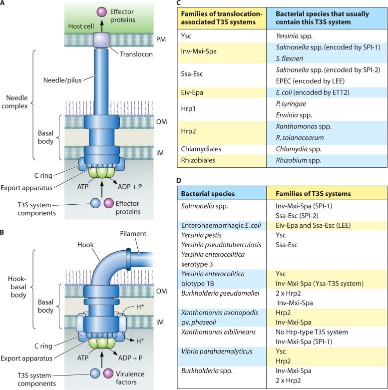
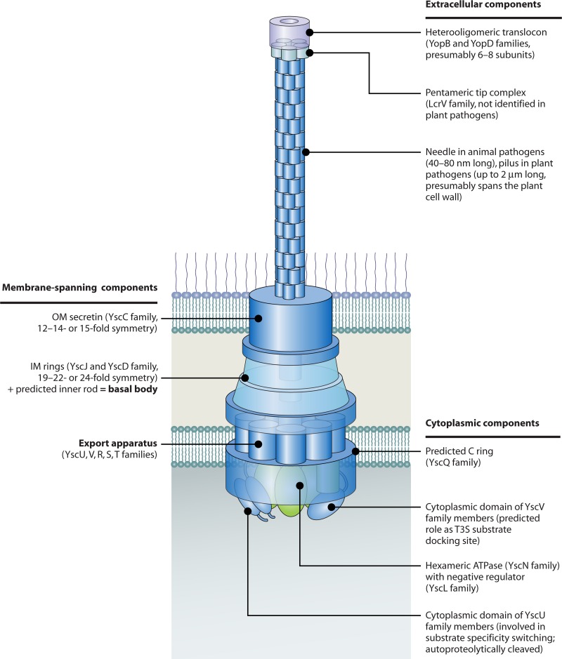
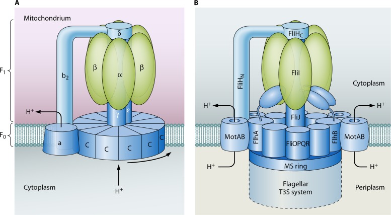
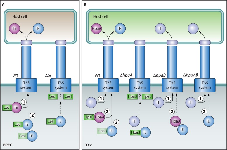
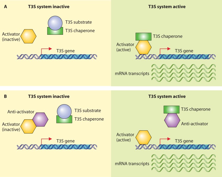
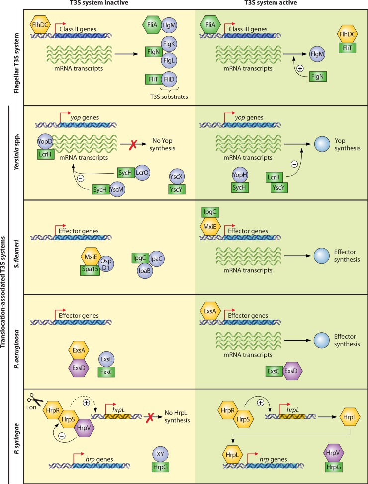
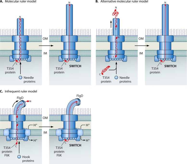
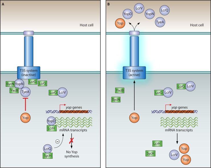
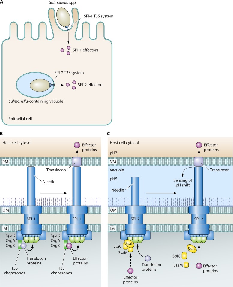
Flagellar and translocation-associated type III secretion (T3S) systems are present in most gram-negative plant- and animal-pathogenic bacteria and are often essential for bacterial motility or pathogenicity. The architectures of the complex membrane-spanning secretion apparatuses of both systems are similar, but they are associated with different extracellular appendages, including the flagellar hook and filament or the needle/pilus structures of translocation-associated T3S systems. The needle/pilus is connected to a bacterial translocon that is inserted into the host plasma membrane and mediates the transkingdom transport of bacterial effector proteins into eukaryotic cells. During the last 3 to 5 years, significant progress has been made in the characterization of membrane-associated core components and extracellular structures of T3S systems. Furthermore, transcriptional and posttranscriptional regulators that control T3S gene expression and substrate specificity have been described. Given the architecture of the T3S system, it is assumed that extracellular components of the secretion apparatus are secreted prior to effector proteins, suggesting that there is a hierarchy in T3S. The aim of this review is to summarize our current knowledge of T3S system components and associated control proteins from both plant- and animal-pathogenic bacteria.


Figures

Fig 1
Fig 1
Overview of translocation-associated and flagellar T3S systems from animal- and plant-pathogenic bacteria. (A) Model of the translocation-associated T3S system. The basal body of the T3S system spans the bacterial IM and OM and consists of ring structures that are presumably connected by a periplasmic rod. The basal body is associated via an extracellular needle (animal-pathogenic bacteria) or pilus (plant-pathogenic bacteria) with a channel-like translocon in the host plasma membrane. The basal body and the needle from animal-pathogenic bacteria are referred to as the needle complex. The energy for the docking and unfolding of T3S substrates, including extracellular components of the T3S system and effector proteins, is probably provided by a cytoplasmic ATPase (shown in green) associated with the T3S system. Note that the cytoplasmic C ring is predicted only for translocation-associated T3S systems. A more detailed representation of single components of translocation-associated T3S systems is given in Fig. 2. (B) Model of the flagellar T3S system. The flagellar basal body is associated via an extracellular hook with the flagellar filament, which is 10 to 20 μm long and is the main bacterial motility organelle. The basal body is surrounded by 8 to 11 stator complexes that drive flagellar rotation and contain proton-conducting channels. The flagellar basal body and the hook are referred to as the hook-basal body. A detailed description of individual components of the flagellar T3S systems is provided in Fig. 3. (C) Summary of different families of translocation-associated T3S systems from bacterial pathogens and symbionts of plants or animals. The SPI-1-like Eiv-Epa T3S system encoded by the ETT2 gene cluster from E. coli is active in only a few strains. Note that P. syringae strains belonging to the phylogenetic subgroup 2c appear to encode an unusual T3S system that is only distantly related to the Hrp1-type T3S system (97). (D) Examples of bacterial species that possess more than one translocation-associated T3S system. Please note that most species contain an additional flagellar T3S system that is not listed in this table.
Fig 2
Fig 2
Schematic representation of individual components of translocation-associated T3S systems from animal- and plant-pathogenic bacteria. Conserved membrane-spanning components of the T3S system include the OM secretin (YscC family) and constituents of the IM ring (YscD and YscJ family) and the export apparatus (YscU, -V, -R, -S, and -T families). The IM ring and the export apparatus are associated with the predicted C ring (presumably a multimer of members of the YscQ family) and the hexameric ATPase (depicted in green), which might provide the energy to facilitate the docking and entry of T3S substrates into the inner channel of the secretion system. Additional cytoplasmic components of the T3S system are the predicted regulator of the ATPase (YscL family) and the cytoplasmic domains of YscU and YscV family members, which are probably involved in substrate docking. Note that the composition of the export apparatus and the cytoplasmic parts of the secretion system is speculative and that multiple copies of a single substituent (e.g., members of the YscV protein family) can be involved in the assembly of the T3S system. Extracellular components of the T3S system include the needle (animal-pathogenic bacteria) and pilus (plant-pathogenic bacteria), which differ in length and serve as transport channels for secreted proteins at the host-pathogen interface. Translocation of effector proteins across the host plasma membrane is mediated by the channel-like translocon, which is a hetero-oligomeric protein complex and is connected to the needle via a tip complex that consists of members of the LcrV protein family. Tip complexes have so far been identified and/or characterized only for animal-pathogenic bacteria.
Fig 3
Fig 3
Schematic representation of components of the flagellar T3S system. The membrane-spanning basal body consists of two OM rings (L and P rings) that are connected via a distal and proximal rod to the IM ring (MS ring). The MS ring is surrounded by 8 to 11 stator complexes in the IM that provide proton-conducting channels and is associated with the export apparatus, the C ring, the ATPase, and the regulator of the ATPase. The architecture of the export apparatus and of the cytoplasmic components of the flagellar T3S system is probably similar to that in translocation-associated T3S systems (see Fig. 2). Structures that are different from those in translocation-associated T3S systems are indicated.
Fig 4
Fig 4
Similarities between FoF1-ATPases and T3S-associated ATPases. (A) Model of the FoF1-ATPase. The FoF1-ATPase consists of a membrane-embedded Fo domain and a catalytic F1 domain. The F1 domain is composed of an α3β3 hexamer and is associated via the central stalk (consisting of the γ and ε subunits) and the peripheral stalk (composed of δ, b2, and a subunits) with the Fo domain. The Fo domain contains a and b (not shown) subunits and 12 c subunits that form proton-conducting channels. The energy provided by the proton influx drives the rotation of the Fo domain and ATP synthesis. (B) Model of the flagellar T3S-associated ATPase FliI and its interaction partners. FliI presumably forms a hexameric complex that is associated with the regulator of the ATPase, FliH, which shares structural similarity with the peripheral stalk of the FoF1-ATPase. Structural similarity was also reported for the chaperone-binding protein FliJ and components of the central stalk of the FoF1-ATPase (235). FliI associates with the export apparatus of the T3S system, which is connected to the MS ring in the IM and is surrounded by stator complexes (also see Fig. 2 and 3). Note that the organization of the ATPase complex is speculative and that the central position of FliJ in the ATPase ring has not been confirmed experimentally. The cytoplasmic C ring is not shown in this model.
Fig 5
Fig 5
Predicted functions of class IB chaperones during control of effector protein secretion in EPEC and X. campestris pv. vesicatoria. (A) Model for the function of the class IB T3S chaperone CesT from EPEC. CesT promotes the secretion and translocation of Tir, which is the first effector protein (E) that is translocated into the host cell. It is assumed that CesT binds preferentially to Tir to promote Tir secretion after assembly of the T3S system. In the absence of Tir, uncomplexed CesT might block the secretion of effector proteins by the T3S system. The question mark indicates that it is still unknown whether the inhibitory activity of CesT is linked to its potential association with components of the T3S system. Dashed arrows indicate reduced secretion and/or translocation rates. (B) Hypothetical mode of action of the class IB T3S chaperone HpaB from X. campestris pv. vesicatoria. HpaB binds to and promotes the secretion of the effector protein HpaA and additional effector proteins (E) after the secretion of translocon proteins (T). Similar to Tir, HpaA is presumably the first effector protein that is bound to the chaperone and travels the T3S system. In the absence of HpaA, the efficient secretion of effector, pilus, and translocon proteins is suppressed, probably by uncomplexed HpaB that binds to components of the secretion apparatus. The question mark indicates that it is unknown whether the association of HpaB with the T3S system contributes to its inhibitory activity. Interestingly, in the absence of HpaB, translocon proteins are secreted and even translocated, suggesting that they contain a functional translocation signal that is suppressed by HpaB. Translocon proteins are also efficiently secreted in an hpaAB double deletion mutant, in which T3S is not suppressed by uncomplexed HpaB.
Fig 6
Fig 6
Control of T3S gene expression by regulatory chaperones. (A) Proposed mode of action of T3S chaperones that act as coactivators of transcriptional regulators. When the T3S system is inactive, T3S chaperones are bound by their cognate substrates in the bacterial cytosol. The activation of the T3S system leads to the secretion of T3S substrates and thus to the liberation of corresponding T3S chaperones, which can subsequently interact with transcriptional activators and promote T3S gene expression. T3S chaperones are represented by green rectangles, and T3S substrates are represented by circles. Transcriptional activators are depicted in yellow. (B) Model for the activity of T3S chaperones that act as antiantiactivators. When the T3S system is inactive, T3S chaperones are bound by their corresponding T3S substrates. The induction of T3S leads to the liberation of T3S chaperones, which can subsequently bind to an antiactivator that suppresses the activity of a transcriptional activator. Upon interaction with the T3S chaperone, however, the antiactivator is released from the transcriptional activator, and the latter can induce T3S gene expression.
Fig 7
Fig 7
Schematic representation of the regulatory mechanisms that control T3S gene expression in flagellar and translocation-associated T3S systems. The expression of effector genes or genes encoding substrates and components of the flagellar T3S system is controlled by transcriptional regulators and regulatory chaperones and depends on the activity of the T3S system. Flagellar gene expression is regulated by the transcriptional activators FlhDC, which are encoded by class I genes and activate the expression of class II genes. Class II gene products include the anti-σ28 factor FlgM, the hook-filament junction proteins FlgK and FlgL, and the filament cap protein FliD, which bind to the corresponding chaperones FliA, FlgN, and FliT, respectively. The secretion of FlgM leads to the release of the σ28 factor FliA, which activates the expression of class III genes. The liberated T3S chaperone FliT binds to FlhC and thus inhibits the expression of class II genes, whereas FlgN positively regulates the translation of flgM class III mRNA. In Yersinia spp., translation of yop mRNAs is suppressed by a YopD-LcrH complex when the T3S system is inactive. LcrQ-SycH and YscM-SycH complexes might act as additional repressors. Activation of Yop secretion leads to relief of the YopD-LcrH-mediated repression of yop mRNA translation and the liberation of SycH and LcrH upon secretion of YopD. The SycH chaperone presumably promotes YopH secretion after its release from the secreted regulator YscM. LcrH might suppress yop gene expression when bound to the T3S chaperone YscY. Effector gene expression in S. flexneri depends on the transcriptional activator MxiE and its coactivator, IpgC. Upon activation of T3S, MxiE and IpgC are released from their secreted interaction partners, i.e., Spa15, OpsD1, IpaC, and IpaD, and can activate effector gene expression. In P. aeruginosa, effector gene expression is controlled by the activator ExsA, which interacts with the antiactivator ExsD when the T3S system is inactive. The T3S chaperone ExsC, which binds to the T3S substrate ExsE, acts as an antiantiactivator when released from ExsE after activation of the T3S system. The interaction of ExsC with ExsD leads to the liberation of ExsA, which subsequently activates effector gene expression. In the plant-pathogenic bacterium P. syringae, expression of hrp (hypersensitive response and pathogenicity) genes that encode the T3S system is induced by HrpR, HrpS, and HrpL. HrpR and HrpS interact with each other and activate the expression of the alternative sigma factor HrpL, which binds to the promoters of hrp genes. HrpR-, HrpS-, and HrpL-dependent activation of hrp gene expression is counteracted by the negative regulator HrpV, which interacts with HrpS, and by the Lon protease, which degrades HrpR. Under T3S-inducing conditions, HrpV interacts with the chaperone-like protein HrpG, which interferes with the negative regulatory activity of HrpV and might also bind to a secreted but so far unknown interaction partner (XY). T3S chaperones are represented in green, and T3S substrates are represented by circles. Transcriptional regulators are depicted in yellow.
Fig 8
Fig 8
Proposed modes of action of T3S4 proteins from animal-pathogenic bacteria. (A) Molecular ruler model. According to the molecular ruler model, the N terminus of the T3S4 protein is attached to the tip of the growing needle. Once the T3S4 protein is stretched, the C-terminal region signals the substrate specificity switch via interaction with the C-terminal domain of a member of the YscU/FlhB family. N, N-terminal region; C, C-terminal region. (B) Alternative molecular ruler model. This model predicts that T3S4 proteins are constantly secreted during needle assembly and thereby measure needle length. The interaction between the C-terminal region of the T3S4 protein and the C-terminal domains of members of the YscU/FlhB family leads to a switch in the substrate specificity and occurs only when the needle has reached a certain length. (C) Infrequent ruler model proposed for flagellar T3S systems. During hook assembly, the T3S4 protein FliK is intermittently secreted and temporarily interacts with hook components such as the hook-capping protein FlgD. However, the rapid secretion of FliK does not allow a productive interaction of the C-terminal domain of FliK with the C-terminal domain of FlhB. After the hook has reached its physiological length of approximately 55 nm, the N-terminal region of FliK interacts more frequently with hook subunits and the reduced secretion rate of FliK allows an interaction of the C-terminal region of FliK with the C-terminal domain of FlhB, and thus the induction of the substrate specificity switch.
Fig 9
Fig 9
Model of the YopN-mediated control of effector protein secretion in Yersinia spp. (A) A complex of YopN, TyeA, and the YopN-specific chaperones SycN and YscB blocks the transit of T3S substrates (represented by circles) through the secretion apparatus. The T3S chaperone LcrG, which interacts with the tip protein LcrV, acts as an additional negative regulator of Yop secretion. YopD-LcrH and LcrQ-SycH complexes suppress yop gene expression when the T3S is inactive (see Fig. 7). (B) Activation of Yop secretion. The activation of the T3S system leads to the secretion of YopD, YopN, TyeA, LcrQ, and LcrV. This relieves the negative effect of YopN and TyeA on Yop secretion as well as of YopD and LcrQ on yop gene expression and thus leads to increased synthesis of Yops, including LcrV. Increased numbers of LcrV proteins bind to LcrG and presumably suppress the negative influence of LcrG on T3S.
Fig 10
Fig 10
Control of SPI-1- and SPI-2-mediated T3S in Salmonella spp. (A) Infection of epithelial eukaryotic cells by Salmonella spp. The SPI-1-encoded T3S system injects effector proteins into epithelial cells, which leads to cytoskeletal rearrangements and membrane ruffling. Bacteria enter the host cell cytosol via endocytosis and activate the SPI-2-encoded T3S system inside the Salmonella-containing vacuole. (B) Predicted function of the SPI-1-encoded SpaO-OrgA-OrgB complex during control of T3S. The SpaO-OrgA-OrgB complex serves as a docking platform for translocon and effector proteins that are sequentially targeted to this complex by a process that presumably requires the presence of corresponding T3S chaperones. According to the predicted hierarchy in T3S, effector proteins form a queue for docking to the SpaO-OrgA-OrgB complex, while translocon proteins are secreted. (C) Control of SPI-2-dependent secretion of translocon and effector proteins. A complex of SpiC, SsaL, and SsaM controls the secretion of translocon and effector proteins dependent on differences in the external pH. At pH 5 (inside the Salmonella-containing vacuole), the complex blocks the efficient secretion of effector proteins, while translocon proteins are secreted. A shift in the extracellular pH to pH 7 leads to the dissociation of the SpiC-SsaL-SsaM complex and thus induces effector protein secretion. The pH shift is probably sensed by the extracellular components of the T3S system. Note that the architecture of the SPI-2 T3S system is speculative and is proposed according to amino acid sequence similarities between predicted components of SPI-2 T3S systems and known translocation-associated T3S systems. The dashed arrow indicates a reduced secretion and/or translocation rate.

References

    1. Abe A, et al. 1999. Enteropathogenic Escherichia coli translocated intimin receptor, Tir, requires a specific chaperone for stable secretion. Mol. Microbiol. 33:1162–1175 - PubMed
    1. Agrain C, et al. 2005. Characterization of a type III secretion substrate specificity switch (T3S4) domain in YscP from Yersinia enterocolitica. Mol. Microbiol. 56:54–67 - PubMed
    1. Aili M, et al. 2008. Regulation of Yersinia Yop-effector delivery by translocated YopE. Int. J. Med. Microbiol. 298:183–192 - PubMed
    1. Akeda Y, Galan JE. 2005. Chaperone release and unfolding of substrates in type III secretion. Nature 437:911–915 - PubMed
    1. Akopyan K, et al. 2011. Translocation of surface-localized effectors in type III secretion. Proc. Natl. Acad. Sci. U. S. A. 108:1639–1644 - PMC - PubMed

Publication types

MeSH terms

LinkOut - more resources