Review
. 2012 Jun;76(2):331-82.
doi: 10.1128/MMBR.05021-11.

Diversity in genetic in vivo methods for protein-protein interaction studies: from the yeast two-hybrid system to the mammalian split-luciferase system

Affiliations
Review

Diversity in genetic in vivo methods for protein-protein interaction studies: from the yeast two-hybrid system to the mammalian split-luciferase system

Bram Stynen et al. Microbiol Mol Biol Rev. 2012 Jun.

Abstract

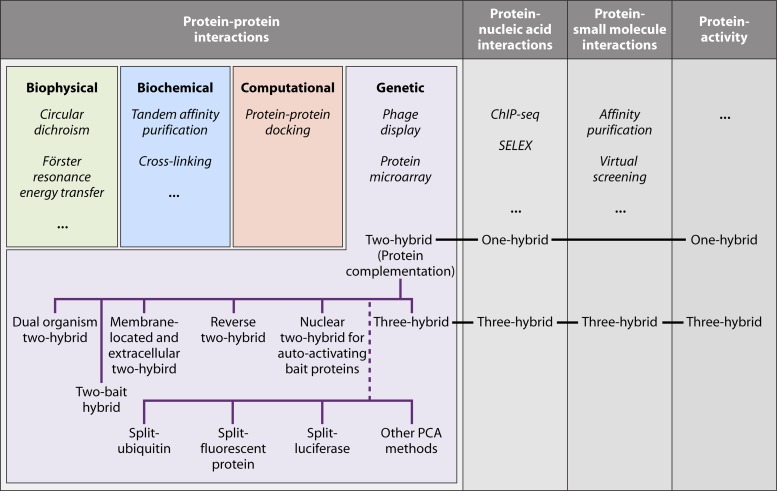
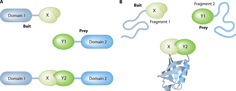
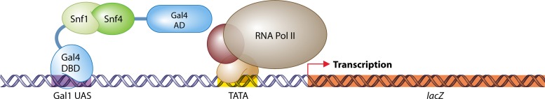
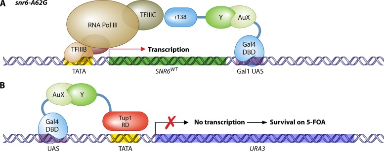
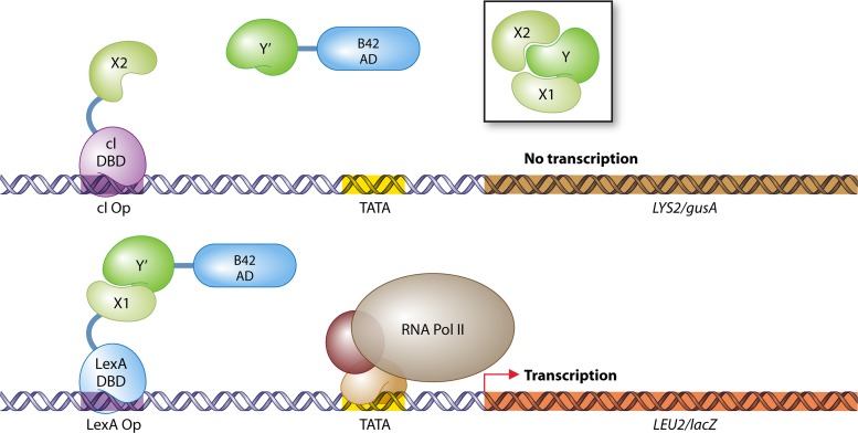
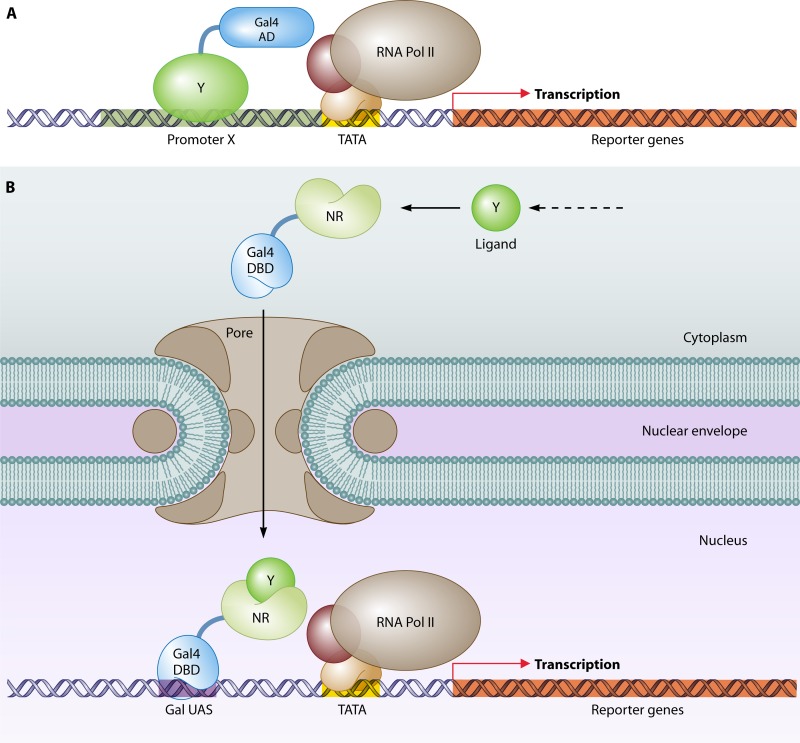
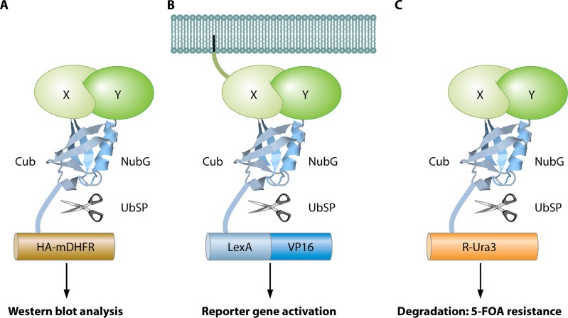
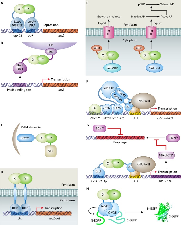
The yeast two-hybrid system pioneered the field of in vivo protein-protein interaction methods and undisputedly gave rise to a palette of ingenious techniques that are constantly pushing further the limits of the original method. Sensitivity and selectivity have improved because of various technical tricks and experimental designs. Here we present an exhaustive overview of the genetic approaches available to study in vivo binary protein interactions, based on two-hybrid and protein fragment complementation assays. These methods have been engineered and employed successfully in microorganisms such as Saccharomyces cerevisiae and Escherichia coli, but also in higher eukaryotes. From single binary pairwise interactions to whole-genome interactome mapping, the self-reassembly concept has been employed widely. Innovative studies report the use of proteins such as ubiquitin, dihydrofolate reductase, and adenylate cyclase as reconstituted reporters. Protein fragment complementation assays have extended the possibilities in protein-protein interaction studies, with technologies that enable spatial and temporal analyses of protein complexes. In addition, one-hybrid and three-hybrid systems have broadened the types of interactions that can be studied and the findings that can be obtained. Applications of these technologies are discussed, together with the advantages and limitations of the available assays.


Figures

Fig 1
Fig 1
Overview of protein-protein interaction technologies and alternative applications for two-hybrid assay-derived methods. ChIP-seq, chromatin immunoprecipitation followed by sequencing; SELEX, systematic evolution by exponential enrichment; PCA, protein fragment complementation assay. Technologies in italics are not discussed in this review.
Fig 2
Fig 2
Two-hybrid systems versus PCAs. (A) Colocalization in two-hybrid systems. Two proteins of interest (X and Y) are each fused to a fixed protein domain, forming the bait and the prey, respectively. In the absence of an interaction (upper part with Y1), the domains remain distant, preventing a detectable output. If the two proteins do interact (lower part with Y2), the bait recruits the prey to a specific cellular location (e.g., reporter gene or plasma membrane), where it can stimulate a detectable output (e.g., gene activation or signal transduction). The domains do not need to be in physical contact. (B) Protein refolding in PCAs. Two proteins of interest (X and Y) are each fused to a fixed protein fragment. If there is no interaction between X and Y (upper part with Y1), the fragments remain unstructured and lack any functional abilities. Upon interaction between the bait and prey (lower part with Y2), the two fragments refold into a fully functional reporter protein (e.g., ubiquitin in the figure). In most cases, the interaction does not need to take place in a specific cellular location, but a minimal time frame of physical contact between the two fragments is necessary to establish complete refolding. The image of the ubiquitin protein is based on the Protein Data Bank (PDB) structure under accession number 1UBQ (671).
Fig 3
Fig 3
The first two-hybrid experiment (178). For the study of the interaction of two proteins of interest (in this case, Snf1 and Snf4), one protein is fused to the DNA-binding domain (DBD) of Gal4 (the bait) and the other protein is fused with the activation domain (AD) of Gal4 (the prey). The bait fusion binds upstream activating sequences (UAS) of the reporter gene lacZ. Association of Snf1 with Snf4 brings the Gal4 AD to lacZ, followed by recruitment of the basal transcriptional machinery, which establishes lacZ transcription, detected by chromogenic analysis.
Fig 4
Fig 4
Nuclear two-hybrid systems for autoactivating bait proteins. (A) RNA Pol III system (511). Through its fusion with the Gal4 DBD, an autoactivating bait (AuX) protein is tethered to the Gal1 upstream activating sequence, located downstream of the reporter gene SNR6. Interaction of the AuX bait with a prey protein (Y) fused to the τ138 subunit of transcription factor III C (TFIIIC) brings the RNA polymerase III holoenzyme to SNR6. Transcription of SNR6 leads to survival under temperature-restrictive (inactive snR6-A62G) conditions. (B) Repressed transactivator system (259). In the repressed transactivator (RTA) system, association of an autoactivating AuX bait with the prey protein Y, attached to the repressor Tup1, inhibits transcription of the reporter gene URA3. This results in survival of the cell on 5-FOA, a substrate for the production of the toxic compound 5-fluorouracil by the gene product of URA3. Pol, polymerase; TF, transcription factor; Y, prey; AuX, autoactivating bait; AD, activation domain; DBD, DNA-binding domain; RD, repressor domain; UAS, upstream activating sequence.
Fig 5
Fig 5
G-protein-based two-hybrid systems. (A) Sos recruitment system (16). A chimeric protein of bait X with hSos is recruited to the plasma membrane upon interaction of X with a membrane protein (Y). Subsequent GDP-GTP exchange of Ras by membrane-localized hSos enables cell growth by virtue of Ras activity in a temperature-sensitive cdc25-2 background strain. (B) Ras recruitment system (64). Similar to the case in the Sos recruitment system, a membrane-bound prey protein Y that interacts with bait X, fused to mammalian Ras (mRas), will bring mRas to the membrane, where it can activate its downstream target adenylate cyclase to establish cell growth at a restrictive temperature (36°C) in a cdc25-2 background strain. (C) G protein fusion system (162). Association of the γ (Ste18) and β pheromone G protein subunits is required for a response to the addition of pheromones. Strong interaction of a membrane bait protein (X) with the prey protein Y, in fusion with Ste18 (Gγ), pulls Gγ away from Gβ. This dissociation hinders the pheromone response in a ste18 strain, which can be detected by growth in the presence of α pheromone (halo assay) or by the lack of gene expression of reporters under the control of pheromone-responsive elements (PREs). (D) Gγ recruitment system (190). Interaction between a membrane-bound prey protein (Y) and a bait protein (X), fused to Gγ lacking a membrane attachment sequence, unites the G protein β and γ subunits to induce the pheromone pathway after addition of α pheromone. Readouts are reporter genes regulated by PREs or the observation of growth in a mating assay with a ste18 strain.
Fig 6
Fig 6
Secretory pathway two-hybrid systems. (A) Secretory pathway. Proteins for secretion are synthesized at the endoplasmic reticulum, transported in vesicles to the Golgi apparatus (optionally for further processing), and secreted into the extracellular fluid by fusion of a secretory vesicle with the plasma membrane. Each compartment hosts one of the available two-hybrid systems. (B) Screening for interactions between extracellular proteins (SCINEX-P) (653). Wild-type Ire1 detects the presence of unfolded proteins in the endoplasmic reticulum. Activation of Ire1 leads to homodimerization, trans-phosphorylation, and, finally, splicing of Hac1 mRNA into correctly translatable mRNA. In SCINEX-P, the bait protein X is fused to an Ire1 variant (Ire1 K702R) that can splice Hac1 mRNA only while the prey protein Y is attached to an Ire1 variant (Ire1 Δtail) that can phosphorylate only its associated Ire1 partner. Upon an X-Y interaction within the lumen of the endoplasmic reticulum, Ire1 Δtail phosphorylates Ire1 K702R, which in turn splices Hac1 mRNA, leading to correct translation of Hac1. Hac1 activity can be sensed by detection of inositol synthesis and by activation of reporters regulated by Hac1-dependent promoters. (C) Golgi two-hybrid system (146). The catalytic (CAT) and membrane-attaching (LOC) domains of the mannosyltransferase Och1 are fused to the bait protein X and prey protein Y, respectively. Interaction of the bait with the prey within the Golgi lumen prevents loss (by secretion) of the Och1 catalytic domain, leading to cell survival at 37°C or at 30°C in the presence of Congo red. (D) Yeast surface two-hybrid system (274). The membrane protein Aga1 keeps the Aga2-bait X fusion at the cellular membrane in the extracellular fluid. Binding of the bait with the prey protein Y, tagged with c-Myc, is detected with anti-c-Myc fluorescent antibodies. Alternatively, both bait and prey proteins are linked with fragments of EGFP. The formation of an X-Y heterodimer results in reassembly of the EGFP fragments and in concomitant fluorescence. The EGFP structure image is based on the PDB structure under accession number 2Y0G (550).
Fig 7
Fig 7
Three-hybrid systems. (A) Identification of target proteins of the E3 ubiquitin ligase complex CBCVHL (41). The pVHL subunit is fused to the LexA DBD, and coexpression of elongins B and C (ELB and ELC) stabilizes the native conformation of pVHL. A human cDNA library in fusion with the Gal4 AD is screened for interactions with pVHL. The human prolyl hydroxylase PHD3 delivers the hydroxyl group to interacting prey proteins, which is essential for recognition by pVHL. (B) Protein–small-molecule interactions (94). O6-Alkylguanine-DNA alkyltransferase (AGT) is fused to the Gal4 DBD and can bind covalently with the small molecule O6-benzylguanine (BG) in vivo. A library of BG small-molecule heterodimers, produced in vitro, can be screened for interactions with a prey protein Y attached to the Gal4 AD. (C) Detection of enzymatic substrate recognition (21). A chimeric protein of LexA and DHFR binds the promoter region of the reporter gene lacZ and interacts with a tripartite small molecule through association of DHFR with Mtx. This small-molecule trimer further consists of a bait linker X and dexamethasone (Dex). Dexamethasone interacts with a fusion of the rat glucocorticoid receptor (RGR) and the B42 AD. The whole complex stimulates expression of lacZ. An enzyme Y that targets and cleaves linker X can be identified by disruption of the transcription activating complex and the loss of lacZ expression. (D) RNA-protein interactions (265). The LexA DNA-binding domain is fused to a head-to-tail dimer of the RNA-binding protein MS2. This hook protein associates with an RNA dimer of bacteriophage MS2 RNA and a bait RNA stretch (X). Interaction of this RNA X with a protein Y fused to the Gal4 AD is detected by stimulation of reporter gene expression. DBD, DNA-binding domain; AD, activation domain; UAS, upstream activating sequence; Op, operator; Pol, polymerase.
Fig 8
Fig 8
Reverse two-hybrid systems. (A) Repression of toxic genes (228, 373, 667). Interaction between bait X and prey Y stimulates transcription of URA3, which results in cell toxicity in the presence of 5-fluoroorotic acid; CYH2, which makes the cells sensitive to cycloheximide; or GAL1, which converts galactose into galactose-1-phosphate, a toxic compound that accumulates in the absence of Gal7. Compounds or mutations (in X or Y) that interfere with the association of the bait with the prey are identified by growth on selective medium. (B) Activation of positive selection markers (589). Interaction between bait X and prey Y leads to expression of tetR, encoding the Tet repressor. Next, TetR represses activation of HIS3, hindering cell growth on medium without histidine. Disruption of the interaction between bait and prey is analyzed by the appearance of histidine prototrophy. DBD, DNA-binding domain; AD, activaion domain; UAS, upstream activating sequence; Pol, polymerase; Op, operator.
Fig 9
Fig 9
The dual-bait system (580). Protein Y interacts with two proteins, X1 and X2 (as shown in the box). Bait proteins X2 and X1 are fused with the DNA-binding proteins cI and LexA, respectively, while a library of mutated prey Y proteins is made in fusion with the B42 activation domain. A mutation in protein Y (Y′) that specifically disables the association with X2 but not X1 is detected by reporter activity of LEU2 and lacZ, without expression of LYS2 or gusA. DBD, DNA-binding domain; AD, activation domain; Op, operator; Pol, polymerase.
Fig 10
Fig 10
One-hybrid systems. (A) DNA-protein interactions (706). A putative DNA-binding protein Y is in fusion with the Gal4 activation domain. Interaction of protein Y with a promoter X stimulates reporter gene expression. This assay can single out proteins that bind a fixed promoter of interest or DNA sequences that are targeted by a fixed DNA-binding protein of interest. (B) Identification of transcriptional activity (696). A chimeric protein with the Gal4 DNA-binding domain and a transcriptional activator (a nuclear receptor [NR] in this example) stimulates expression of a reporter gene under the control of Gal upstream activating sequences. An application of such a system is the identification of ligands that trigger NR nuclear import and activity. AD, activation domain; DBD, DNA-binding domain; Pol, polymerase; UAS, upstream activating sequence.
Fig 11
Fig 11
The split-ubiquitin system. (A) mDHFR-HA readout (312). Bait protein X and prey protein Y are fused to the C-terminal (Cub) and mutated (I13G) N-terminal (NubG) domains, respectively, of ubiquitin. In addition, a chimera of mDHFR and the HA epitope completes the bait construct. Upon interaction of X with Y, a fully reconstituted ubiquitin is recognized by ubiquitin-specific proteases (UbSP) that cleave HA-mDHFR, resulting in a shift on a Western blot using anti-HA antibodies. (B) LexA-VP16 readout (611). Interaction between a membrane-bound bait X and prey Y leads to cleavage of the artificial transcription factor LexA-VP16. Released LexA-VP16 localizes to the nucleus to activate reporter genes. (C) R-Ura readout (372). Interaction between bait X and prey Y induces the release of Ura3 (with an N-terminal arginine) by proteases. Exposed arginine makes Ura3 highly unstable, resulting in its degradation. Due to the strongly reduced concentration of Ura3, the cells become viable on medium with 5-FOA, a prototoxic substrate of Ura3. The image of the ubiquitin protein is based on the PDB structure under accession number 1UBQ (671).
Fig 12
Fig 12
Specific bacterial genetic PPI detection methods. (A) Repressor-based two-hybrid system (137). Interaction between two proteins of interest, X and Y, can be monitored by fusion of each with a variant of the LexA repressor (408 or wild type). Heterodimerization of LexA408 and LexA+, induced by X-Y association, is required for efficient repression of the reporter gene lacZ, under the control of the LexA operators op408 and op+. (B) PhaR two-hybrid system (690). The binding of bait X with prey Y, fused to the DNA-binding domain (DBD) of the repressor PhaR and the PHB granule-associated protein PhaP, respectively, results in lacZ expression by recruitment of PhaR to PHB granules. (C) GFP recruitment system (132). Bait X is situated at cell division sites through the action of its chimeric partner, B. subtilis DivIVA. Interaction of protein X with GFP-tagged prey protein Y results in focused fluorescence at the cell division sites. GFP recruitment systems are also available for the pathogenic fungus Candida albicans, for C. elegans, and for mammalian cells. (D) ToxR two-hybrid system (357). The V. cholerae ToxR transcriptional activator requires dimerization of its periplasmic domain for full reporter transcription activation. In the ToxR two-hybrid system, the periplasmic domain is replaced by two proteins of interest, X and Y. An interaction between these two proteins results in efficient ctx promoter binding of the truncated ToxR protein (ToxR′) and subsequent gene expression of lacZ or the chloramphenicol acetyltransferase gene (cat). (E) Tat two-hybrid system (621). The Tat signal sequence (ss) peptide tethers bait protein X to the periplasm. A chimeric fusion of prey Y with the maltose-binding protein without a signal sequence (ΔssMBP) localizes to the periplasm only upon interaction of X with Y. This translocation is required for growth on medium with maltose as the sole carbon source. Alternatively, the prey protein Y is fused to a localization-deficient DsbA enzyme (ΔssDsbA), which catalyzes the formation of active alkaline phosphatase (AP). Active AP converts p-nitrophenyl phosphate (pNPP) to yellow p-nitrophenol (pNP). (F) Bacterial two-hybrid system for DNA-protein interactions (314). To increase sensitivity in the search for zinc finger-DNA associations, the binding of a zinc finger motif X to its target DNA sequence, Y (zinc finger binding motif Zfbm Y), is facilitated by inclusion of two fixed zinc fingers from Zif268 and the target DNA sequence Zif268 bm. The zinc finger fusion further consists of the S. cerevisiae Gal11 interaction domain (Gal11 ID), which binds the S. cerevisiae Gal4 dimerization domain (Gal4 ID). The latter domain is fused to the N-terminal domain of the RNA polymerase α subunit for indirect activation of an operon comprising the S. cerevisiae auxotrophic marker HIS3 and aadA, which confers resistance to spectinomycin. (G) Bacterial reverse two-hybrid system (239). Interaction between chimeric proteins of bait X with the bacteriophage λ cI repressor (which binds the λ cI OR2 operator) and prey Y with the N-terminal domain of the RNA polymerase α subunit results in activation of a gene encoding the C-terminal domain of the bacteriophage 186 cI repressor (186 cI CTD). This truncated protein sequesters and inactivates full-length 186 cI, which normally downregulates cytotoxic 186 prophage genes. Resulting cell death can be circumvented by mutations that block the bait-prey interaction. (H) Intein-mediated split-GFP assay (485). Bait protein X is in fusion with the N-terminal fragments of the intein VDE and GFP, while prey protein Y constructs include their respective C-terminal counterparts. Interaction between X and Y reconstitutes VDE, which splices out and covalently reattaches the GFP fragments to create an isolated GFP monomer, detected by fluorescence. The EGFP structure image is based on the PDB structure under accession number 2Y0G (550).
Fig 13
Fig 13
Visualization of PPIs by PCAs. (A) Split-YFP assay in the plant pathogen Magnaporthe grisea (732). Pmk1 and Mst7 kinases, which are components of the MAP kinase pathway essential to appressorium formation and plant infection, were fused to the C-terminal and N-terminal parts of YFP, respectively. Interaction between Pmk1 and Mst7 was observed in vivo in appressorium formations only when the putative docking site of Mst7 was intact. A, appressorium; G, germ tube. (Adapted from reference with permission.) (B) Multicolor split-FP assay in tobacco culture cells. Protoplasts were transfected by direct DNA uptake and visualized using laser scanning confocal microscopy. Simultaneous interactions between Agrobacterium tumefaciens VirE2 (VirE2-cCFP) and the Arabidopsis nuclear transport adapter protein importin α-1 (Impa-1-nCerulean) and between VirE2 and importin α-4 (Impa-4-nVenus) were observed in the cytoplasm (cerulean) and nucleus (Venus), respectively. Nuclear localization was confirmed by colocalization of the nuclear marker mCherry-VirD2NLS. Labels below each image indicate the filter set/channel imaged. DIC, differential interference contrast image. (Adapted from reference with permission.) (C) Visualization of odor-evoked calcium release upon formation of functional heteromeric complexes of odorant receptors (ORs) in Drosophila, using a split-YFP assay. (Top) Complementary N-terminal and C-terminal fragments of YFP [YFP(1) and YFP(2)] were fused to the odorant receptor OR83b. (Left) Dimerization of OR83b in neurons lacking native OR83b, visualized by the split-YFP method. (Right) Dimerization of OR83b is still visible in Gr21a neurons, which do not express any native ORs, suggesting a direct PPI. (Bottom) Complementary N-terminal and C-terminal fragments of YFP [YFP(1) and YFP(2)] were fused to the odorant receptor OR43a. YFP complementation is visible in neurons with OR83b but not in neurons lacking OR83b. This implicates that OR43a dimerization depends on the presence of OR83b and may not be a direct PPI. (Adapted from reference with permission.) (D) In vivo imaging of split Renilla luciferase (RLuc) complementation in living mice (344). The strategy to monitor translocation of a particular protein into the nucleus is based on reconstitution of split RLuc by the intein Dna-E (also see Fig. 12H). RLuc-N (N-terminal part) was fused to DnaE-N and a nuclear localization signal. This chimera localizes mainly to the nucleus. RLuc-C (C-terminal part) was fused to DnaE-C and a protein of interest, the nuclear androgen receptor (AR), which localizes to the cytosol. Translocation of AR into the nucleus was visualized upon addition of 5a-dihydrotestosterone (DHT), which binds AR, in COS-7 cells implanted on the backs of mice. DHT-induced translocation of AR results in reconstitution of the DnaE intein and its splicing-reassembly property. Consequently, the spliced and reconstituted RLuc recovers its bioluminescence activity, which is imaged by using a cooled CCD camera and measured as photons per second per cm2. The differential translocation of AR in the presence (+) or absence (−) of DHT could hence be evaluated quantitatively. (Adapted from reference [copyright 2004, National Academy of Sciences].)
Fig 14
Fig 14
Unique mammalian genetic PPI methods. (A) Mammalian protein-protein interaction trap (MAPPIT) (172). The bait protein is a fusion with a leptin receptor (LR), which contains three Y-to-F mutations so it is unable to activate STATs spontaneously (one representative phenylalanine [F] is shown). The prey fusion contains a domain of gp130 which can recruit STATs. After interaction of the bait with the prey, Janus kinases (JAKs) phosphorylate gp130, which stimulates binding of gp130 with the STATs. The STATs themselves are phosphorylated by the JAKs, which results in the formation of a STAT complex. The STAT complex binds the rat PAP1 promoter (rPAP1p) and activates luciferase transcription. The leptin receptor is further fused with the extracellular domain of EpoR, a receptor of erythropoietin (Epo), and therefore LR complex formation, which is necessary to make the association with the JAKs, is induced by addition of Epo. (B) Reverse MAPPIT (171). For reverse MAPPIT, a functional LR protein is used with one tyrosine (Y) residue that can be phospharylated upon activation. (Left) The prey Y fusion contains a phosphatase (PaseDom) domain which, upon interaction with bait X, removes the phosphate of JAK2, thereby preventing STAT recruitment. (Right) Inhibition of the bait-prey association by competing proteins (Comp) or compounds (C) reestablishes normal JAK-STAT signaling, which ultimately leads to luciferase reporter gene transcription. (C) Split-TEV method (698). Bait protein X and prey protein Y are fused to the N-terminal and C-terminal domains, respectively, of TEV protease. Association of X with Y initiates the reconstitution of a fully functional TEV protease, which cleaves TEV-specific recognition sequences (rsTEV). A membrane-bound or cytoplasmic protein (MCP), linked by an rsTEV to either luciferase or the artificial transcription factor LexA-VP16, prevents strong bioluminescence or LexA-VP16 nuclear localization, respectively. TEV protease activity releases luciferase, for induction of strong luminescence, or LexA-VP16, for reporter gene activation. The image of TEV protease is based on the PDB structure under accession number 1LVM (512).
None
None
None
None

References

    1. Abel S, et al. 2011. Regulatory cohesion of cell cycle and cell differentiation through interlinked phosphorylation and second messenger networks. Mol. Cell 43:550–560 - PMC - PubMed
    1. Abida WM, Carter BT, Althoff EA, Lin H, Cornish VW. 2002. Receptor-dependence of the transcription read-out in a small-molecule three-hybrid system. Chembiochem 3:887–895 - PubMed
    1. Aguilera C, et al. 2011. c-Jun N-terminal phosphorylation antagonises recruitment of the Mbd3/NuRD repressor complex. Nature 469:231–235 - PubMed
    1. Ahmed KS, Saloma NH, Kadah YM. 2011. Improving the prediction of yeast protein function using weighted protein-protein interactions. Theor. Biol. Med. Model. 8:11. - PMC - PubMed
    1. Aho S, Arfman A, Pummi T, Uitto J. 1997. A novel reporter gene MEL1 for the yeast two-hybrid system. Anal. Biochem. 253:270–272 - PubMed

Publication types

LinkOut - more resources