. 2013 Apr 18;32(16):2114-20.
doi: 10.1038/onc.2012.233. Epub 2012 Jun 11.

PAK4 kinase activity and somatic mutation promote carcinoma cell motility and influence inhibitor sensitivity

Affiliations

PAK4 kinase activity and somatic mutation promote carcinoma cell motility and influence inhibitor sensitivity

A D Whale et al. Oncogene. .

Abstract

Hepatocyte growth factor (HGF) and its receptor (c-Met) are associated with cancer cell motility and invasiveness. p21-activated kinase 4 (PAK4), a potential therapeutic target, is recruited to and activated by c-Met. In response, PAK4 phosphorylates LIM kinase 1 (LIMK1) in an HGF-dependent manner in metastatic prostate carcinoma cells. PAK4 overexpression is known to induce increased cell migration speed but the requirement for kinase activity has not been established. We have used a panel of PAK4 truncations and mutations in a combination of overexpression and RNAi rescue experiments to determine the requirement for PAK4 kinase activity during carcinoma cell motility downstream of HGF. We find that neither the kinase domain alone nor a PAK4 mutant unable to bind Cdc42 is able to fully rescue cell motility in a PAK4-deficient background. Nevertheless, we find that PAK4 kinase activity and associated LIMK1 activity are essential for carcinoma cell motility, highlighting PAK4 as a potential anti-metastatic therapeutic target. We also show here that overexpression of PAK4 harbouring a somatic mutation, E329K, increased the HGF-driven motility of metastatic prostate carcinoma cells. E329 lies within the glycine-rich loop region of the kinase. Our data suggest that E329K mutation leads to a modest increase in kinase activity, conferring resistance to competitive ATP inhibitors in addition to promoting cell migration. The existence of such a mutation may have implications for the development of PAK4-specific competitive ATP inhibitors should PAK4 be further explored for clinical inhibition.


Figures
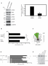
Figure 1
Figure 1
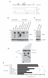
PAK4 is required for HGF-mediated cell motility, but not chemotaxis. (A) For western blot analysis cells were lysed in all experiments as follows. 10 minutes in lysis buffer (0.5% NP-40, 30 mM sodium pyrophosphate, 50 mM Tris-HCl pH 7.6, 150 mM NaCl, 0.1 mM EDTA, 50 mM NaF, 1 mM Na3VO4 and complete mini-EDTA free protease inhibitor (Roche)) then clarification by centrifugation at 14,000 g for 10 minutes. Lysates were immunoblotted according to standard procedures. Blots were developed by enhanced chemiluminescence (ECLplus, GE Healthcare). Control and PAK4 shRNA expressing cell lysates were probed for PAK4/6/1/2, c-Met, tGFP and GAPDH for details of antibodies see supplemental materials and methods. PAK4 expression from cell lysates stably expressing control and PAK4 shRNA from 4 independent experiments were quantified relative to control ± s.d. (B and D) PC3 cells stably expressing control and PAK4 shRNA were maintained in low serum for 24 h, stimulated with HGF and imaged or PC3 cells stably expressing PAK4 shRNA were transfected with plasmids encoding mRFP or mRFP-PAK4r and serum starved for 24 h. Cells were then stimulated with HGF in the presence of either DMSO or LIMKi and imaged. Cell images were collected using an axiovert 100 microscope and Sensicam (PCO Cook) CCD camera, taking a frame every 10 min for 21 h using AQM acquisition software (Kinetic Imaging Ltd, Belfast, U.K.). Subsequently cells were tracked for the whole of the time-lapse sequence using Motion Analysis software (Kinetic Imaging Ltd). Unless indicated, at least 50 cells were tracked over nine separate films from three separate experiments for each experimental condition. Mathematical analysis was then carried out using Mathematica 6.0™ workbooks . Mean track speeds for each condition were compared using the Student’s T-test and statistical significance was accepted for a min P≤0.05. See Supplemental Materials and Methods for details of migration analysis. C) PAK4-depleted cells were maintained in low serum for 24 h, exposed to a gradient of HGF in a Dunn chemotaxis chamber for 12 h and individual cells (n=123) from two separate experiments tracked. A chemotaxis circular histogram was generated to show the proportion of cells with a direction of migration lying within each 18° segment (source of HGF at the top). Red arrow indicates mean direction of cell migration; green segment 99% confidence interval calculated from a Rayleigh test. E) purified GST, GST-PAK4 kinase domain, GST-PAK4Δkinase and GST-PAK4ΔCRIB beads were used to pulldown Myc-LIMK1 from cell lysates. Samples were analysed by anti-Myc immunoblotting and ponceau staining. Representative of three separate experiments. GST-pull down assays were performed according to standard protocols, for details see supplemental Materials and methods.
Figure 2
Figure 2
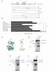
Active PAK4 kinase domain is necessary, but not sufficient for motility (A) Domain structure of PAK4 and GST- and mRFP-tagged shRNA-resistant PAK4 proteins. CRIB: Cdc42 and Rac interactive binding motif; PxxP: Pro-x-x-Pro amino acid sequence motifs. GID: GEF-H1/Gab-1 interaction domain. Dotted line indicates corresponding PAK4 shRNA target region. (B) Expression of shRNA resistant mRFP-PAK4 derivatives in PAK4 depleted cells. lysates were probed for PAK4 expression and GAPDH. A higher exposure of the area contained within the dotted line is shown below to visualise endogenous PAK4 in control shRNA cell lysates. (C) Control and PAK4 shRNA expressing PC3 cell lysates transfected with mRFP-PAK4 Kinase domain were probed for mRFP expression and GAPDH. (D) Mean migration speed ±s.e.m of cells stably expressing PAK4 or control shRNA transiently transfected with plasmids encoding mRFP or mRFP-PAK4 derivatives in response to HGF stimulation. n= ≥40 cells for each population over 3 separate experiments see figure 1 for image capture and tracking analysis details. Statistical significance was calculated using Student’s t-test, ***p<0.0001. (E) GST, or GST-tagged PAK4 derivatives were used to pulldown co-overexpressed HA-Cdc42G12V from 293 cell lysates. Blot is representative of 3 separate experiments.
Figure 3
Figure 3
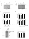
PAK4 somatic mutation E329K enhances PC3 cell motility. (A) Domain structure of PAK4 and GST/mRFP-tagged PAK4 proteins used in overexpression experiments (Abbreviated as in Fig 2). (B) Mean migration speed ±s.e.m of cells transiently transfected with plasmids encoding either mRFP or mRFP-tagged PAK4 derivatives in response to HGF stimulation. n= ≥48 cells for each population (except cells expressing mRFP-kinase (n=30) and -PAK4K350, 351M (n=33)) over 3 separate experiments. Statistical significance was calculated using Student’s t-test, ***p<0.0001, **p<0.001, *p<0.05. (C) (left) Ribbon diagram of the kinase domain structure of human PAK4 (2X4Z) bound to PF-3758309 ATP analog (not shown) 22 generated using Rasmol. Glycine-rich loop region (G-loop) shown in yellow. (right top) Highlighted G-loop showing basic (Lysine 326) and acidic (Glutamic acid 329) side chains preceeding the G-loop motif (right bottom) Rotated view of G-loop showing electrostatic interactions and distance between K326 and E329 (D, E and F) GST, GST-PAK4 or GST-PAK4E329K were used to pulldown co-overexpressed HA-Cdc42G12V, HA-Gab-1 or Myc-LIMK1 from 293 cell lysates. Blots are representative of 3 separate experiments.
Figure 4
Figure 4
E329K is an active kinase. Kinase assays were performed as previously described (43) (A -D) GST-PAK4 and GST-PAK4E329K kinase activity was assayed without inhibitor (Lanes 3 and 6) or with either Purvalanol A ((A): 0.5μM, lanes 4 and 7; 10μM, lanes 5 and 8) or PAK4i ((C): 1μM, lanes 4 and 7; 5μM, lanes 5 and 8). For inhibitor experiments, 0.5μM or 10μM Purvalanol A or 1μM or 5μM PAK4i was added to the kinase buffer. The reaction was stopped by adding SDS-page loading buffer. Autoradiographs and western blots were quantified using Andor IQ software (Belfast, UK) and the level of phosphorylation normalised to GST-protein levels. Phosphorylation of histone and autophosphorylation are indicated by arrows. Retained purified PAK4 proteins were subjected to GST immunoblotting to monitor expression levels (input). (A and C) PAK4 autophosphorylation and histone phosphorylation relative to wildtype PAK4 without Purvalanol A ±s.e.m. (B) or without PAK4i (D) ±s.e.m from 5 and 4 separate experiments, respectively. Statistical significance was calculated using student t-test, *p<0.05.

Similar articles

Cited by

References

    1. Cecchi F, Rabe DC, Bottaro DP. Targeting the HGF/Met signalling pathway in cancer. Eur J Cancer. 2010;46:1260–1270. - PMC - PubMed
    1. Knudsen BS, Woude GV. Showering c-MET-dependent cancers with drugs. Curr opin Genetics Dev. 2008;18:87–96. - PubMed
    1. Lai AZ, Abella JV, Park M. Crosstalk in Met receptor oncogenesis. Trends Cell Biol. 2009;19:542–551. - PubMed
    1. Paliouras GN, Naujokas MA, Park M. Pak4, a novel Gab1 binding partner, modulates cell migration and invasion by the Met receptor. Mol Cell Biol. 2009;29:3018–3032. - PMC - PubMed
    1. Abo A, Qu J, Cammarano MS, Dan C, Fritsch A, Baud V, et al. PAK4, a novel effector for Cdc42Hs, is implicated in the reorganization of the actin cytoskeleton and in the formation of filopodia. Embo J. 1998;17:6527–6540. - PMC - PubMed

Publication types

MeSH terms