. 2012 Aug 2;24(29):3981-7.
doi: 10.1002/adma.201200776. Epub 2012 Jun 12.

Multivalent porous silicon nanoparticles enhance the immune activation potency of agonistic CD40 antibody

Affiliations

Multivalent porous silicon nanoparticles enhance the immune activation potency of agonistic CD40 antibody

Luo Gu et al. Adv Mater. .

Abstract

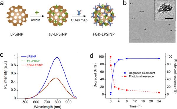
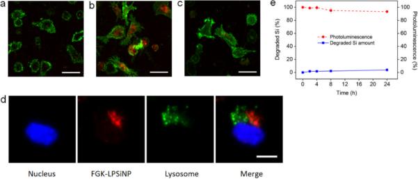
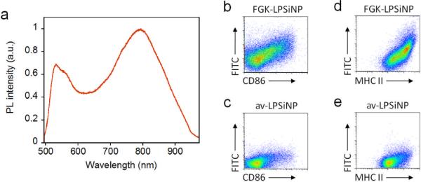
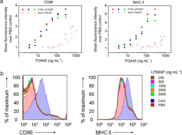
One of the fundamental paradigms in the use of nanoparticles to treat disease is to evade or suppress the immune system in order to minimize systemic side effects and deliver sufficient nanoparticle quantities to the intended tissues. However, the immune system is the body's most important and effective defense against diseases. It protects the host by identifying and eliminating foreign pathogens as well as self-malignancies. Here we report a nanoparticle engineered to work with the immune system, enhancing the intended activation of antigen presenting cells (APCs). We show that luminescent porous silicon nanoparticles (LPSiNPs), each containing multiple copies of an agonistic antibody (FGK45) to the APC receptor CD40, greatly enhance activation of B cells. The cellular response to the nanoparticle-based stimulators is equivalent to a 30-40 fold larger concentration of free FGK45. The intrinsic near-infrared photoluminescence of LPSiNPs is used to monitor degradation and track the nanoparticles inside APCs.


Figures

Figure 1
Figure 1. Preparation and characterization of FGK45 loaded luminescent porous silicon nanoparticles (FGK-LPSiNP)
a) Schematic representation of the preparation of FGK-LPSiNP. LPSiNP was first coated with avidin by physisorption (av-LPSiNP). Biotinylated FGK45 was then conjugated to the nanoparticles through biotin-avidin binding to form FGK-LPSiNP. b) Transmission electron microscope image of FGKLPSiNP (inset shows the porous nanostructure of one of the nanoparticles). Scale bar is 1 μm (100 nm for the inset). c) Photoluminescence (PL) spectra of LPSiNP, av-LPSiNP and FGK-LPSiNP. PL was measured using UV excitation (λex= 370 nm). d) Appearance of dissolved silicon in solution (by ICP-OES) and decrease in photoluminescence intensity from a sample of FGK-LPSiNP (50 μg mL−1) incubated in PBS solution at 37°C as a function of time.
Figure 2
Figure 2. Dendritic cell uptake of FGK-LPSiNPs
Fluorescence microscope images of mouse bone marrow-derived dendritic cells (BMDC) incubated with a) LPSiNPs or b) FGK-LPSiNPs for 1.5 h at 37 °C. c) free FGK45 inhibits uptake of FGK-LPSiNPs. BMDC were blocked with free FGK45 for 30 min and then incubated with FGK-LPSiNPs for 1.5 h at 37 °C. BMDC were detected by staining with Alexa Fluor 488 conjugated CD11c antibody (green). FGK-LPSiNPs were detected by their intrinsic visible/near-infrared photoluminescence (red, λex= 405 nm and λem= 700 ± 50 nm). The scale bars are 40 μm. d) FGK-LPSiNPs distribution in BMDC. BMDC were incubated with FGK-LPSiNP for 1.5 h at 37 °C. The lysosomes (green) of the cells were stained with LysoTracker (Invitrogen). Blue and red indicate the cell nucleus and FGK-LPSiNPs, respectively. The scale bar is 10 μm. e) Degradation of LPSiNPs (50 μg mL−1) in pH 4 buffer solution at 37 °C as a function of time.
Figure 3
Figure 3. Interaction of FGK-LPSiNPs with B cells
a) Photoluminescence spectrum of LPSiNPs coated with FITC-labeled avidin, showing the emission bands from both the FITC label (λmax ~ 520 nm) and porous silicon (λmax ~790 nm). b–e) Flow cytometry data quantifying the level of expression of the B cell activation markers CD86 (b, c) and MHC II (d, e) after incubation with 5 μg mL−1 of FGK-LPSiNPs (b, d) or av-LPSiNPs (c, e) for 42 h. The nanoparticles used in this experiment were coated with FITC-labeled avidin. The FITC signal from the cells is plotted against the expression level of CD86 (b, c) or MHC II (d, e) after stimulation. FGK-LPSiNPs used here contain 36 μg of FGK45 per milligram of nanoparticles. Note the quantity of FGK45 loaded is smaller when LPSiNPs are coated with FITC conjugated avidin compared with non-labeled native avidin.
Figure 4
Figure 4. Amplified activation potency of FGK-LPSiNPs compared to free FGK45
a) Flow cytometry analysis of the expression of B cell activation markers CD86 and MHC II, represented as the relative mean fluorescence intensity of the marker staining, after incubation with either FGK-LPSiNPs or free FGK45 for 42 h at 37 °C. The concentration of FGK45 on the FGK-LPSiNP constructs is reported based on the total loading of FGK45 on the nanoparticles (58 μg of FGK45 per mg of nanoparticles). Data are from independent experiments. b) Flow cytometry histograms of B cell activation markers CD86 and MHC II after incubation with various concentrations of LPSiNPs for 42 h at 37 °C. PBS (red shaded) and CpG (blue shaded) were used as negative and positive controls, respectively.

Similar articles

Cited by

References

    1. Park J-H, Gu L, Maltzahn G. v., Ruoslahti E, Bhatia SN, Sailor MJ. Nature Mater. 2009;8:331. - PMC - PubMed
    1. Tasciotti E, Liu XW, Bhavane R, Plant K, Leonard AD, Price BK, Cheng MMC, Decuzzi P, Tour JM, Robertson F, Ferrari M. Nat. Nanotechnol. 2008;3:151. - PubMed
    1. Salonen J, Laitinen L, Kaukonen AM, Tuura J, Bjorkqvist M, Heikkila T, Vaha-Heikkila K, Hirvonen J, Lehto VP. J. Control. Release. 2005;108:362. - PubMed
    1. Low SP, Voelcker NH, Canham LT, Willams KA. Biomaterials. 2009;30:2873. - PubMed
    1. Xiao L, Gu L, Howell SB, Sailor MJ. ACS Nano. 2011;5:3651. - PMC - PubMed

Publication types