

Targeting of Rac GTPases blocks the spread of intact human breast cancer
- PMID: 22689141
- PMCID: PMC3442288
- DOI: 10.18632/oncotarget.520
Targeting of Rac GTPases blocks the spread of intact human breast cancer
Abstract
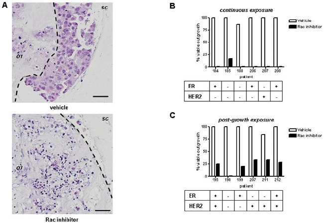
High expression of Rac small GTPases in invasive breast ductal carcinoma is associated with poor prognosis, but its therapeutic value in human cancers is not clear. The aim of the current study was to determine the response of human primary breast cancers to Rac-based drug treatments ex vivo. Three-dimensional organotypic cultures were used to assess candidate therapeutic avenues in invasive breast cancers. Uniquely, in these primary cultures, the tumour is not disaggregated, with both epithelial and mesenchymal components maintained within a 3-dimensional matrix of type I collagen. EHT 1864, a small molecule inhibitor of Rac GTPases, prevents spread of breast cancers in this setting, and also reduces proliferation at the invading edge. Rac1+ epithelial cells in breast tumours also contain high levels of the phosphorylated form of the transcription factor STAT3. The small molecule Stattic inhibits activation of STAT3 and induces effects similar to those seen with EHT 1864. Pan-Rac inhibition of proliferation precedes down-regulation of STAT3 activity, defining it as the last step in Rac activation during human breast cancer invasion. Our data highlights the potential use of Rac and STAT3 inhibition in treatment of invasive human breast cancer and the benefit of studying novel cancer treatments using 3-dimensional primary tumour tissue explant cultures.
Conflict of interest statement
All authors declare no conflicting interests.
Figures


Similar articles
-
Inhibition of the Rho GTPase, Rac1, decreases estrogen receptor levels and is a novel therapeutic strategy in breast cancer.Endocr Relat Cancer. 2011 Feb 23;18(2):207-19. doi: 10.1677/ERC-10-0049. Print 2011 Apr. Endocr Relat Cancer. 2011. PMID: 21118977 Free PMC article.
-
Specificity and mechanism of action of EHT 1864, a novel small molecule inhibitor of Rac family small GTPases.J Biol Chem. 2007 Dec 7;282(49):35666-78. doi: 10.1074/jbc.M703571200. Epub 2007 Oct 11. J Biol Chem. 2007. PMID: 17932039
-
Characterization of EHT 1864, a novel small molecule inhibitor of Rac family small GTPases.Methods Enzymol. 2008;439:111-29. doi: 10.1016/S0076-6879(07)00409-0. Methods Enzymol. 2008. PMID: 18374160
-
Characterization of a Dual Rac/Cdc42 Inhibitor MBQ-167 in Metastatic Cancer.Mol Cancer Ther. 2017 May;16(5):805-818. doi: 10.1158/1535-7163.MCT-16-0442. Mol Cancer Ther. 2017. PMID: 28450422 Free PMC article.
-
Rac1 and Rac3 isoform activation is involved in the invasive and metastatic phenotype of human breast cancer cells.Breast Cancer Res. 2005;7(6):R965-74. doi: 10.1186/bcr1329. Epub 2005 Sep 30. Breast Cancer Res. 2005. PMID: 16280046 Free PMC article.
Cited by
-
Identification of p130Cas/ErbB2-dependent invasive signatures in transformed mammary epithelial cells.Cell Cycle. 2013 Aug 1;12(15):2409-22. doi: 10.4161/cc.25415. Epub 2013 Jun 28. Cell Cycle. 2013. PMID: 23839042 Free PMC article.
-
RHOA Therapeutic Targeting in Hematological Cancers.Cells. 2023 Jan 28;12(3):433. doi: 10.3390/cells12030433. Cells. 2023. PMID: 36766776 Free PMC article. Review.
-
CD147 promotes Src-dependent activation of Rac1 signaling through STAT3/DOCK8 during the motility of hepatocellular carcinoma cells.Oncotarget. 2015 Jan 1;6(1):243-57. doi: 10.18632/oncotarget.2801. Oncotarget. 2015. PMID: 25428919 Free PMC article.
-
The crosstalk between breast carcinoma-associated fibroblasts and cancer cells promotes RhoA-dependent invasion via IGF-1 and PAI-1.Oncotarget. 2017 Dec 28;9(12):10375-10387. doi: 10.18632/oncotarget.23735. eCollection 2018 Feb 13. Oncotarget. 2017. PMID: 29535813 Free PMC article.
-
A new role of the Rac-GAP β2-chimaerin in cell adhesion reveals opposite functions in breast cancer initiation and tumor progression.Oncotarget. 2016 May 10;7(19):28301-19. doi: 10.18632/oncotarget.8597. Oncotarget. 2016. PMID: 27058424 Free PMC article.
References
-
- Gillet JP, Calcagno AM, Varma S, Marino M, Green LJ, Vora MI, Patel C, Orina JN, Eliseeva TA, Singal V, Padmanabhan R, Davidson B, Ganapathi R, Sood AK, Rueda BR, Ambudkar SV, et al. Redefining the relevance of established cancer cell lines to the study of mechanisms of clinical anti-cancer drug resistance. Proceedings of the National Academy of Sciences of the United States of America. 2011;108(46):18708–18708. - PMC - PubMed
-
- Leeper AD, Farrell J, Williams LJ, S. TJ, Dixon JM, Wedden SE, Harrison DJ, Katz E. Determining tamoxifen sensitivity using primary breast cancer tissue in collagen-based three-dimensional culture. Biomaterials. 2012;33(3):907–907. - PubMed
Publication types
MeSH terms
Substances
Grants and funding
LinkOut - more resources
Full Text Sources
Medical
Molecular Biology Databases
Research Materials
Miscellaneous
